SUMMARY
BACKGROUND AND OBJECTIVES:
Out-of-hos-pital cardiac arrest is a major cause of death with survival rates as low as 5% to 35%. A large number of patients who survive resuscitation will face sig-nificant neurological damage, as a result of the ischemia that occurs both during cardiac arrest and reperfusion. However understanding of the mecha-nisms responsible for brain damage has not resulted in prognostic improvement. Therapeutic hypother-mia after resuscitation may be a valid option associ-ated to reduction of neurological damage. The pur-pose of this study was to review scientific evidence related to a therapy for patients resuscitated from cardiac arrest.
CONTENTS: Description and analysis of the main risk factors associated with neurological damage after re-suscitation from cardiac arrest as well as prognostic criteria was carried out. A non-systematic search was conducted in the PubMed data base for papers on a therapeutic approach for patients resuscitated from cardiac arrest. Bibliographic references of reviewed papers were also analyzed. Practical rules were drafted for such an approach.
Care of Patient Resuscitated
from Cardiac Arrest*
Abordagem do Paciente Reanimado, Pós-Parada Cardiorrespiratória
João Carlos Ramos Gonçalves Pereira1
1. Hospital Assistant for Internal Medicine Sub-Specialist in Intensive Care of the Medical Intensive Care Unit, Medical Services III
*Received from the Hospital de São Francisco Xavier, Centro Hospi-talar de Lisboa Ocidental, Lisboa, Portugal
Submitted on January 29, 2008 Accepted for publication on May 2, 2008
Address for correspondence: João Gonçalves Pereira, M.D. Rua dos Soeiros, 307, 8º Andar 1500-580 Lisboa, Portugal Phone: 00-351-962441546 E-mail: [email protected]
©Associação de Medicina Intensiva Brasileira, 2008
CONCLUSIONS: Patients resuscitated from cardiac
arrest face a high level of risk of neurological damage. Therapeutic hypothermia and control of physiological parameters to optimise brain perfusion, may improve prognosis.
Key Words: Cardiac arrest, Hypothermia, Postanoxic brain damage
RESUMO
JUSTIFICATIVA E OBJETIVOS: A parada
cardiorres-piratória (PCR) ocorrida em ambulatório tem elevada mortalidade, sendo a sobrevida entre 5% e 35%. Dos pacientes que são reanimados uma percentagem ele-vada i ca com déi cits neurológicos, resultantes das lesões ocorridas, tanto no período de ausência de cir-culação ou durante a reperfusão. No entanto a com-preensão dos mecanismos da lesão cerebral não tem traduzido na melhoria do prognóstico. A hipotermia terapêutica após a reanimação parece ser uma opção válida associada à diminuição destas seqüelas neuro-lógicas. O objetivo deste estudo foi rever a evidência cientíi ca relativa à abordagem do paciente reanimado após PCR.
CONTEÚDO: Descrição e abordagem dos principais
fatores de risco associados à lesão neurológica após PCR, bem como dos seus critérios de prognóstico. Feita pesquisa não sistemática na base de dados Pub-Med dos artigos referentes à abordagem terapêutica dos pacientes reanimados de parada cardíaca. As re-ferências bibliográi cas dos artigos de revisão foram igualmente analisadas. Elaboradas normas práticas para essa abordagem.
CONCLUSÕES: Os pacientes que sobrevivem à PCR
têm elevado risco de i car com lesões neurológicas graves. A hipotermia terapêutica e o controle das variá-veis i siológicas, com otimização da perfusão cerebral, podem melhorar o seu prognóstico.
INTRODUCTION
Cardiac arrest (CA), notwithstanding the underlying causes has high morbidity and mortality rates. Survival is less than 40% if it takes place in-hospital and lower than 10% if it takes place out-of-hospital, a percentage that has remained unchanged during the last years1.
Many of the patients who survive remain with neurolo-gical damage2.
During CA, absence of circulation causes brain hypo-perfusion, especially of the sub-cortical areas and of the threshold territories between cerebral arteries that, due to lower perfusion, are more subject to ischemia (hemodynamic infarctions). Areas with former ischemic injury are especially affected.
After cardiac resuscitation, reperfusion also contributes to ischemia and cerebral edema3, activating
biochemi-cal cascades responsible for migration of intracellular calcium, production as well as local release of free oxygen radicals and excitatory amino acids, (notably glutamate), mechanisms that contribute for apoptosis. Likewise local production of lactate and thrombosis of the micro-circulation increase the risk of ischemia3.
These phenomena last for about 48-72 hours after re-covery of the cardiac rhythm and of circulation4.
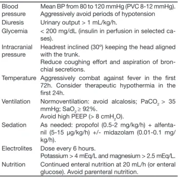
Identii cation of these physiopathological processes has contributed to the development of treatments that, with the possible exception of hypothermia, have not shown to be benei cial (Table 1).
Table 1 – Care of Patients in the First 48 hours after Cardiac Arrest Blood
pressure
Mean BP from 80 to 120 mmHg (PVC 8-12 mmHg). Aggressively avoid periods of hypotension Diuresis Urinary output > 1 mL/kg/h.
Glycemia < 200 mg/dL (insulin in perfusion in selected ca-ses).
Intracranial pressure
Headrest inclined (30º) keeping the head aligned with the trunk.
Reduce coughing effort and aspiration of bron-chial secretions.
Temperature Aggressively combat against fever in the i rst 72h. Consider therapeutic hypothermia in the i rst 24h.
Ventilation Normoventilation: avoid alcalosis; PaCO2 > 35 mmHg; SaO2 ≥ 92%.
Avoid high PEEP (> 8 cmH2O).
Sedation As needed: propofol (0.5-2 mg/kg/h) + alfenta-nil (5-15 μg/kg/h) +/- midazolam (0.01-0.1 mg/ kg/h).
Electrolites Dose every 6 hours.
Potassium > 4 mEq/L and magnesium > 2.5 mEq/L. Nutrition Continued enteral nutrition at 20 mL/h (or enteral
glucose). Avoid parenteral nutrition.
OVERALL MEASURES
Treatment of post-cardiac arrest aims to preserve orga-nic functions (especially the brain), avoiding perfusion pressure on various vascular territories. This strategy complements the diagnostic and therapeutic approach to the cause of CA and potential complications, par-ticularly eventual i brinolisys, coronary intervention or conversion of cardiac dysrhythmias.
The initial approach must include an electrocardiogram (to identify the cause of CA and of intercurrent dys-rhythmia), chest X-ray (for exclusion of iatrogenies as-sociated with resuscitation maneuvers such as pneu-mothorax and rib fractures) and blood gas analysis (with dosing of electrolytes and lactic acid).
Blood Pressure
In the healthy individual, cerebral perfusion pressure (CPP) is independent from systemic blood pressure (SBP). This is the function of brain self-regulation with adjustment of its vascular tonus to the systemic pres-sure variations. This capacity is altered by lack of cir-culation5.
After CA there is a period of brain hyperthermia that lasts about 15-30 minutes followed by vasodilation and consequent decrease of CPP. Under these conditions any decrease of BP may cause hypoperfusion and di-ffuse cerebral ischemia5.
Indeed, in the absence of central self-regulation the CPP is equal to the difference between systemic BP and intracranial pressure (ICP), the latter normally ran-ging between 5 and 20 mmHg. Because the required CPP is 60 mmHg6,7, the mean BP must be above 80
mmHg (especially in the i rst 72 hours after cardiac ar-rest), to maintain an adequate brain perfusion. This is assured by early administration of volume and vaso-pressor amines, as well as by conversion of dysrhyth-mias to avoid prolonged periods of hypotension8.
After CA, another common hemodynamic phenome-non is myocardial depression9, even when there is no
acute or chronic coronary disease. This disease is a consequence of cardiac hypoperfusion and of resusci-tation maneuvers (above all that of electric cardiover-sion), normally reverting in 24 hours. Dobutamine and combined insulin, glucose and potassium therapy10
may contribute to minimize this phenomenon.
It is not clear that arterial hypotension, even when se-vere, contributes to brain injury6 Nevertheless, since it
Intracranial Pressure
After a cardiac arrest, even a temporary increase of ICP may contribute to brain injury. ICP increases with obstruction of blood drainage along the internal jugular veins and catheterization of these veins must be avoi-ded or cervical rotation maintained. Ideally, the head must remain aligned with the trunk and the headrest inclined at a 30º.
Because sedoanalgesics decrease sympathetic res-ponse and because neuromuscular blockers reduce the coughing rel ex and the respiratory effort, they may mi-nimize elevation of thoracic pressure related to aspira-tion of bronchial secreaspira-tion, with the alveolar recruitment maneuvers11 and with high PEEP (above 8 cmH
2O).
In this framework of cardiac arrest, invasive monito-ring of ICP does not seem clinically advisable, as the clearly pathological values (above 20 mmHg) which as such would have a therapeutic indication) reveal diffu-se cytotoxic brain edema, with irreversible dysfunction, whose treatment is pointless12.
Blood Glucose
Glycemia indices are often high in patients admitted at the ICU. This alteration is multifactorial, probably rela-ted to endocrine response to stress13.After CA
hypo-glycemia, at in-hospital admission as well as during the i rst 24 hours, worsens independently of prognosis8,14.
It remains unclear if this alteration contributes to the neurological injury or if it is in itself an indicator of se-verity. Nevertheless, experimentally, increase of glyce-mia eases accumulation of lactate in the brain tissues during hypoxia13 and this alteration may contribute to
brain injury.
In a clinical trial carried out in a single center, in an ICU with surgical patients15 control of capillary glycemia
with a cut off level of 110 mg/dL, reduced mortality as well as incidence of neuropathy. This result however was not reproduced in other studies.
Although control of capillary glycemia seems to be as-sociated to a better prognosis of the critical patients (especially if not diabetic)15, the ideal cut off level
re-mains unclear. There seems to be sufi cient evidence to recommend maintaining glycemia below 200 mg/dL, (ideally at a lower level)13.The recommended value also
depends upon experience in the ICU and its monitoring capacity, because aggressive use of insulin to maintain low levels of glycemia is associated with a higher risk of hypoglycemia and neuroglycopenia that may, them-selves worsen neurological injury.
In general, glucose solutions (especially in the i rst 24
hours after admission) should be avoided as well as parenteral nutrition, considering the possibility of low doses of enteral nutrition in hemodynamically stable patients.
Temperature
Cerebral temperature normally is about 0.5º C above systemic temperature. In the injured brain this differen-ce is signii cantly higher and may reach up to 3º C. In acute cerebral ischemia there is also a regional increa-se of temperature, asymmetric to the rest of the brain mass16.
Animal studies have shown that increment of cerebral temperature is accompanied by neurological deterio-ration and that its controlled decrease reduces these injuries.
Indeed in mice submitted to asphyxia (where sponta-neous hypothermia is noted), forced rewarming incre-ases mortality and histological injuries in the various brain areas during the i rst 72 hours17. In dogs
submit-ted to experimental ventricular i brillation (20 minutes period of absence of circulation), induction of hypo-thermia reduces the observed neurological dei cits, as well as encephalic histological alterations18.
On the other hand, also in the experimental ischemic BVA, cerebral cooling decreased the volume of the in-fracted region16.
After CA, increase of the systemic temperature can be noted and consequent cerebral hyperthermia. Upon ad-mission or in the i rst 24 hours, such an event worsens prognosis, although it remains unclear if this change is an indicator of severity of neurological disease or the determinant of clinical worsening16,19.
Thus, in the presence of acute brain injury, diffuse or not, after CA or after any other etiology, fever must be aggressively avoided20 in the i rst 72 hours of
evo-lution.
Recently two studies, an European and an Australian, showed independently that therapeutic hypothermia after CA that occurred in an out-of-hospital facility sig-nii cantly reduced neurological damage and one of the studies found a signii cant decrease in mortality3,21.
55%) p = 0.012, RR of 0.74 and NNT of 721.
In the Australian study there was also a more frequent neurological recovery in the group of patients submit-ted to hypothermia (49% versus 26%, RR de 1.85 with NNT of 4)3.
In both studies inclusion criteria were very restricted. In the i rst, about 90% of the evaluated patients were excluded; therefore it remained doubtful if the benei t of this therapeutic strategy was not overestimated.
Therapeutic hypothermia consists of reducing the central temperature to 32º C - 33º C, to interrupt the physiopathological cascade responsible for the neuro-logical reperfusion injury. It must be maintained for 12 hours and seemingly it is benei cial to prolong up to 24 hours. It must be considered for all patients in whom there is indication for active treatment. This is regard-less of the heart rate at the moment of the CA (IIa indi-cation of the ILCOR for CA post ventricular i brillation occurred in out-of-hospital environment and indication IIb for CA at any rhythm, and any place) whenever there is no contraindication22 (Table 2).
Table 2 – Contraindication for Therapeutic Hypothermia
Time of sustained hypotension (Systolic BP < 80 mmHg or mean AP < 45 mmHg) over 30 min after resuscitation
Time of medically non-assisted CA more than 10 minutes. Resuscitation during more than 45 min.
Time after CA longer than 12h CA secondary to trauma
Primary coagulopahy (but no oral anticoagulation)
CA acknowledged as secondary to aortic dissection, intracra-nial hemorrhage or massive hemorrhage
Terminal disease or indication of no resuscitation
Cooling can be internal or external, that is to say, with infusion of iced solution, ice packs, ventilators, thermal blankets or plaques as well as appropriate intravascu-lar catheters19,23.Extracorporeal circulation can also be
used with external cooling of the blood.
To increase tolerance to cold and reduce production of heat (by tremors and shivers) sedoanalgesia may be used, magnesium sulphate in perfusion24 and
neuro-muscular blockers, preferably administered in an inter-mittent form.
Cooling must be early and aggressive to rapidly decre-ase central temperature avoiding periods of hyperther-mia, and later can be slower (about 1º C/h). Rewarming must always be slow and passive (not above 0.5º C/h) to prevent worsening of the injury and brain edema associated to hypothermia rebound, frequent in such circumstances17.
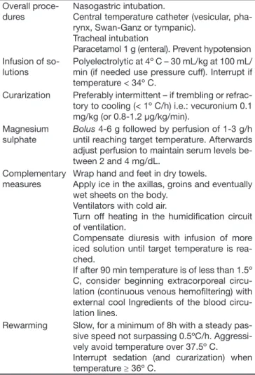
Infection, heart rate instability (especially bradydys-rhythmias), coagulation, pressure sores and cold burns as well as hyperglycemia and hypomagnesemia are potential complications of hypothermia19 (Table 3).
Table 3 – Therapeutic Hypothermia Overall
proce-dures
Nasogastric intubation.
Central temperature catheter (vesicular, pha-rynx, Swan-Ganz or tympanic).
Tracheal intubation
Paracetamol 1 g (enteral). Prevent hypotension Infusion of
so-lutions
Polyelectrolytic at 4º C – 30 mL/kg at 100 mL/ min (if needed use pressure cuff). Interrupt if temperature < 34º C.
Curarization Preferably intermittent – if trembling or refrac-tory to cooling (< 1º C/h) i.e.: vecuronium 0.1 mg/kg (or 0.8-1.2 μg/kg/min).
Magnesium sulphate
Bolus 4-6 g followed by perfusion of 1-3 g/h until reaching target temperature. Afterwards adjust perfusion to maintain serum levels be-tween 2 and 4 mg/dL.
Complementary measures
Wrap hand and feet in dry towels.
Apply ice in the axillas, groins and eventually wet sheets on the body.
Ventilators with cold air.
Turn off heating in the humidii cation circuit of ventilation.
Compensate diuresis with infusion of more iced solution until target temperature is rea-ched.
If after 90 min temperature is of less than 1.5º C, consider beginning extracorporeal circu-lation (continuous venous hemoi ltering) with external cool Ingredients of the blood circu-lation lines.
Rewarming Slow, for a minimum of 8h with a steady pas-sive speed not surpassing 0.5ºC/h. Aggressi-vely avoid temperature over 37.5º C.
Interrupt sedation (and curarization) when temperature ≥ 36º C.
Indicated for patients with cardiocirculatory recovery maintained during at least 5 min with the Glasgow coma score ≤ 9 or abnormal agitation (attributed to neurological function). In cardiac arrest in the presence of physicians consi-der the possibility of waiting for 60 min to evaluate eventual early neurological recovery.
Ventilation
Mechanical ventilation after CA must be adjusted to the patient’s clinical condition and to his gas exchange for prevention of hypoxemia and maintenance of normo-capnia.
Hypoxemia may worsen prognosis especially due to a higher risk of a second episode of CA20.Therefore, PaO
2
must remain above 65 mmHg and the SaO2 higher than 92%. Because hypothermia increases oxygen afi nity to hemoglobin during this procedure, a higher mini-mum saturation is required.
respi-ratory alkalosis must be avoided as they can trigger brain vasoconstriction and consequent decrease of global perfusion and diffuse ischemia25,26. Indeed,
pa-radoxically hyperventilation sometimes used to reduce cerebral edema may induce worsening of the clinical condition.
PHARMACOLOGICAL SUPPORT
Sedation
Post CA sedation facilitates patient adjustment to the ventilatory prostheses and/or realization of therapeutic maneuvers, especially hypothermia. Benzodiazepinics or propofol may be used, preferentially according to se-dation scales, to avoid a cumulative effect27. Utilization
of short life drugs allows for neurological intermittent evaluation.
Opioids are elected for control of the automatic res-piratory stimuli, normally sustained after a CA, which interferes with mechanical ventilation and contributes to hypercapnia and alkalosis, often found in patients with central neurological injuries. Further, they are more effective for prevention of muscle tremor associated with hypothermia.
There is no evidence that maintenance of this sedoa-nalgesia for a pre-established period of time inl uences neurological preservation, therefore it should be inter-rupted, if not needed.
Electrolytes
Electrolytic disorders are common after CA, due to lack of circulation and of resuscitation maneuvers, including administration of solutions and adrenalin28.
Decrease of the potassium concentrations, also ag-gravated by intracellular migration of this cation du-ring hypothermia, is associated to increased dys-rhythmias and its concentration should remain at 4 and 4.5 mEq/L.
Similarly, hypomagnesiemia worsens neurological prognosis29. Its infusion facilitates therapeutic
hypo-thermia24 and decreases incidence of dysrhythmias.
Anticoagulation
Utilization of thrombolytic therapy during a refractory CA increases the number of patients with sustained he-modynamic recovery30.
This, together with evidence of the pro-thrombotic conditions after resuscitations infers that anticoagula-tion may have therapeutic benei ts as it reduces risk of thrombus. Indeed, after cardiac resuscitation there is a
greater pro-thrombotic activity and overall decrease of anticoagulant factors (antithrombine II, protein C and S). These alterations are pronounced in patients who die in the i rst two days31.
Anticoagulation may contribute to decrease the risk of another CA, more so after myocardial infraction or pul-monary embolism20.
However, this theoretical benei t was not documented in in vivo clinical studies.
Anticonvulsants
After CA, convulsions and myoclonias are frequent and found in about 30% of the patients. They do not have a signii cant prognostic worsening when the event is isolated. On the contrary, the state of epileptic illness alone worsens neurological injury and must be aggres-sively managed (phenyltoin, phenobarbital and seda-tives)32 with eventual continued electroencephalic
mo-nitoring. Levatiracetam and sodium valproate do not have a signii cant action on the stage of the illness, but can be used as chronic maintenance therapy after cli-nical stabilization.
This stage of epileptic illness may take place without motor manifestation (non-convulsive stage of disease) however, in view of an unknown cause of persistent coma; an ECG must be made33.
Conversely, the stage of myoclonic disease describes extensive brain injury, usually irreversible34-36. The
the-rapeutic approach includes sodium valproate and clo-nazepan, although these drugs to not seem to inl uen-ce clinical evolution.
This is different from the Lance Adams syndrome whe-re thewhe-re awhe-re generalized myoclonies, together with pwhe-re- pre-servation of vigility and level of consciousness. Use of pyracetam may improve such manifestations.
Evaluation of the Prognosis
Early assessment of the neurological prognosis is fun-damental for stratii cation of the therapeutic interven-tion, especially to identify patients that do not benei t from intensive care.
Form the clinical point of view, in the non-sedated pa-tient, absence of papillary rel ex and response to pain on the 3rd day of evolution after CA, is an independent
factor of poor prognosis, with a specii city of over 95%35.
Al-though the auditory evoked potentials do not increase this specii city, they are useful to coni rm the integrity of the conduction paths of the cerebral trunk (essential for assessment of SSEP)37.
The pattern of burst-suppression at ECG, although less specii c, also portrays very severe brain injury34.
Its prognostic usefulness increases with time after CA, that is why this exam must not be performed before the 3rd day38.
Enolase (Neuron-Specii c Enolase) dosed in blood or in cephalorachidian l uid correlates to cerebral injury and to the prognosis. However, thresholds for clinical as-sessment are not yet dei ned39,40.
Final Considerations
Therapeutic interventions intended to preserver life and organic functions after CA improve prognosis, but at the same time increase the survival of patients with neurological injuries and a cognitive commitment with severe sequels.
The option to implement extraordinary measures for life support in these circumstances therefore has social implications and must be discussed in view of the le-gislation in force, of the sensibility of society and of the medical community, as well as that of the families invol-ved. This will help to decide which therapeutic options are to be carried out or omitted, especially in extreme cases.
CONCLUSION
Cardiac arrest is a dramatic event with a high mortality. In patients that survive, the period of absence of circu-lation and the reperfusion injuries may lead to severe neurological damage.
Although there are no efi cient pharmacological thera-pies for such a situation, minimizing risk factors (tension, hyper or hypoglycemia, hypoxemia or hypo-capnia, hyperthermia, electrolytic disorder), optimizing of CA and therapeutic hypothermia may improve prog-nosis.
Acronyms
CA - Cardiac arrest
CPP – Cerebral perfusion pressure BP – Blood pressure
ICP – Intracranial pressure
PEEP – Positive end expiratory pressure ICU - Intensive care unit
BVA - Brain vascular accident
ILCOR – International Liaison Committee on Resusci-tation
PaO2 – Partial arterial pressure of oxygen SaO2 – Hemoglobin arterial saturation PaCO2 – Carbon dioxide arterial pressure EEG – Electroencephalogram
SSEP - Somatosensitive evoked potentials
REFERENCES
01. Rea TD, Eisenberg MS, Becker LJ, et al. Temporal trends in sudden
car-diac arrest: a 25-year emergency medical services perspective. Circula-tion, 2003;107:2780-2785.
02. Fischer M, Fischer NJ, Schuttler J - One-year survival after
out-of-hospi-tal cardiac arrest in Bonn city: outcome report according to the ‘Utstein style’. Resuscitation, 1997;33:233-243.
03. Bernard SA, Buist M - Induced hypothermia in critical care medicine: a
review. Crit Care Med, 2003;31:2041-2051.
04. Siesjo BK - Mechanisms of ischemic brain damage. Crit Care Med,
1988;16:954-963.
05. Buunk G, van der Hoeven JG, Meinders AE - Cerebrovascular
reac-tivity in comatose patients resuscitated from a cardiac arrest. Stroke, 1997;28:1569-1573.
06. Mullner M, Sterz F, Binder M, et al. Arterial blood pressure after human
cardiac arrest and neurological recovery. Stroke, 1996;27:59-62.
07. Langhelle A, Tyvold SS, Lexow K, et al. In-hospital factors associated
with improved outcome after out-of-hospital cardiac arrest. A compari-son between four regions in Norway. Resuscitation, 2003;56:247-263.
08. Skrifvars MB, Pettila V, Rosenberg PH, et al. A multiple logistic
regres-sion analysis of in-hospital factors related to survival at six months in patients resuscitated from out-of-hospital ventricular i brillation. Resus-citation, 2003;59:319-328.
09. Laurent I, Monchi M, Chiche JD, et al. Reversible myocardial
dysfunc-tion in survivors of out-of-hospital cardiac arrest. J Am Coll Cardiol, 2002;40:2110-2116.
10. Angelos MG, Murray HN, Gorsline RT, et al. Glucose, insulin and
potas-sium (GIK) during reperfusion mediates improved myocardial bioener-getics. Resuscitation, 2002;55:329-336.
11. Bein T, Kuhr LP, Bele S, et al. Lung recruitment maneuver in patients with
cerebral injury: effects on intracranial pressure and cerebral metabolism. Intensive Care Med, 2002;28:554-558.
12. Gueugniaud PY, Garcia-Darennes F, Gaussorgues P, et al. Prognostic
signii cance of early intracranial and cerebral perfusion pressures in post-cardiac arrest anoxic coma. Intensive Care Med, 1991;17:392-398.
13. Marik PE, Raghavan M - Stress-hyperglycemia, insulin and
immunomod-ulation in sepsis. Intensive Care Med, 2004;30:748-756.
14. Mullner M, Sterz F, Binder M, et al. Blood glucose concentration after
cardiopulmonary resuscitation inl uences functional neurological re-covery in human cardiac arrest survivors. J Cereb Blood Flow Metab, 1997;17:430-436.
15. van den Berghe G, Wouters P, Weekers F, et al. Intensive insulin therapy
in the critically ill patients. New Engl J Med, 2001;345:1359-1367.
16. Polderman KH - Application of therapeutic hypothermia in the ICU:
op-portunities and pitfalls of a promising treatment modality. Part 1: Indica-tions and evidence. Intensive Care Med, 2004;30:556-575.
17. Hickey RW, Ferimer H, Alexander HL et al - Delayed, spontaneous
hypo-thermia reduces neuronal damage after asphyxial cardiac arrest in rats. Crit Care Med, 2000;28:3511-3516.
18. Sterz F, Safar P, Tisherman S, et al. Mild hypothermic cardiopulmonary
resuscitation improves outcome after prolonged cardiac arrest in dogs. Crit Care Med, 1991;19:379-389.
19. Polderman KH - Application of therapeutic hypothermia in the
inten-sive care unit. Opportunities and pitfalls of a promising treatment mo-dality. Part 2: Practical aspects and side effects. Intensive Care Med, 2004;30:757-769.
20. Nolan JP, Deakin CD, Soar J, et al. European Resuscitation Council
21. Mild therapeutic hypothermia to improve the neurologic outcome after cardiac arrest. N Engl J Med, 2002;346:549-556.
22. Nolan JP, Morley PT, Hoek TL, et al. Therapeutic hypothermia after
cardi-ac arrest. An advisory statement by the Advancement Life support Task Force of the International Liaison committee on Resuscitation. Resusci-tation, 2003;57:231-235.
23. Green RS, Howes DW - Stock your emergency department with ice
packs: a practical guide to therapeutic hypothermia for survivors of car-diac arrest. CMAJ, 2007;176:759-762.
24. Zweil er RM, Voorhees ME, Mahmood MA, et al. Magnesium sulfate in-creases the rate of hypothermia via surface cooling and improves com-fort. Stroke, 2004;35:2331-2334.
25. Laffey JG, Kavanagh BP - Hypocapnia. N Engl J Med, 2002;347:43-53.
26. Menon DK, Coles JP, Gupta AK, et al. Diffusion limited oxygen delivery
following head injury. Crit Care Med, 2004;32:1384-1390.
27. Sessler CN - Sedation scales in the ICU. Chest, 2004;126:1727-1730.
28. Buylaert WA, Calle PA, Houbrechts HN - Serum electrolyte
disturbanc-es in the post-rdisturbanc-esuscitation period. The Cerebral Rdisturbanc-esuscitation Study Group. Resuscitation, 1989;17:(Suppl17):S189-S206.
29. Meloni BP, Zhu H, Knuckey NW - Is magnesium neuroprotective
follow-ing global and focal cerebral ischaemia? A review of published studies. Magnes Res, 2006;19:123-137.
30. Bottiger BW, Bode C, Kern S, et al. Efi cacy and safety of thrombolytic
therapy after initially unsuccessful cardiopulmonary resuscitation: a pro-spective clinical trial. Lancet, 2001;357:1583-1585.
31. Adrie C, Monchi M, Laurent I, et al. Coagulopathy after successful
car-diopulmonary resuscitation following cardiac arrest: implication of the
protein C anticoagulant pathway. J Am Coll Cardiol, 2005;46:21-28. 32. Walker M - Status epilepticus: an evidence based guide. BMJ,
2005;331:673-677.
33. Benbadis SR, Tatum WO 4th - Prevalence of nonconvulsive status
epilep-ticus in comatose patients. Neurology, 2000;55:1421-1423.
34. Zandbergen EG, de Haan RJ, Stoutenbeek CP, et al. Systematic review
of early prediction of poor outcome in anoxic-ischaemic coma. Lancet, 1998;352:1808-1812.
35. Thomke F, Marx JJ, Sauer O, et al. Observations on comatose
survi-vors of cardiopulmonary resuscitation with generalized myoclonus. BMC Neurol, 2005;18:5:14.
36. Wijdicks EF, Parisi JE, Sharbrough FW - Prognostic value of myo-clonus status in comatose survivors of cardiac arrest. Ann Neurol, 1994;35:239-243.
37. Tiainen M, Kovala TT, Takkunen OS, et al. Somatosensory and brainstem
auditory evoked potentials in cardiac arrest patients treated with hypo-thermia. Crit Care Med, 2005;33:1736-1740.
38. Berek K, Jeschow M, Aichner F - The prognostication of cerebral hypoxia after out-of-hospital cardiac arrest in adults. Eur Neurol, 1997;37:135-145.
39. Rech TH, Vieira SR, Nagel F, et al. Serum neuron-specii c enolase as
ear-ly predictor of outcome after in-hospital cardiac arrest: a cohort study. Crit Care, 2006;10:R133.
40. Tiainen M, Roine RO, Pettila V, et al. Serum neuron-specii c enolase