structures by reproducing it in 10 knees of five cadavers. Results: This approach was seen to make it easy to locate the entry point, with lesions only occurring in the Hoffa fat. In three of our cases, there were lesions of the chondral surface, which is an obstacle that is difficult to overcome. Conclusion: There is a need to develop specific material to minimize in-jury to intra-articular structures when using this route.
Keywords –Tibial Fractures; Orthopedic Procedures; Frac-ture Fixation, Intramedullary
04 artigo 525
ORIGINAL ARTICLE
1 – Resident in Orthopedics and Traumatology, Institute of Orthopedics and Traumatology, HC-FMUSP, São Paulo, SP, Brazil.
2 – Associate Professor in the School of Medicine, University of São Paulo, and Head of the Hand and Microsurgery Group, Institute of Orthopedics and Traumatology, HC-FMUSP, São Paulo, SP, Brazil.
3 – Attending Physician and Head of the Traumatology Group, Institute of Orthopedics and Traumatology, HC-FMUSP, São Paulo, SP, Brazil. 4 – Attending Physician in the Traumatology and Reconstruction Group, Institute of Orthopedics and Traumatology, HC-FMUSP, São Paulo, SP, Brazil. 5 – Head of the Trauma Service, Henry Ford Hospital, Detroit, USA.
Work performed at the Medical Investigation Laboratory for the Musculoskeletal System (LIM41), Department of Orthopedics and Traumatology, FMUSP. Correspondence: Rua Dr. Ovídio Pires de Campos 333, Cerqueira Cesar, 05403-010 São Paulo, SP, Brazil. E-mail: [email protected]
Work received for publication: May 25, 2011; accepted for publication: September 19, 2011.
ANATOMICAL STUDY ON THE LATERAL SUPRAPATELLAR ACCESS
ROUTE FOR LOCKED INTRAMEDULLARY NAILS
IN TIBIAL FRACTURES
Italo Scanavini Cerqueira1, Pedro Araujo Petersen1, Rames Mattar Júnior2, Jorge dos Santos Silva3, Paulo Reis4,
Guilherme Pelosini Gaiarsa4, Massimo Morandi5
AbSTRACT
Objective: Intramedullary nails are the gold standard for treating tibial shaft fractures. Knee pain is a frequent com-plication after the procedure. Alternative routes such as the suprapatellar approach for nail insertion are seen as an op-tion for avoiding late postoperative knee pain. The quesop-tion is whether this approach might give rise to any injury to intra-articular structures of the knee. Methods: This study analyzed the suprapatellar approach and the risk to adjacent
Rev Bras Ortop. 2012;47(2):169-72
INTRODUCTION
Intramedullary nails are currently considered to be the gold standard for treating tibial shaft fractures. One of the most frequent complications that has to be combated is knee pain after the procedure, and even more chronically, after consolidation. According to some authors, chronic knee pain may affect more than 50% of the cases(1-4).
With the aim of avoiding this symptom, alternative routes for inserting the nail have been used, including by means of the lateral patellar paratendon, medial patellar paratendon or transtendon. However, these alternatives seem not to make any difference regarding the incidence of post-treatment pain, and even removal of the nail often does not improve this complication(5).
One possibility is to change the access route so as to avoid such a close relationship with the patellar
tendon. Lesions of this tendon are associated at di-fferent levels with knee pain after implantation of an intramedullary nail(6-8).
Because suprapatellar routes do not injure the tendon, they consequently lead to lower levels of chronic knee pain after implant placement, or even absence of pain.
Such routes have also been used for other reasons, such as for religious patients, especially Muslims, for whom the scar in the patellar tendon region makes the act of kneeling to pray difficult, and among afro--descendents, in whom formation of chloride in inci-sions close to the tendon occurs more readily, which restricts the range of motion of the affected knee(9).
Other questions that remain are whether, with this new route, there is any injury to the intra-articular structures of the knee; and what consequences to the
The authors declare that there was no conflict of interest in conducting this work
170
future clinical conditions these lesions would cause, in relation to the route generally used (patellar transten-don). Moreover, it needs to be known whether these consequences are of low enough importance to justify using this route, in order to diminish or do away with knee pain caused by patellar tendon injuries.
Tornetta and Collins(10), Tornetta et al(11) and
McConnell et al(12) described an expanded lateral
parapatellar access, with good access to the trochlear fossa for implanting an intramedullary nail with the leg extended, this making it possible to use nails in fractures that are more proximal, with less displacement due to muscle forces. They reported that in the first case, there was a small cartilage lesion with this procedure, but with good results subsequently.
In 2010, Morandi et al(13) described a lateral
supra-patellar route in semi-extension, as an option for these fractures, and for patients for whom an anterior scar in the knee would be problematic, such as religious individuals who spend much time kneeling, patients with a tendency to form cheloids or other special ca-ses of wounds in the region of the access route.
ObJECTIVES
The objective of this study was to evaluate the possibility of using this suprapatellar surgical access to introduce the intramedullary nail for the tibia, with comparisons between a variety of data, such as access to the correct entry point, the safety zone and difficulties in correctly positioning the guidewire for the intramedullary nail; and to identify the structures that may become injured with this access route, which could be the joint cartilage of the femur, or the posterior face of the patella, or the patellar tendon itself. Thus, the study aimed to indirectly predict whether post-implantation knee pain could be diminished or even done away with, through using this alternative route, without producing new complications due to intra-articular lesions.
MATERIALS AND METHODS
Ten knees from five cadavers of differing ages and sex were dissected at the Death Investigation Ser-vice of the University of São Paulo (SVOC-USP), in which the cause of death did not involve fracture of the lower limbs or local deformities, in order to obtain data to use regarding this access route.
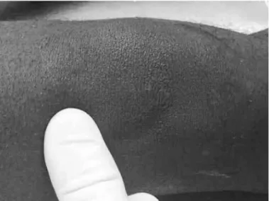
By means of a lateral suprapatellar access route of 2.5 cm (Figure 1) a previously cut 10 cm3 syringe was
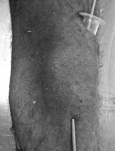
inserted to serve as a cannulated guide. This was placed under the patella, going in the direction of the tibia. A Steinmann wire of 3 mm in thickness was passed through it and was attached to the tibia at the point of entry im-mediately laterally to the projection of the anterior crest of the tibia, without using radioscopy (Figure 2).
Following this, after careful dissection, an access route was constructed going from the lateral apex of the patella to the insertion of the patellar tendon, to reach the knee joint under direct viewing. The guide was viewed anteriorly to the femoral trochlea and was placed under the patella (Figure 3).
The following were evaluated:
• Precision of blind positioning of the point of entry; • Structures at risk during this procedure, such as the
Hoffa fat, patellar tendon, menisci and joint cartilage;
• Whether any chondral lesion was caused by the gui -de during its insertion;
• Anatomical repairs that might make using this route
difficult.
RESULTS
In the 10 knees from the five cadavers, the distance from the suprapatellar incision to the point of entry of the guidewire ranged from 94 to 110 mm. The anato-mical repairs that caused difficulty in introducing the guide were noted: the trochlea and the Hoffa fat were highlighted in eight knees, while there were only two knees without resistance from any tissues. The point of entry was not reached in two of the knees, and the dis-tance from the guide insertion to the correct location of the entry point ranged from 0 to 4 mm (Table 1).
Figure 1 – Position of the incision: 2.5 cm above the superolateral corner of the patella.
171
Figure 2 – Soft-tissue protector positioned using guidewire, de-monstrating the ease of locating the entry point, and external wire showing direction of the tibial medullary canal.
Figure 3 – Guidewire at the point of entry and area of the trochlea at the start of the cartilage: a point that was difficult to surmount with a malleable protector (not shown in this figure).
DISCUSSION
Since the first description of intramedullary nails by Kuntscher(14) and the modification by Grosse et
al(15) that created locked nails, infrapatellar access
routes have been used. These could be medial, lateral
Table 1 – List of entry points with local anatomical structures.
Case Side Entry point Structures crossed
Distance from incision to
entrance
Repairs
1 R anteriorly4 mm HF 105mm HF
1 L 3 mm medially HF 104mm HF
2 R Correct HF 110mm Trochlea
2 L Correct HF 110mm Trochlea
3 R Not reached PA 95mm None
3 L Not reached PA 94mm None
4 R posteriorly2 mm HF 103mm Trochlea
4 L 2 mm medially HF 103mm Trochlea
5 R 4 mm laterally HF and PT 110mm HF
5 L 3 mm laterally HF and PT 110mm HF PT – patellar tendon; HF – Hoffa fat; PA – Pes anserinus.
or patellar transtendon routes.
The discussion on the best access route and the incidence of anterior knee pain has been extensive and has given rise to controversy.
Studies by Keating et al(16) demonstrated that there
is a high correlation between the transtendon route and anterior knee pain, while Väistö et al(4,6) did not
find any relationship between anterior pain and the access route. No author has been able to correlate nail protrusion in the proximal cortical bone with anterior knee pain, and this has led several authors to conclude that the cause of the pain was related to local surgical manipulation and lesions of the infrapatellar nerve.
Court-Brown et al(17) correlated anterior pain with
age. They showed that it is much more frequent in young and active patients, and that it causes great difficulty in kneeling, with pain presented even at rest.
In 1996, concerned about the antecurvatum defor-mity that occurs through using intramedullary nails in fractures that are more proximal in the tibia, Tornetta and Collins(10) and Tornetta and Ryan(18) described a
semi-extended position with a wide lateral parapa-tellar route, using the trochlea as a guide for locating the point of entry. In 2007, the same authors described a percutaneous route through a lateral suprapatellar incision, using a cannula and a trocar. This route ena-bled easy access to the safe point of entry described previously, and reduced the risks of perforating the posterior cortical bone and misaligning the fracture through quadriceps tension while inserting the nail.
In 2010, Morandi et al(13) described a lateral ANATOMICAL STUDY ON THE LATERAL SUPRAPATELLAR ACCESS ROUTE FOR LOCKED INTRAMEDULLARY
NAILS IN TIBIAL FRACTURES
172
suprapatellar route that they considered to be simpler than the medial routes, with easier access to the safe point of entry in the tibia. They indicated this not only for very proximal fractures but also for patients for whom the act of kneeling was important, or for whom there had been multiple trauma with injury to the soft tissue surrounding the patellar tendon, or furthermore, for multiple trauma patients for whom this route reduced the manipulation of other fractures, such as the femur and pelvis, during the treatment.
This new access route, of great interest in principle because it seems to reduce the incidence of anterior knee pain, presents risks to the intra-articular struc-tures. These risks have not been properly studies in papers in the literature and, for this to be done, spe-cific instruments with special dimensions and charac-teristics for this route firstly need to be developed, so that an assessment can then be made regarding which lesions might be caused by these instruments and, finally, a true indication for this new route can be made, in order to be able to use it safely.
The authors cited used adaptations from endosco-pic cannulae and trocars, cut-down syringes and a va-riety of protectors until reaching the models currently used, which were developed from previous attempts and not from specific projects and analysis.
In the present study, we observed that it was very easy to locate the entry point through this route, even without using radioscopy. Positioning the guidewire totally blindly only injured the Hoffa fat in most ca-ses. This structure is always crossed when using this
routeoffa fatH. Only in one case was the guidewire positioned anteriorly to the entry point, thereby da-maging the pes anserinus.
However, in four of the five cadavers studied, the start of the chondral surface of the femur became an obstacle that was difficult to overcome because of the very simple and flexible sleeve used, which was a syrin-ge tube that was cut obliquely without the embolus.
In three knees, this elevated chondral surface could not be surmounted and, in another three knees, it su-ffered injury.
Thus, we defined this chondral surface of the an-terior face of the knee as a limiting structure, and this should be used as a safety parameter when new studies are conducted using specific instruments to examine the lateral suprapatellar route for inserting a locked intramedullary nail in the tibia.
Such instruments need to be developed for subse-quent in vivo use for follow-up regarding postopera-tive pain on the anterior face of the knee.
The lateral suprapatellar access route for place-ment of an intramedullary nail in tibial fractures is an alternative that may be viable with adequate ins-truments, which need to be developed through more controlled studies.
CONCLUSION
We conclude that the lateral suprapatellar access made it easy to locate the entry point for the guidewire of the intramedullary nail.
Rev Bras Ortop. 2012;47(2):169-72
REFERENCES
1. Bhattacharyya T, Seng K, Nassif NA, Freedman I. Knee pain after tibial nailing: the role of nail prominence. Clin Orthop Relat Res. 2006;449:303-7. 2. Cartwright-Terry M, Snow M, Nalwad H. The severity and prediction of anterior
knee pain post tibial nail insertion. J Orthop Trauma. 2007;21(6):381-5. 3. Toivanen JA, Väistö O, Kannus P, Latvala K, Honkonen SE, Järvinen MJ.
Anterior knee pain after intramedullary nailing of fractures of the tibial shaft. A prospective, randomized study comparing two different nail-insertion techni-ques. J Bone Joint Surg Am. 2002;84(4):580-5.
4. Väistö O, Toivanen J, Kannus P, Järvinen M. Anterior knee pain after intrame-dullary nailing of fractures of the tibial shaft: an eight-year follow-up of a pros-pective, randomized study comparing two different nail-insertion techniques. J Trauma. 2008;64(6):1511-6.
5. Althausen PL, Neiman R, Finkemeier CG, Olson SA. Incision placement for intra-medullary tibial nailing: an anatomic study. J Orthop Trauma. 2002;16(10):687-90. 6. Väistö O, Toivanen J, Paakkala T, Järvelä T, Kannus P, Järvinen M. Anterior
knee pain after intramedullary nailing of a tibial shaft fracture: an ultrasound study of the patellar tendons of 36 patients. J Orthop Trauma. 2005;19(5):311-6. 7. Kakar S, Tornetta P 3rd. Open fractures of the tibia treated by immediate in-tramedullary tibial nail insertion without reaming: a prospective study. J Orthop Trauma. 2007;21(3):153-7.
8. Laflamme GY, Heimlich D, Stephen D, Kreder HJ, Whyne CM. Proximal ti-bial fracture stability with intramedullary nail fixation using oblique interlocking screws. J Orthop Trauma. 2003;17(7):496-502.
9. Bong MR, Koval KJ, Egol KA. The history of intramedullary nailing. Bull NYU Hosp Jt Dis. 2006;64(3-4):94-7.
10. Tornetta P 3rd, Collins E. Semiextended position of intramedullary nailing of the proximal tibia. Clin Orthop Relat Res. 1996;(328):185-9.
11. Tornetta P 3rd, Riina J, Geller J, Purban W. Intraarticular anatomic risks of tibial nailing. J Orthop Trauma. 1999;13(4):247-51.
12. McConnell T, Tornetta P 3rd, Tilzey J, Casey D. Tibial portal placement: the radio-graphic correlate of the anatomic safe zone. J Orthop Trauma. 2001;15(3):207-9. 13. Morandi M, Banka T, Gaiarsa GP, Guthrie ST, Khalil J, Hoegler J, et al. Intrame-dullary nailing of tibial fractures: review of surgical techniques and description of a percutaneous lateral suprapatellar approach. Orthopedics. 2010;33(3):172-9. 14. Kuntscher GB. The Kuntscher method of intramedullary fixation. J Bone Joint
Surg Am. 1958;40(1):17-26.
15. Grosse A, Kempf I, Lafforgue D. [Treatment of fragments, loss of bony subs-tance and pseudarthrosis of femur and tibia using screw fixation (40 cases)]. Rev Chir Orthop Reparatrice Appar Mot. 1978;64(Suppl 2):33-5.
16. Keating JF, Orfaly R, O’Brien PJ. Knee pain after tibial nailing. J Orthop Trauma. 1997;11(1):10-3.
17. Court-Brown CM, Gustilo T, Shaw AD. Knee pain after intramedullary tibial nai-ling: its incidence, etiology, and outcome. J Orthop Trauma. 1997;11(2):103-5. 18. Tornetta P 3rd, Ryan S. Tibial metaphyseal fractures: nailing in extension. In:
Paper presented at: Orthopaedic Trauma Association 24th Annual Meeting: