WORM PV ETAL.
828 REV ASSOC MED BRAS 2016; 62(9):828-830
IMAGE IN MEDICINE
Giant scalp arteriovenous malformation
PAULO VALDECI WORM1, LEONARDO GILMONE RUSCHEL2*, MARCELO ROSA ROXO3, RAFAEL CAMELO3
1Neurosurgeon at Hospital São José, Santa Casa de Misericórdia de Porto Alegre, Porto Alegre, RS, Brazil 2Resident of the Neurosurgery Program at Instituto de Neurologia de Curitiba (INC), Curitiba, PR, Brazil 3Neurosurgery Resident, Hospital São José, Santa Casa de Misericórdia de Porto Alegre, Porto Alegre, RS, Brazil
S
UMMARYStudy conducted at Hospital São José, Santa Casa de Misericórdia de Porto
Alegre, Porto Alegre, RS, Brazil
Article received: 7/24/2015
Accepted for publication: 7/27/2015
*Correspondence:
Address: Rua Carlos Trein Filho, 550/403 Porto Alegre, RS – Brazil Postal code: 90450-120 [email protected]
http://dx.doi.org/10.1590/1806-9282.62.09.828
Arteriovenous malformations (AVMs) of the scalp are rare lesions. The clinical picture presents with complaints of increased scalp, scalp disigurement, pain and neurological symptoms. Its origin can be congenital or traumatic. We pres-ent a case of giant scalp AVMs and its managempres-ent, followed by a brief literature review on the subject. The diagnosis of scalp AVMs is based on physical exam-ination and conirmed by internal and external carotid angiography or comput-ed tomographic angiography (CTA). Surgical excision is especially effective in scalp AVMs, and is the most frequently used treatment modality.
Keywords: scalp, arteriovenous malformations, vascular diseases.
I
NTRODUCTIONExtracranial scalp arteriovenous malformations (AVMs) are relatively rare, accounting for only 8.1% of cases of AVMs.1 They are vascular aberrations that develop during
the fetal period and result from failure of embryonic vas-culature to differentiate into arteries and veins.2 Although
controversy exists regarding the cause of these lesions, it is generally accepted that they may be either congenital or traumatic in origin.3
The clinical picture is characterized by increased scalp, scalp deformation, pulsatile mass, headache, tinnitus, convulsion, bleeding and dizziness, but it can often be asymptomatic. Diagnosis of AVMs is based on physical examination and conirmed by internal and external ca-rotid angiography, or computed tomographic angiogra-phy (CTA).4 Treatment options include endovascular
occlusion, direct percutaneous injection of sclerosing agents and, in selected cases, surgical resection.5
We present a case of scalp AVM and its management at our institution. We provide below a brief literature review and some commentaries regarding the clinical and radiological features of this rare entity.
C
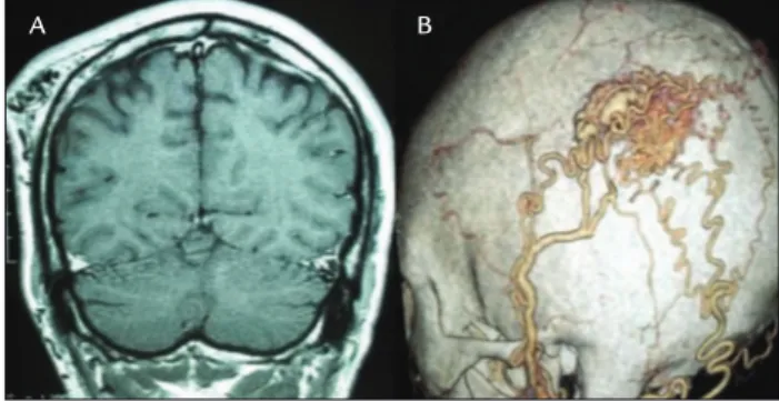
ASE REPORTWe present the case of a 33-year-old male with a complaint of a pulsatile and increasingly bigger mass within the left temporo-occipital region of scalp for 3 years. There was no previous trauma. We identiied an area of focal thickening
of the scalp, above the lateral and occipital skull surfaces, soft and pulsatile on palpation. There was no audible bruit or any abnormalities on neurologic examination. The CTA demonstrated a lesion of vascular nature, presumably an arteriovenous malformation beneath the scalp. There was a nidus fed by branches from ipsilateral supericial temporal and occipital arteries (Figure 1). There was no communica-tion with the intracranial circulacommunica-tion. The treatment of choice was surgical, with ligation of the feeder branches without scarifying parent vessels, with total excision of the malforma-tion. Scalp irrigation was preserved (Figure 2). Postoperative period was uneventful, and postoperative CTA showed no signiicant and/or pathologic vessels left (Figure 3).
FIGURE 1 A. Coronal T1-W MRI showing the scalp AVM on the left parieto-occipital region. B. Markedly dilated supericial temporal and occipital arteries supplied the nidus; a draining vein can also be seen.
GIANTSCALPARTERIOVENOUSMALFORMATION
REV ASSOC MED BRAS 2016; 62(9):828-830 829
D
ISCUSSIONAVMs of the scalp are abnormal istulous connection between the feeding arteries and draining veins, without an intervening capillary bed within the subcutaneous layer. Their rarity and unpredictable behavior render sys-tematic studies, assessment of prognosis, and treatment controversies.6 Different names have been used to describe
these lesions, and the most common are cirsoid aneurysm, plexiform angioma, scalp arteriovenous istula, arterio-venous aneurysm and arterioarterio-venous malformation.7
Almost all patients present with a scalp swelling that has gradually increased in size from birth or after head trauma. Rapid increases in size have been reported to occur at puberty, during menstruation and during pregnancy. Their clinical signs are associated with the size of AVMs. Associated symptoms and signs include pain, throbbing headaches and bruit,3,8,9 hemorrhage,8,10 seizures and
psy-chomotor retardation.11 Large lesions have also been
asso-ciated with scalp necrosis.8 AVMs cause direct shunting of
high-volume arterial blood through low-resistance arterio-venous istulae, often resulting in arterio-venous hypertension, hypoperfusion of vessels and tissue downstream, and re-duced cerebral perfusion, known as the steal phenomenon.12
About 10 to 20% of scalp AVMs develop following pene-trating or non-penepene-trating trauma to the scalp.3,13
Total excision of a scalp AVM requires comprehensive knowledge of the feeding arteries, draining veins and nidus. Catheter angiography has been the gold standard for diagnosis14 but CTA can also provide a very high
im-aging resolution and the observation of related bony structures, which may be important for surgical planning, as in the case of our patient.4
Treatment of scalp AVMs allows many possibilities and individualization is key. Surgical excision is especially effective in selective cases, when endovascular treatment is dificult or not feasible.3,5,15 Moreover, successful
endovas-cular treatment of scalp AVMs has been reported, but has often been found to be insuficient for those with more extensive istulae or with other complicating components.16
In relation to surgical treatment, common complica-tions consist of hemorrhage, necrosis of the scalp, and sepsis caused by wound infection.3,17 Hemorrhage may be
prevented with preoperative embolization, clamping, and suturing of feeding vessels. Several authors recommend preoperative endovascular treatment to reduce blood supply of large lesions, as to facilitate surgery.18,19
R
ESUMOMalformação arteriovenosa gigante de escalpo
Malformações arteriovenosas (MAV) do couro cabeludo são lesões raras. O quadro clínico apresenta-se com queix-as de aumento do couro cabeludo, desiguração do couro cabeludo, dor e sintomas neurológicos. A origem pode ser congênita ou traumática. Apresentamos um caso de MAV gigante de couro cabeludo e o tratamento adotado, seguindo-se uma breve revisão da literatura. O diagnósti-co das MAV de diagnósti-couro cabeludo baseia-se no exame físidiagnósti-co e é conirmado pela angiograia carótida interna e ex-terna ou angiograia por tomograia computadorizada. A excisão cirúrgica é especialmente eicaz em MAV de
couro cabeludo e é a modalidade de tratamento mais frequentemente utilizada.
Palavras-chave: couro cabeludo, malformações arterio-venosas, doenças vasculares.
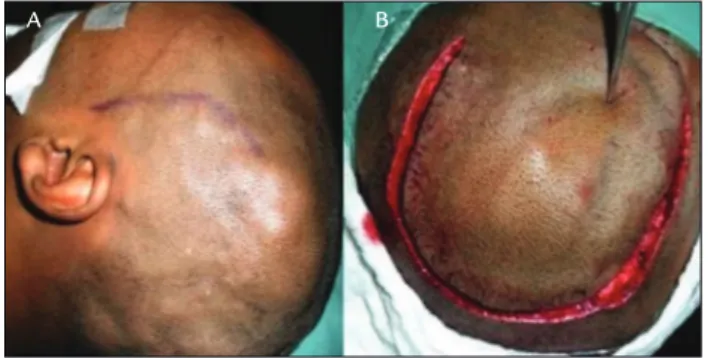
FIGURE 2 A. Soft thickened scalp can be seen above the left
temporal and occipital skull surfaces, corresponding to the scalp AVM. B. A horseshoe incision was made with a view to preserving scalp irrigation, critical to the success of the surgery.
FIGURE 3 A. The scalp AVM shown after en bloc resection.
B. Postoperative CT showing no signiicant and/or pathologic vessels relative to the AVM.
A
A
B
WORM PV ETAL.
830 REV ASSOC MED BRAS 2016; 62(9):828-830
11. Mohanty S, Rao CJ. A large cirsoid aneurysm of the scalp associated with epilepsy. J Neurol Neurosurg Psychiatry. 1976; 39(9):835-6.
12. Mast H, Mohr JP, Osipov A, Pile-Spellman J, Marshall RS, Lazar RM, et al. ‘Steal’ is an unestablished mechanism for the clinical presentation of cerebral
arteriovenous malformations. Stroke. 1995; 26(7):1215-20.
13. Matsushige T, Kiya K, Satoh H, Mizoue T, Kagawa K, Araki H. Arteriovenous malformation of the scalp: case report and review of the literature. Surg Neurol. 2004; 62(3):253-9.
14. Wilkinson HA. Recurrence of vascular malformation of the scalp 18 years following excision. Case report. J Neurosurg. 1971; 34(3):435-7.
15. Massimi L, De Bonis P, Esposito G, Novegno F, Pettorini B, Tamburrini G, et al. Vertex scalp mass as presenting sign of a complex intracranial vascular malformation. J Neurosurg Pediatr. 2009; 3(4):307-10.
16. Lanzino G, Passacantilli E, Lemole GM Jr., McDougall C, Spetzler RF. Scalp arteriovenous malformation draining into the superior sagittal sinus associated with an intracranial arteriovenous malformation: just a coincidence? Case report. Neurosurgery. 2003; 52(2):440-3.
17. Muthukumar N, Rajagopal V, Manoharan AV, Durairaj N. Surgical management of cirsoid aneurysms. Acta Neurochir (Wien). 2002; 144(4):349-56.
18. Komiyama M, Nishikawa M, Kitano S, Sakamoto H, Imai K, Tsujiguchi K, et al. Non-traumatic arteriovenous fistulas of the scalp treated by a combination of embolization and surgical removal. Neurol Med Chir (Tokyo). 1996; 36(3):162-5.
19. Tanaka T, Hasegawa Y, Kanki T, Abe T. [Combination of intravascular surgery and surgical operation for occipital subcutaneous arteriovenous istula in a patient with neuroibromatosis type I]. No Shinkei Geka. 2002; 30(3):309-13.
R
EFERENCES1. Huber P. Krayenbühl/Ya argil Cerebral angiography. 2. ed. New York: Thieme; 1982.
2. Schultz RC, Hermosillo CX. Congenital arteriovenous malformation of the face and scalp. Plast Reconstr Surg. 1980; 65(4):496-501.
3. Fisher-Jeffes ND, Domingo Z, Madden M, de Villiers JC. Arteriovenous malformations of the scalp. Neurosurgery. 1995; 36(4):656-60.
4. Teksam M, McKinney A, Truwit CL. Multi-slice CT angiography in evaluation of extracranial-intracranial bypass. Eur J Radiol. 2004; 52(3):217-20. 5. Barnwell SL, Halbach VV, Dowd CF, Higashida RT, Hieshima GB.
Endovascular treatment of scalp arteriovenous istulas associated with a large varix. Radiology. 1989; 173(2):533-9.
6. Kohout MP, Hansen M, Pribaz JJ, Mulliken JB. Arteriovenous malformations of the head and neck: natural history and management. Plast Reconstr Surg. 1998; 102(3):643-54.
7. Khodadad G. Arteriovenous malformations of the scalp. Ann Surg. 1973; 177(1):79-85.
8. Nagasaka S, Fukushima T, Goto K, Ohjimi H, Iwabuchi S, Maehara F. Treatment of scalp arteriovenous malformation. Neurosurgery. 1996; 38(4):671-7.
9. Dandy WE. Arteriovenous aneurysms of the scalp and face. Arch Surg. 1946; 52:1-32.