Critical limb ischemia in a young patient with cystic disease
of the popliteal artery
Isquemia crítica em membro inferior em paciente jovem com doença cística de artéria poplítea
Michel Nasser1, Luca Giovanni Antonio Pivetta2, José Luis Teixeira Filho2, Evelin Schueteze Rocha2, Ana Eliza Botta2
PART I
Case description
The patient is a 45-year-old male physician, non-di-abetic, non hypertensive, non-smoker and athlete, who was admitted to the Emergency Room complaining of severe pain on the left lower limb for 4 hours, decreased temperature, absence of left popliteal and distal pulse, accompanied by decreased sensitivity in the left foot, but
with the motor function preserved. The patient reported bilateral calf intermittent claudication that had began one year earlier and had worsened to approximately 120 m in the last days.
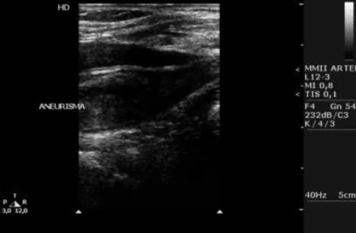
Ankle-brachial index (ABI) measurements showed an, ABI=0.40 at the let and ABI=0.85 at the right lower limb. Color Doppler ultrasound showed a thrombosed let popliteal artery aneurysm and approximately 50% steno-sis of the right popliteal artery (Figures 1 and 2). he phy-sician performing the examination raised the suspicion
Abstract
Cystic adventitial disease of the popliteal artery is a rare vascular disease of unknown etiology in which a mucin-containing cyst may compromise the artery and, because of the compression, develops stenosis or occlusion of afected artery. We report the case of a 45-year-old male with cystic adventitial disease of the left popliteal artery, after admission for an acute limb ischemia in his left leg, diagnosed non-invasively with EcoDoppler vascular dual scan, magnetic resonance angiography, angiography. Performed complete removal of the cyst with arteriectomy and venous replacement. he pathologic result ware consistent with the diagnostic of cystic adventitial disease. he patient is in ambulatory follow up and was afected by cystic disease in the contralateral limb. he authors described the best methods of diagnosis and treatment for cystic adventitial disease, discussing if the urgency procedure was the most appropriate and whether there was an alternative treatment for the case. Finally, questioning what would be the best procedure to be performed in the other limb.
Keywords: intermittent claudication; cyst; popliteal artery.
Resumo
Degeneração cística da artéria poplítea é uma doença vascular rara, de etiologia desconhecida, na qual um cisto com conteúdo mucinoide compromete a artéria e desenvolve estenose ou oclusão do vaso. Apresentamos um caso de um paciente do sexo masculino, 45 anos, admitido na emergência hospitalar, que apresentava quadro de isquemia aguda no membro inferior esquerdo. Submetido à EcoDoppler, angiorressonância magnética e angiograia pré-operatória. Realizada arterectomia com enxerto fêmoro-poplíteo infrainguinal de safena invertida. O exame patológico foi consistente com o diagnóstico de doença cística da artéria poplítea. O paciente encontra-se em acompanhamento ambulatorial e foi acometido pela doença cística no membro contralateral. Os autores descreveram os melhores métodos de diagnóstico e tratamento para a doença, discutindo se o procedimento de urgência foi o mais adequado e se haveria opções terapêuticas alternativas para o caso, além de qual seria a melhor conduta a ser realizada no membro contralateral também acometido pela doença.
Palavras-chave: claudicação intermitente; cisto; artéria poplítea.
Study carried out at the Universidade Federal de São Carlos (UFSCar) – São Carlos (SP), Brazil.
1 Professor at the Medical Department of Universidade Federal de São Carlos (UFSCar) – São Carlos (SP), Brazil. 2 Medical student at UFSCar – São Carlos (SP), Brazil.
Financial support: none.
Conlict of interest: Luca Giovanni Antonio Pivetta has a scholarship of Conselho Nacional de Desenvolvimento Cientíico e Tecnológico (CNPq), and works in a Project of literature review on cystic degeneration of the popliteal artery.
Acute ischemia: cystic artery disease - Nasser M et al. J Vasc Bras 2012, Vol. 11, Nº 2 145
of periarterial cyst. Color Doppler imaging showed ste-nosis and external compression – possibly a cyst – of the right popliteal artery (Figure 3). Knee lexion maneuvers were performed bilaterally, with absence of bilateral distal
pulses. Color Doppler low showed alterations at maneu-vers of hyperextension of the feet.
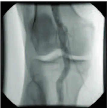
MR angiography showed let arterial occlusion and no signs of right arterial occlusion. he patient also underwent preoperative catheter angiography (Figure 4), and then was sent to the operating room.
PART II
With acute arterial ischemia of the let lower limb for 6 hours, the patient underwent surgical repair, and tissue induration was observed on the popliteal artery wall, thus occluding the vessel lumen. Tissue samples were collected and sent for analysis.
he patient underwent popliteal artery resection and replacement by a femoropopliteal infragenicular bypass grat with reversed saphenous vein, with good results in the immediate postoperative period. Anatomopathological examination showed the adventitial layer with thicken-ing of the intima in the same proportion and cystic spaces (Figure 5). Multinucleated giant cells, neutrophils, eosino-phils, and crystals were not detected, thus excluding other forms of arteritis (Figure 6). he indings were suggestive of cystic disease of the popliteal artery.
hirty days ater operation, the patient presented re-current claudication for a distance of 70 m on the right lower limb. Ninety days later, the claudication distance was 120 m, so the patient was given vasodilator and antiplatelet treatment. At the day 180, claudication distance was 150 m,
Figure 1. B-mode echography showing thrombosed aneurysm at the left.
Figure 2. B-mode echography showing thrombosed aneurysm at the left. Image indicating adequately the existence of a thrombus.
Figure 3. 50% stenosis of the right popliteal artery, suspected periarterial cyst.
A. FEMORAL SUPERF
so angiography (Figure 7) and CT angiography (Figures 8 and 9) imaging were performed, showing cystic disease af-fecting the right lower limb.
Was the initial procedure adequate?
Do we have alternative treatment options for this case? What would be best to perform in the contralateral limb that is also afected by the disease?
Discussion
Arterial cystic disease (ACD) is characterized by the presence of cysts, oten multiple, located in the arterial wall. he cysts, which have a gelatinous, viscous content, may compress and distort the adventitia and tunica media,
Increased pressure within the cysts can also cause steno-sis and occlusion of the afected artery. It is also known as cystic degeneration of adventitia1, cystic disease of the
ad-ventitia2,3, adventitia colloid cyst4, mucinous cystic disease5,
adventitial cyst of the popliteal artery6,7, and other
desig-nations8-10. Both cystic degeneration of the adventitia and
adventitial cyst disease are restricted designations, for they do not encompass all cases, because cysts may be located on the outer layers of the media4. More comprehensive names,
such as cystic disease, or degeneration of the arterial wall,
Figure 5. Anatomopathological study (mucicarmim) showing the ad-ventitious layer with tickneing of the intima in the same proportion and few cystic spaces.
Figure 6. Hematoxylin-eosin staining showing no multinucleated giant cells, neutrophils, eosinophils, and crystals, difering from other arteritis.
Figure 7. Angiography of the contralateral limb showing cistos multi-loculados.
Acute ischemia: cystic artery disease - Nasser M et al. J Vasc Bras 2012, Vol. 11, Nº 2 147
or even degeneration or cystic disease of the popliteal ar-tery, are more appropriate11,12.
he pathogenesis of the disease remains unknown, but four theories have tried to explain it: microtrauma13;
system-ic disease4; embryonic origin14; true synovial cysts1,12,15,16.
he incidence of cystic disease of the popliteal artery is 1 case per 1,200 cases of intermittent claudication, or 1 case in 1,000 femoral angiographies performed14. his
dis-ease afects men more frequently, at a ratio men:women of 4:117, and is more commonly observed in individuals in the
fourth to the sixth decades of life.
Mafei et al.18 suggest that non-recognition and lack of
report may be the causes of low incidence of this disease, and that the number cases diagnosed is likely to increase with increasing levels of suspicion.
hus, in younger adults with intermittent claudica-tion and with few or no risk factors for atherosclerosis, one should consider the following diferential diagnosis: popliteal artery entrapment syndrome (PAES), cystic ad-ventitial disease (CAD), ibromuscular dysplasia, the ex-ternal Iliac Artery Endoibrosis (common among cyclists), arteritis, arterial embolism, and pseudoxanthoma elasti-cum (PXE)-related arterial occlusive lesions.
Currently, there are several imaging methods for di-agnosis and surgical treatment planning. It is essential to deine the number of cysts, their location, extent and situ-ation. A Doppler ultrasound imaging may be suicient for diagnosis19, but a proper surgical planning requires other
imaging methods.
Computed tomography (CT) scan20 is still used for
diagnosis, but there are better methods such as magnetic
resonance imaging (MRI) or angiography (MRA)21. Helical
CT angiography associated with three-dimensional re-construction seems to provide information similar MR or MRA, but it requires contrast injection22.
Maged et al.23 recently showed good results using High
Spatial Resolution Magnetic Resonance Imaging (MR-HSR). hey identiied connections between the cysts and the adjacent joint, which helped in the ligation of these con-nections during surgery, avoiding recurrence of the cysts in some cases.
Ortiz et al.24 reported that diferent therapeutic
ap-proaches result in variable recurrence rates. he resection of the afected artery with a vein grat is indicated in cases of complete arterial occlusion and weakening of the arterial wall, or in cases of secondary intimal ulceration.
Some authors25,26 show that cyst resection with
saphe-nous vein grat interposition is followed by a recurrence rate of about 5%, and that cyst draining, even with its re-moval from the wall, is followed by a recurrence rate of 6%. On the other hand, percutaneous aspiration of cyst alone presents a recurrence rate of 34%. Some other methods are rarely used due to high recurrence rates, including endo-vascular treatment with angioplasty or endoendo-vascular stent placement, which have been reported with a recurrence rate of up to 100%.
Maged et al.27 described a case of recurrent popliteal
ar-tery cystic disease ater surgical enucleation of the cyst. his case was successfully treated with angioplasty, and symp-toms were solved in a 24-month follow-up.
Do et al.26 reported the successful treatment of 7
pa-tients (6 men) who had symptomatic cystic adventitial dis-ease (CAD) of the popliteal artery without occlusion by percutaneous image-guided cyst aspiration. In their series, no patient developed complications, and the procedure was technically and clinically successful in all cases. Duplex ul-trasonography follow-up lasted 1 to 32 months (mean 14.8 months), and, ater the procedure, no patient developed signiicant stenosis. However, the authors acknowledge that their successful results with the percutaneous aspiration tech-nique have not been reproduced in the literature and that this technique has not gained much recognition worldwide24,28.
Conclusions
Cystic disease of the popliteal artery is a rare entity, but physicians should be attentive to its diagnosis. All young patients complaining of intermittent claudication with no risk factors for atherosclerosis should be managed with the hypothesis of cystic disease of the popliteal artery. Diagnosis must be initially performed using non-invasive methods,
such as color Doppler ultrasound. Bilateral knee lexion maneuvers (Ishikawa), as well as feet hyperextension ma-neuvers should be part of the clinical examination.
hree-dimensional helical CT scan appears to provide images comparable to those of MRI or MRA, but it requires contrast injection. If possible, high spatial resolution MRI should be used. Although there are several alternative di-agnosis methods, one must never forget that angiography is the gold standard before revascularization and it should be, whenever necessary.
Various therapeutic methods can be used for the ment of popliteal artery cystic disease. Endovascular treat-ment with angioplasty or intravascular stent placetreat-ment is not a good choice in such cases, because this disease afects arteries in areas of lexion, and the published data of Ortiz et al.24 showed an occlusion rate of almost 100%.
Image-guided cyst aspiration is a less invasive tech-nique, but it was successful only in the hands of Do et al. Maged et al.23 and even Do et al.26, in a literature review,
showed a recurrence rate close to 34%.
Most studies addressing cyst resection with homologous grat implantation20,23,24 and cyst draining with its complete
removal from the arterial wall, are the treatments of choice, since they present similar recurrence rates. However, the re-ports by Melliere et al.29 suggest that the technique of artery
resection and homograt implantation provides better short and long-term results, compared to cyst draining alone, even when it is removed from the arterial wall.
herefore, surgical resection and homograt implantation are the best treatment choices for the popliteal artery cystic dis-ease. Regarding endovascular treatment, it needs need further systematic studies before being indicated; however, it could be considered an adjunctive therapy option in cases of recurrent stenosis ater cyst resection and grat placement.
In the current case, since the patient had no ischemia of the right lower limb, we chose to manage him the contralat-eral lim conservatively, with prescription of exercises aim-ing at the strengthenaim-ing of the muscles, use of peripheral vasodilator and anti-platelet aggregation agent and clini-cal follow-up. In case of ischemia in the future, the best ap-proach would be resection of the afected arterial segment, with autologous vein grat interposition.
References
1. Alder VW, Zwicker H. [he diagnosis of cystic degeneration of the adventitia]. Rofo. 1977;126(4):331-4. German.
2. Bergan JJ. Adventitial cystic disease of the popliteal artery. In: Rutherford RD, ed. Vascular Surgery. Philadelphia: WB Saunders Company, 1977. p. 569.
3. Terry JD, Schenken JR, Lohf MR, Neis DD. Cystic adventitial disease. Hum Pathol. 1981;12(7):639-42.
4. Lassalle C, Paquis M, Florquet J, Mathieu P, Frisch R. Kyste colloide de I’adventice de l’aertère poplitée. A propos de trois cas. Ann Med Nancy. 1978;17:575-7.
5. Müller M, Rodriguéz J. Obstrucción de lá arteria poplitea por enfermedad quística mucinosa de la pared arterial. Angiologia. 1975;27(1):1-6
6. Blum L, Giron F. Adventitial cyst of the popliteal artery with secondary inlammatory entirapment. Mt Sinai J Med Ny. Mt Sinai J Med. 1976;43(4):471-5.
7. Rollo HA, Gama JCFD, Lastória S,Yoshida WB, Mafei FH. Cisto de adventícia em artéria poplítea. Relato de dois casos. AMB Rev Ass Med Bras. 1982; 28:79-81.
8. Bakstrom CG, Linell F, Östberg B. Cystic, mixomatous adventitial degeneration of the radial artery with development of ganglion in the connective tissue. Report of two cases. Acta Chir Scand. 1965;129:447-51
9. Miranda Junior F, Francisco Jr J, Burihan E. Cisto de artéria poplítea. Rev Bras Cir. 1982;72:4:221-2.
10. Albernaz DTS, Albernaz LFL, Eggers EE. Doença cística da artéria poplítea: relato de caso. J Vasc Bras. 2010;9(3):168-72.
11. Lie JT, Juergens JL. Degenerative arterial disease other than atherosclerosis. In: J.L. Juergens JL, Spittell Jr JA, Fairbairn II JF, editors. Allen-Barker-Hines Peripheral Vascular Diseases. Philadelphia: WB Saunders Company, 1980. p. 237-53.
12. Shute K, Rothnie NG. he aetiology of cystic arterial disease. Br J Surg. 1973;60:397-400.
13. Mark TM, Rywlin AM, Unger H. Cystic adventitial degeneration of the popliteal artery. Its occurence in a patient with the nail-patella syndrome. Arch Pathol Lab Med. 1983;107:186-8.
14. Flanigan DP, Burnharn SJ, Goodreau JJ, Bergan JJ. Summary of cases of adventitial cystic disease of the popliteal artery. Ann Surg. 1979;189:165-75.
15. Bourke BM, Appleberg M, Reeve TS, Hollings RM. Cystic adventitial disease of the popliteal artery: A report of two cases and a review of the literature. Aust N Z J Surg. 1982;52(2):171-3
16. Lewis GJT, Douglas DM, Reid W, Watt JK. Cystic adventitial disease of the popliteal artery. Br Med. 1967;60:411-5.
17. Ishikawa K. Cystic adventitial disease of the popliteal artery and of other stem vessels in the extremities. Jpn J Surg. 1987;17:221-9.
18. Mafei FHA, Barbosa AG, Rollo HA, Neser A, Lastoria S. Adventitial cystic disease of the popliteal artery in Brazil: additional data on the geographical distribution of the disease. Angiology. 1982;33:339-42.
19. Stapf M, Zoller W, Spengel F. Image-directed Doppler ultrasound indings in adventitial cystic disease of the popliteal artery. J Clin Ultrasound. 1989;17:689-91.
Acute ischemia: cystic artery disease - Nasser M et al. J Vasc Bras 2012, Vol. 11, Nº 2 149
21. Crolla RMPH, Steyling JF, Hennipman A, Slootweg PJ, Taams A. A case of cystic adventitial disease of the popliteal artery demonstrated by magnetic resonance imaging. J Vasc Surg. 1993;18:1052-5.
22. Miller A, Salenius J, Sacks BA, Gupta SK, Shoukimas GM. Noninvasive vascular imaging in the diagnosis and treatment of adventitial cystic disease of the popliteal artery. J Vasc Surg. 1997;26:715-20.
23. Maged IM, Turba UC, Housseini AM, Kern JA, Kron IL, Hagspiel KD. High spatial resolution magnetic resonance imaging of cystic adventitial disease of the popliteal artery. J Vasc Surg. 2010;51:471-4.
24. Ortiz M WR, Lopera JE, Giménez CR, Restrepo S, Moncada R, Castañeda-Zúñiga WR. Bilateral adventitial cystic disease of the popliteal artery: a case report. Cardiovasc Intervent Radiol. 2006;29:306-10.
25. Rai S, Davies RS, Vohra RK. Failure of endovascular stenting for popliteal cystic disease. Ann Vasc Surg. 2009;23:410.e1-e5
26. Do DD, Braunschweig M, Baumgartner I, Furrer M, Mahler F. Adventitial cystic disease of the popliteal artery: percutaneous US-guided aspiration. Radiology. 1997;203:743-6.
27. Maged IM, Kron IL, Hagspiel KD. Recurrent cystic adventitial disease of the popliteal artery: successful treatment with
percutaneous transluminal angioplasty. Vas Endovasc Surg. 2009;43:399-402.
28. Sieunarine K, Lawrence-Brown MM, Kelsey P. Adventitial cystic disease of the popliteal artery: early recurrence after CT guided percutaneous aspiration. J Cardiovasc Surg. 1991;32(5):702-4.
29. Mellière D, Ecollan P, Kassab M, Becqemin JP. Adventitial cystic disease of the popliteal artery: treatment by cyst removal. J Vasc Surg. 1988;8(5):638-42.
Correspondence
Michel Nasser Avenida Venicio Bruneli 287 – Quinta dos Oitis CEP 14808-000 – Araraquara (SP), Brazil E-mail: [email protected]
Author’s contributions