Arquivos Brasileiros de Cardiologia - Volume 82, Nº 6, Junho 2004
561
flow occurred across the left ventricular outflow tract with a peak gradient of 57.2 mmHg. Systolic anterior motion (SAM) could not be demonstrated by M mode echocardiography.
It was suspected that the severe mitral insufficiency was secon-dary to the hyperdynamic left ventricular function and severe volu-me depletion. During echocardiographic monitoring, 1000 mL of normal saline was rapidly infused over a period of 30 minutes. A progressive slowing of the heart rate from 96 to 64 beats per minute, a decrease in outflow gradient (from 57.2 to 10.1 mmHg), and total resolution of the mitral insufficiency were documented (Fig.1a, 1b, 2a, and 2b).
Discussion
The effect of volume depletion on the mitral valvular apparatus has been previously reported 1,2. Volume depletion may cause left ventricular outflow tract obstruction, SAM, and mitral valve
dys-function 3. Our patient had a demonstrable gradient across the
outflow tract and severe mitral regurgitation during the
hyperdy-Case Report
Severe Volume Depletion Leading to Mitral
Insufficiency
Alexandre C. Ferreira, Arley Arrais Peter, Simon Chakko, Howard Willens,
Eduardo de Marchena
Miami, Florida, USA
Department of Medicine, Division of Cardiology, Cardiovascular Center, University of Miami, Miami, Florida, USA
Mailing address: Arley A. Peter - Rua Fausto Cabral, 266 - 60155-410 - Papicu – Fortaleza, CE – E-mail: [email protected]
Received: 3/18/2003 Accepted: 7/20/03
We report a patient with profound hypovolemia who deve-loped dynamic left ventricular outflow tract obstruction and severe mitral regurgitation. Both the outflow tract obstruction and mitral regurgitation resolved with volume replacement. Unlike previous reports of dynamic left ventricular outflow obstruction and mitral regurgitation, the degree of mitral regurgitation was severe. Possible mechanisms are discussed.
A hyperdynamic cardiac state secondary to severe volume de-pletion or catecholamine surge causes dynamic outflow obstruction even in the absence of septal hypertrophy 1. In previous reports of such dynamic outflow obstruction 2, the severity of mitral insuffi-ciency, when present, was usually mild. We describe a patient with severe volume depletion in whom mitral insufficiency was severe and resolved with rapid intravenous saline infusion.
Case Report
A 67-year-old woman with a history of chronic hepatitis C and liver cirrhosis, who had undergone combined liver-kidney trans-plant 4 months prior to admission. After her liver transtrans-plant, she recovered the function of her native kidneys, which led to massive diuresis. The patient also developed a high-flow biliary fistula as a complication of her liver transplant. Despite normal oral intake, she presented with dizziness and weakness. The echocardiogram obtained prior to the liver transplant was normal, and a noninvasive ischemia workup was negative.
Physical examination revealed a blood pressure of 95/60 mmHg and a heart rate of 98 beats per minute. The apical impulse was normal. A new apical systolic murmur with an intensity of 4/6, radiating to the axilla, was heard. The electrocardiogram showed an intermittent rate related left bundle-branch block. Echocardio-graphy revealed a normal mitral valvular apparatus, left ventricle, and left atrium. Left ventricular wall thickness was normal, and the estimated ejection fraction was 70%. No septal hypertrophy was present. On Doppler, severe mitral insufficiency was revealed with evidence of flow-reversal in the pulmonary veins. Turbulent
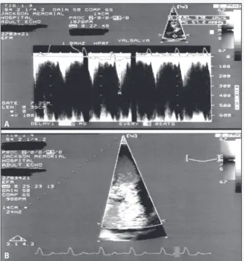
Fig. 1 - a) Outflow tract velocity showing a peak gradient of 57.2 mmHg at baseline; b) Severe mitral insufficiency demonstrated by color Doppler prior to infusion of normal saline.
A
Arquivos Brasileiros de Cardiologia - Volume 82, Nº 6, Junho 2004
562
Volume depletion leading to mitral insufficiency
1. Levisman J. Systolic Anterior motion of the mitral valve due to hypovolemia and anemia. Chest 1976; 70: 687-8.
2. Mintz GS, Kotler MN, Segal BL, Parry WR. Systolic anterior motion of the mitral valve in the absence of septal hypertrophy. Circulation 1978; 57: 256. 3. Kessler KM, Anzola E, Sequeira R, Serafini AN, Meyrburg RJ. Mitral Valve
prolapse and systolic anterior motion: a dynamic spectrum. Am Heart J 1983; 105: 685-7.
4. Lefebvre XP, Yoganathan AP, Levine RA. Insights from in vitro flow visualization into the mechanism of systolic anterior motion of the mitral valve in
hypertro-References
phic cardiomyopathy under steady flow conditions. J Biomed Eng 1992; 114: 406-13.
5. Sherrid MV, Chu CK, Delia E, et al. An echocardiografic study of the fluid mecha-nics of obstruction in hypertrophic cardiomiopathy. J AM Coll Cardiol 1993; 22: 816-25.
6. Fischer SD, Eichelberger JP, Pomerantz R, Delehanty J. Transformation of mitral valve prolapse to dynamic left ventricular outflow tract obstruction and back again in a patient with acute transient myocardial depression. J Am Doc Echocardiog 2000; 13: 319-21.
namic state secondary to hypovolemia. Although SAM could not be demonstrated, volume replacement led to both a decrease in the gradient and complete resolution of the mitral insufficiency.
The mechanism of the mitral insufficiency in our patient is not clear. We postulate that the functional impairment of the mitral valve was due to the increased outflow tract velocities, which affected leaflet coaptation 4-6. Increased left ventricular systolic pressure causing an increased transvalvular gradient may also have contributed to the degree of mitral regurgitation. Left bundle-branch block or ischemia could conceivably cause mitral insufficiency. However, we believe these are unlikely causes in our patient because the degree of mitral regurgitation had no relationship to the intermittent left bundle-branch block, and a prior noninvasive ischemia workup was negative.
In summary, this unusual case illustrates the hemodynamic effects of volume depletion on mitral valve dynamics. In the pre-sence of severe volume depletion, particularly when an outflow gradient is present, functional mitral insufficiency may be seen. It can easily be resolved with fluid replacement.
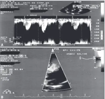
Fig. 2 - a) Outflow tract Doppler velocity demonstrating a significant reduction in the gradient to 10 mmHg after infusion of normal saline; b) Resolution of the mitral insufficiency after the saline infusion.
A