Arquivos Brasileiros de Cardiologia - Volume 8 2 , Nº 6 , Junho 2 0 0 4
519
The association of liver disease and pulmonary vascular dilations has been emphasized by several authors for years, allowing for greater knowledge of the pathophysiological aspects of the arterial hypoxemia found in some patients with the chronic liver disease called hepatopulmonary syndrome. This clinical condition is cha-racterized by the triad of liver dysfunction, intrapulmonary vascular dilation, and hypoxemia1 -1 1. Pulmonary capillary vasodilation is an
extrahepatic complication of severe liver disease, probably due to the vasoactive mediation of nitric oxide2 ,7 ,1 1 -1 3, causing the
oc-currence of an intrapulmonary right-to-left shunt with a conse-quent alteration in alveolar-capillary diffusion and pulmonary ven-tilation/perfusion imbalance1 ,9 ,1 1 ,1 4 ,1 5. In advanced stages of liver
disease, both arterial vasodilation and true pulmonary arteriovenous communications may be present1 ,1 5. The patients may have
nor-mal arterial blood gas analysis or arterial hypoxemia in 9 -2 9 % of the cases, which may be severe and associated with cyanosis and dyspnea1 ,3 ,7 ,1 6 ,1 7. Hemodynamic conditions with elevated cardiac
output, low systemic and pulmonary vascular resistances, and a reduction in the mixed arterial and venous oxygen content may also be present1 ,5 -7 ,1 0 -1 2 ,1 8.
Contrast echocardiography, lung perfusion scan with techne-tium9 9 -labeled macroaggregated albumin, and pulmonary angio-graphy are some of the diagnostic methods used for identifying intrapulmonary vascular alterations in patients with chronic liver disease. Contrast echocardiography is considered the gold standard for the diagnosis of this condition with numerous advantages as compared with other methods, allowing the detection of intra-pulmonary shunts in patients with normal angiographic study or arterial blood gas analysis, or both. Recent studies have emphasized the diagnostic superiority of contrast transesophageal echocar-diography in the research for pulmonary vascular alterations in this group of patients3 ,6 ,1 2 ,1 6 -1 9 -2 6.
The objective of this study was to compare the results of con-trast echocardiography in the transthoracic and transesophageal moda-lities, in addition to determining its importance in the diagnosis of intrapulmonary vascular dilations in candidates for liver transplantation.
M ethods
Contrast echocardiography was consecutively performed in 7 6 patients with severe and advanced liver diseases, who had been included in the protocol for liver transplantation. Patients diagnosed
Original Article
Cont rast Echocardiography in t he Diagnosis of
Int rapulm onary Vascular Dilat ions in Candidat es
f or Liver Transplant at ion
Paulo Robert o Pavarino, Hélio August o do Reis Corbucci, Carlos Henrique de M archi,
Paula Fernanda da M at a, M oacir Fernandes de Godoy
São José do Rio Pret o, SP - Brazil
Hospital de Base de São José do Rio Preto and Faculdade de Medicina de São José do Rio Preto
Mailing address: Paulo Roberto Pavarino – Rua José Felipe Antonio, 3 0 3 /3 2 – Bl.0 7 – São José do Rio Preto, São Paulo, SP, Brazil Cep 1 5 0 9 0 -4 3 0 – E-m a il: pa va rino@ ca rdiol.br
Received: 3 /1 0 /0 3 Accepted: 1 0 /2 0 /0 3
English version by Stela Maris Costalonga
Objective
To determine the importance of contrast echocardiography in the diagnosis of intrapulmonary vascular dilations in patients with severe liver disease, who are candidates for liver transplantation.
M ethods
The study comprised 76 patients with chronic liver disease and no evidence of intrinsic pulmonary disease, heart failure, or congenital heart disease with intracardiac communications, who underwent transthoracic echocardiography with second har-monic imaging. Thirty-two of them underwent consecutive tran-sesophageal study. The result of contrast echocardiography was considered positive when the presence of contrast was detected in the left cardiac chambers with a delay of 4 to 6 cardiac cycles after initial opacification of the right cardiac chambers.
Results
The prevalence of intrapulmonar y vascular dilations was 53.9% (41/76 patients). The sensitivity, specificity, positive and negative predictive values, and accuracy of transthoracic cardiography as compared with those of transesophageal echo-cardiography for confirming pulmonary vascular abnormalities in patients with liver disease were, respectively, 75%, 100%, 1 0 0 %, 8 0 %, and 8 7 .5 %. The degree of arterial oxygenation showed no correlation with the occurrence of a positive echo-cardiographic study. Arterial hypoxemia (PaO2 < 70 mm Hg) was observed in 9 (15.9%) of the 76 patients. The echocardio-graphic study was positive in 37 (55.2%) of the 67 nonhypoxemic patients and in 4 (44.4%) of the 9 hypoxemic ones.
Conclusion
Contrast echocardiography proved to be effective, easy, and safe to use in candidates for liver transplantation. Transthoracic echocardiography may be used in the diagnostic routine of in-trapulmonary vascular dilations, the transesophageal study being reserved for inconclusive cases with clinical suspicion.
Key w ords
Arquivos Brasileiros de Cardiologia - Volume 8 2 , Nº 6 , Junho 2 0 0 4
520
Contrast echocardiography in the diagnosis of intrapulmonary vascular dilations
with chronic pulmonary diseases, heart failure, and congenital heart diseases with intracardiac communications were excluded from the study. The study was conducted after an individual ex-planation about the objectives of the investigation was provided and informed written consent was obtained. The research protocol was evaluated and approved by the Committee on Ethics in Re-search of the institution.
The patients’ mean age was 4 4 ± 1 4 .6 years, 5 9 (7 7 .6 %) were males and 1 8 (2 2 .4 %) were females. Of the 7 6 patients with advanced liver disease, 7 2 had been diagnosed with liver cirrhosis and 4 with liver fibrosis. Of the patients with hepatic cell damage, 1 2 had alcoholic cirrhosis, 9 had hepatitis B, 1 6 had hepatitis C, 3 had hepatitis B and C, 1 5 had mixed cirrhosis (hepati-tis B or C, or both, associated with alcohol), 9 had cryptogenic cirrhosis, 2 had autoimmune cirrhosis, 3 had biliary cirrhosis (primary biliary obstruction), 1 had hemochromatosis, 1 had nonalcoholic steatohepatitis, and 1 had Wilson’s disease. Of the patients with intrahepatic or extrahepatic fibrosis, 1 had schistosomiasis, 1 had paracoccidiodomycosis, and 2 had veno-occlusive diseases (1 throm-bosis of the portal vein and 1 Budd-Chiari syndrome).
For obtaining 2 -dimensional images, an ATL device (Advanced Technology Laboratories Inc., Bothel, WA, USA) was used accor-ding to previously established techniques and sections, with the patient in the left lateral decubitus position 2 7. Contrast
transtho-racic echocardiography was performed in an HDI 5 0 0 0 device with an electronic broadband phased-array transducer with a fre-quency of 2 to 4 MHz. Second harmonic imaging was used in all examinations to reduce imaging artifacts and to increase contrast resolution. Contrast transesophageal echocardiography was per-formed in a CX 2 0 0 Apogee device with the introduction of a multiplanar esophageal probe of 5 .0 MHz at a depth of approxi-mately 3 0 cm from the superior dental arch after local anesthesia of the pharynx. The use of 4 -chamber echocardiographic view allowed for the simultaneous visualization of the atria and, when possible, of the left and right superior pulmonary veins2 8 ,2 9.
The following measurements were taken: diameter of the left atrium, diastolic and systolic dimensions of the left ventricle, the left ventricular ejection fraction using cubed diameters, and systolic pressure of the right ventricle estimated based on tricuspid regur-gitation using the modified equation of Bernoulli2 8 ,3 0 -3 3.
The echocardiographic study was developed following the me-thods reported by Krowka et al1 1 and Aller et al3. The microbubbles
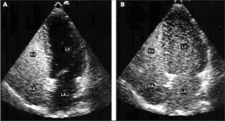
were manually produced by transferring 1 0 to 1 5 times 1 0 mL of saline solution from 1 syringe to another connected in a 3 -way device, and, then, rapidly administered in a peripheral venous route. The study was considered positive when the abnormal pre-sence of contrast was detected in left cardiac chambers with a delay of 4 to 6 cardiac cycles, after initial opacification of the right cardiac cham bers (fig. 1 ). Three injections were usually performed to determine the reproducibility. The results were re-corded in videocassette tapes and analyzed by 2 observers. The subsequent injections were initiated only after complete removal of the microbubbles from the right and left cavities. According to the opacification of the left atrium, a semiquantitative analysis of the microbubbles was performed according to the criteria establi-shed by Aller et al3. The simultaneous comparison of the maximum
intensity of the echocardiographic images produced by the mi-crobubbles between the right and left cardiac cavities provided
the following classification: degree 1 - absence of microbubbles; degree 2 passage of a few isolated microbubbles; degree 3 -passage of numerous isolated microbubbles; degree 4 - -passage of numerous microbubbles resulting in an increase in echogenicity; degree 5 - opacification of the left atrium, but to a lesser extent as compared with that of the right atrium; degree 6 - complete opacification of the left atrium similar to that of the right atrium. Degrees 1 and 2 were considered normal or the absence of pul-monary vasodilations; degree 3 was considered to represent mild pulmonary vasodilations; and finally, degrees 4 to 6 were considered significant or important pulmonary vasodilations.
The arterial oxygen partial pressure (PaO2) was determined in samples of arterial blood collected from the radial artery under normal conditions, as close as possible to the day of
echocardio-graphic study (1 -3 days). A PaO2 value lower than 7 0 mm Hg
was considered an indication of arterial hypoxemia.
The continuous quantitative variables were analyzed with the
aid of the Student t test, and Tukey correction was used when
necessary. The frequencies were compared using the chi-square test or Fisher exact test. An alpha level of 5 % was admitted, and P values ≤ 0 .0 5 were considered significant.
Results
All diagnostic procedures were well tolerated, and transeso-phageal echocardiography was performed with no complications. The use of transthoracic and transesophageal contrast echocar-diography showed the presence of pulmonary vascular dilations in 5 3 .9 % (4 1 /7 6 ) of the patients. In the transthoracic study in iso-lation, prevalence of intrapulmonary vascular dilations was observed in 4 8 .7 % (3 7 /7 6 ) of the cases. Of the 3 2 patients consecutively undergoing transesophageal study, 1 6 (5 0 %) had a positive contrast echocardiography (P = 1 .0 ; Fisher exact test). Four patients with an initially inconclusive transthoracic study underwent transeso-phageal echocardiography and were considered positive. Hepato-pulmonary syndrome was observed in only 4 (5 .3 %) patients. Ac-cording to the criteria established by Aller et al3, 1 6 (2 1 %)
pa-tients had mild pulmonary vasodilations, 2 5 (3 3 %) papa-tients had significant pulmonary vasodilations, and 3 5 (4 6 %) had normal echocardiograms. Transthoracic echocardiography had a sensitivity of 7 5 %, specificity of 1 0 0 %, positive predictive value of 1 0 0 %, negative predictive value of 8 0 %, and accuracy of 8 7 .5 % for the
Arquivos Brasileiros de Cardiologia - Volume 8 2 , Nº 6 , Junho 2 0 0 4
521
Contrast echocardiography in the diagnosis of intrapulmonary vascular dilationsdiagnosis of intrapulmonary vascular dilations as compared with transesophageal echocardiography, which is considered the gold standard.
Analyzing the etiology of liver disease, 2 8 (4 6 .7 %) of the 6 0 patients with cirrhosis due to hepatic cell destruction, 8 (8 8 .9 %) of the 9 patients with cryptogenic cirrhosis, 3 with biliary cirrhosis, and 2 with cirrhosis due to veno-occlusive diseases had a positive echocardiogram. Comparing the results of the patients with cryp-togenic cirrhosis and pulmonary vascular dilations with those of patients with cirrhosis due to all the other etiologies, by using the chi-square test, no significant statistical difference was found between the groups (P = 0 .0 5 9 ). However, comparing cryptogenic cirrhosis in isolation with those with hepatic cell destruction, the difference was statistically significant (P = 0 .0 4 4 ).
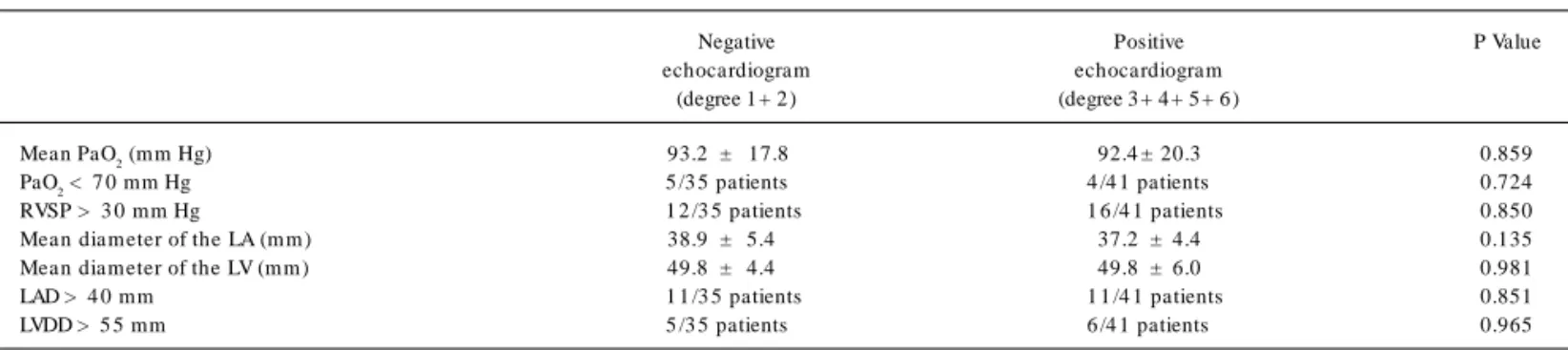
The variables of blood gas analysis and of Doppler echocardio-graphy are shown in table I. Arterial hypoxemia was present in 9 (1 5 .9 %) of 7 6 patients, but only 4 (4 4 .4 %) had evidence of pulmonary vascular dilations. Of the 6 7 patients with no arterial hypoxemia, 3 7 (5 5 .2 %) had a positive echocardiogram.
Discussion
The pulmonary vascular changes found in patients with chronic liver disease are disseminated vasodilations with diameters ranging from 1 5 to 1 5 0 µm, which are more prominent at the capillary level and close to gas exchange areas. The deviation of pulmonary blood flow to the dilated capillaries avoids the functioning alveolar units, impairing the pulmonary diffusion-perfusion relation with a consequent decrease in arterial oxygen saturation1 ,5 ,7 ,9 -1 1.
The intrapulmonary vascular abnormalities are not routinely detected because of their uncommon presentation in the general population and because of their unespecific appearance on routine examinations3 4. Studies1 ,3 ,4 ,7 ,1 1 ,1 6 ,1 7 ,3 5 ,3 6 with contrast
echocardio-graphy in patients with severe liver cirrhosis have revealed the existence of intrapulmonary vascular dilations in 1 3 to 4 7 % of the patients, even in normal angiographic studies. In our study, the identification of pulmonary vascular dilations using contrast echo-cardiography was possible in 4 1 of 7 6 patients studied, ie, 5 3 .9 % of the cases, a result similar to those in the literature.
Studies by Vedrinne et al2 4 and Aller et al3 showed the
superio-rity of contrast transesophageal echocardiography for diagnosing intrapulmonary vascular dilations in candidates for liver transplan-tation3 ,2 4 ,2 5. Transesophageal echocardiography, considered the
gold standard for diagnosing intrapulmonary vascular dilations,
allo-wed dem onstra tion of the presence of tha t condition in 5 0 % (1 6 /3 2 ) of our cases. In 4 patients with previously inconclusive transthoracic study, due to an inadequate acoustic window, intra-pulmonary vascular dilations could only be demonstrated after using transesophageal echocardiography. In our study, the use of second harmonic imaging in transthoracic echocardiography significantly contributed to the obtainment of satisfactory results similar to those in the transesophageal study. Comparing the proportion of individuals with pulmonary vascular dilations diagnosed on trans-thoracic and transesophageal echocardiography using the Fisher test, no statistical difference was found (P= 1 ), showing that the efficacy of the 2 diagnostic methods is equivalent. The comparison of the results of contrast transthoracic and transesophageal echo-cardiography consecutively performed in 3 2 patients showed the following results: sensitivity, 7 5 %; specificity, 1 0 0 %; positive pre-dictive value, 1 0 0 %; negative prepre-dictive value, 8 0 %; and accuracy, 8 7 .5 %. This validates contrast transthoracic echocardiography with second harmonic imaging as a rapid, safe, noninvasive, re-liable, and inexpensive diagnostic test for studying these patients. Hepatopulmonary syndrome, usually reported in 9 to 2 9 % of liver failure cases1 ,3 ,7 ,1 6 ,1 7, was found in 4 (5 .3 %) of our patients.
In the present study, the degree of arterial oxygenation had no statistical correlation with the occurrence of a positive echocar-diography. These findings are similar to those of Krowka et al1 8,
who also did not report a correlation between pulmonary vascular abnormalities and blood gases in patients with a positive echo-cardiogram (1 3 .2 % of the cases) as compared with those with a normal echocardiogram. Mimidis et al4 also found normal arterial
blood gas analysis in 5 6 cirrhotic individuals, 8 (1 4 .3 %) of whom had a positive contrast echocardiogram. Vedrinne et al2 4, however,
found hypoxemia in 5 6 % and 3 3 % of the patients with intrapul-monary shunts diagnosed on transesophageal and transthoracic echocardiography, respectively. In regard to the mean PaO2 values found in this study, they were similar in the different degrees of opacification of the left cardiac cavities (P= 0 .8 5 9 ), despite the results by Hopkins et al3 6, which were significantly lower in
in-dividuals with greater opacification of the left cavities (P < 0 .0 1 ). Based on our results, one may state that occasional abnormalities in arterial oxygenation of patients with chronic liver disease should not be considered indicators of intrapulmonary shunts, and, in isolation, they do not indicate that condition.
In the face of a probable hyperdynamic circulatory condition existing in individuals with intrapulmonary vascular shunts, which could cause alterations in the diameter and volume of the left
Table I - Variables of blood gas analysis and Doppler echocardiography
Negative Positive P Value
echocardiogram echocardiogram
(degree 1 + 2 ) (degree 3 + 4 + 5 + 6 )
Mean PaO2 (mm Hg) 93.2 ± 17.8 92.4 ± 20.3 0.859
PaO2 < 7 0 mm Hg 5 /3 5 patients 4 /4 1 patients 0.724
RVSP > 3 0 mm Hg 1 2 /3 5 patients 1 6 /4 1 patients 0.850
Mean diam eter of the LA (m m ) 38.9 ± 5.4 37.2 ± 4.4 0.135
Mean diam eter of the LV (m m ) 49.8 ± 4.4 49.8 ± 6.0 0.981
LAD > 4 0 mm 1 1 /3 5 patients 1 1 /4 1 patients 0.851
LVDD > 5 5 mm 5 /3 5 patients 6 /4 1 patients 0.965
PaO2 - arterial oxygen partial pressure; RVSP - right ventricular systolic pressure; LA - left atrium ; LV - left ventricle; LAD - left atrial diam eter; LVDD - left ventricular
Arquivos Brasileiros de Cardiologia - Volume 8 2 , Nº 6 , Junho 2 0 0 4
522
Contrast echocardiography in the diagnosis of intrapulmonary vascular dilations
1. Muller C, Schenk P. Hepatopulm onary syndrom e. Review article. Wien Klin
Wo-chenschr 1 9 9 9 ; 1 1 1 : 3 3 9 -4 7 .
2. Barbosa WF, Kondo M. Alterações vasculares pulm onares na hipertensão porta.
Rev Soc Cardiol Estado de São Paulo 2 0 0 0 ; 1 0 : 6 0 9 -2 0 .
3. Aller R, Moya JL, Moreira V, et al. Diagnosis of hepatopulm onary syndrom e with
contrast transesophageal echocardiography. Advantages over contrast transtho-racic echocardiography. Dig Dis Sci 1 9 9 9 ; 4 4 : 1 2 4 3 -8 .
4. Mim idis KP, Vassilakos PI, Mastorakou, et al. Evaluation of contrast
echocardio-graphy and lung perfusion scan in detecting intrapulm onary vascular dilation in normoxemic patients with early liver cirrhosis. Hepatogastroenterology 1 9 9 8 ; 4 5 : 2303-07.
5. Silva AO, D’Albuquerque LAC. Doenças do Fígado. Vol I. Rio de Janeiro: Revinter,
2 0 0 1 : 6 6 0 -3 .
6. Krowka MJ, Cortese DA. Hepatopulmonary syndrome: an evolving perspective in
the era of liver transplantation. Hepatology 1 9 9 0 ; 1 1 : 1 3 8 -4 2
7. Castro M, Krowka MJ. Hepatopulmonary Syndrome: A pulmonary vascular
com-plication of liver disease. Clinics Chest Med 1 9 9 6 ; 1 7 : 3 5 -4 8 .
8. El-Gam al M, Stoker JB, Spiers EM, Whitaker W. Cyanosis com plicating hepatic
cirrhosis. Report of a case due to m ultiple pulm onary arteriovenous fistulas. Am J Cardiol 1 9 7 0 ; 2 4 : 4 9 0 -4 .
9. Berthelot P, Walker JG, Sherlock S, Reid L. Arterial changes in the lungs in
cirrho-sis of the liver-lung spider nevi. N Engl J Med 1 9 6 6 ; 2 7 4 : 2 9 1 -8 .
10. Krowka MJ, Cortese DA. Pulmonary aspects of liver disease and liver transplanta-tion. Clin Chest Med 1 9 8 9 ; 1 0 : 5 9 3 -6 1 6 .
11. Krowka MJ, Cortese DA. Pulm ona ry a spects of ch ronic liver disea se a nd liver transplantation. Mayo Clin Proc 1 9 8 5 ; 6 0 : 4 0 7 -1 8 .
12. Aboussouan LS, Stoller JK. The hepatopulmonary syndrome. Baillière’s Best Pract Res Clin Gastroenterol 2 0 0 0 ; 1 4 : 1 0 3 3 -4 8 .
13. Oh KS, Bender TM, Bowen A, Ledesma-Medina J. Plain radiographic, nuclear me-dicine and angiographic observations of hepatogenic pulmonary angiodysplasia. Pediatr Radiol 1 9 9 3 ; 1 3 : 1 1 1 -5 .
14. Ciappi G, Chiesa A, Chiandussi L, et al. Study of the causes of the hypoxem ia in hepatic cirrhosis. Relative importance of pulmonary and extra-pulmonary shunt. Minerva Med 1 9 6 6 ; 5 7 : 3 5 3 3 -6 .
15. Krowka MJ, Cortese DA. Hepatopulmonary syndrome – Current concepts in diag-nostic and therapeutic considerations. Chest 1 9 9 4 ; 1 0 5 : 1 5 2 8 -3 7 .
16. Hourani JM, Bellamy PE, Tashkin DP, Batra P, Simmons MS. Pulmonary disfunc-tion in advanced liver disease: frequent occurrence of an abnormal diffusing capa-city. Am J Med 1 9 9 1 ; 9 0 : 6 9 3 -7 0 0 .
17. Auletta M, Oliviero U, Iasiuolo L, Scherillo G, Antoniello S. Pulmonary hypertension associated with liver cirrhosis: An echocardiography study. Angiology 2 0 0 0 ; 5 1 : 1013-20.
18. Krowka MJ, Tajik J, Dickson R, Wiesner RH, Cortese DA. Intrapulmonary vascular dilatations (IPVD) in liver transplant candidates. Screening by two-dim ensional contrast-enhanced echocardiography. Chest 1 9 9 0 ; 9 7 : 1 1 6 5 -7 0 .
References
19. Cerdeña IL, Ojeda FB, Trujillo DA, et al. Pulmonary arteriovenous fistulas. Diagno-sis using contrast echocardiography and advantages of the real-time bidimensio-nal technic. Rev Esp Cardiol 1 9 8 3 ; 3 6 : 4 4 3 -6 .
20. Barzilai B, Waggoner AD, Spessert C, Picus D, Goodenberg D. Two-dim ensional contrast echocardiography in the detection and follow-up of congenital pulmonary arteriovenous malformations. Am J Cardiol 1 9 9 1 ; 6 8 : 1 5 0 7 -1 0 .
21. Shub C, Ta jik AJ, Sewa rd JB, Dines DE. Detecting intra pulm ona ry right-to-left shunt with contrast echocardiography: observations in a patient with diffuse pul-monary arteriovenous fistulas. Mayo Clinic Proc 1 9 7 6 ; 5 1 : 8 1 -4 .
22. Kuram ochi T, Izum i S, Nakayam a, et al. Contrast echocardiography detection of arteriovenous shunt in a hypoxemic patient with liver cirrhosis. J Cardiol 1 9 9 4 ; 2 4 : 155-60.
23. Hind CR, Wong CM. Detection of pulmonary arteriovenous fistulae in patient with cirrhosis by contrast 2 D echocardiography. Gut 1 9 8 1 ; 2 2 : 1 0 4 2 -5 .
24. Vedrinne JM, Duperret S, Bozollon T, et al. Com parison of transesophageal and tra ns th ora cic contra s t ech oca rdiogra p h y for detection of a n intra p ulm ona ry shunt in liver disease. Chest 1 9 9 7 ; 1 1 1 : 1 2 3 6 -4 0 .
25. Ho WJ, Chu PH, Chiang SY, Chiang CW. Localizing intrapulmonary shunt in hepa-topulmonary syndrome by transesophageal echocardiography. Jpn Heart J 1 9 9 9 ; 4 0 : 3 6 9 -7 4 .
26. Pilatis ND, Jacobs LE, Rerkpattanapipat P, et al. Clinical predictors of pulmonary hypertension in patients undergoing liver transplant evaluation. Liver Transpl 2 0 0 0 ; 6 : 8 5 -9 1 .
27. Tajik AJ, Seward JB, Hagler DJ, Mair DD, Lie JT. Two-dimensional real time ultra-sonic imaging of the heart and great vessels. Technique, image orientation, struc-ture identification, and validation. Mayo Clin Proc 1 9 7 8 ; 5 3 : 2 7 1 -3 0 3 . 28. Morcef FAP. Ecocardiografia Uni-Bidim ensional, Transesofágica e Doppler. 2 nd
ed. Rio de Janeiro: Revinter, 1 9 9 6 .
29. Assef JE, Belém L, Castro-Lima A, Torreão JAM. Ecocardiografia Transesofágica: Atlas-Texto. Rio de Janeiro: Revinter, 2 0 0 0 .
30. Sahn DJ, DeMaria A, Kisslo J, Weyman A. Recommendations regarding quantita-tion in M-m ode echocardiography: results of a survey of echocardiography m ea-surements. Circulation 1 9 7 8 ; 5 8 : 1 0 7 2 -8 3 .
31. Pom bo JF, Troy BL, Russel RO. Left ventricular volum es and ejection fraction by echocardiography. Circulation 1 9 7 8 ; 4 2 : 4 8 0 -9 0 .
32. Feingenbaum H. Echocardiography. 5 th ed. Philadelphia: Lea & Febiger, 1 9 9 4 . 33. Ortiz J, Silva CES, Gh efter CGM, et a l. O Ecoca rdiogra m a no Apoio à Decisã o
Clínica. 2 th ed. Rio de Janeiro: Revinter; 1 9 9 7 .
34. Gianesella RB, Rossi Filho RI, Zielinsky P. Diagnóstico e terapêutica da fístula ar-teriovenosa pulm onar na infância. Descrição de caso e revisão da literatura. Arq Bras Cardiol 2 0 0 1 ; 7 7 : 2 7 4 -7 .
35. Murakami JW, Rosembaum DM. Right-to-left pulmonary shunting in pediatric he-patopulmonary syndrome. Clin Nucl Med 1 9 9 9 ; 2 4 : 8 9 7 .
36. Hopkins WE, Waggoner AD, Barzilai B. Frequency and significance of intrapulmona-ry right-to-left shunting in end-stage hepatic disease. Am J Cardiol 1 9 9 2 ; 7 0 : 5 1 6 -9 .
cavities or in pulmonary vascular bed pressure, the present study did not find any correlation between these variables and the diag-nosis of intrapulmonary vascular dilations on contrast echocardio-graphy. In regard to the findings of the etiology of liver disease, although interesting, there is no pathophysiological support for the statement that pulmonary vascular disorders are more frequent in certain groups of patients with chronic liver disease.
Briefly, contrast transthoracic echocardiography with