31
Original Article
Contribution to the Diagnosis and Treatment of
Pulmonary Arteriovenous Fistulae after a
Bidirectional Glenn Operation
Maria Virgínia Tavares Santana, Paulo Paredes Paulista, Sérgio Cunha Pontes Júnior,
César Augusto Esteves, Valmir Fernandes Fontes, José Eduardo Moraes Rego Sousa
São Paulo - SP, Brazil
Objective
To determine the incidence of pulmonary arteriovenous fistulae (PAVFs) after the bidirectional Glenn operation and the possible independent variables that could influence their appearance; to confirm the use of microbubble contrast echocardiography for the detection of PAVFs; and to test the sensitivity and specificity of pulmonary angiography.
Methods
From March 1990 to December 1995, 59 patients were opera-ted upon. Their ages ranged from 2 to 132 months (mean, 32.7± 33.6). All underwent clinical and laboratory examinations, mi-crobubble contrast echocardiography, and cardiac catheterization.
Results
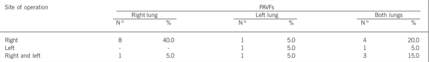
Of the 54 survivors, 20 (37.0%) had PAVFs. The ages ranged from 2 to 132 months (mean, 29.6±29.7). In 13 (65%) patients, the bidirectional Glenn operation was performed on the right-hand side; in 2 (10.0%) patients, on the left-hand side; and in 5 (25%) patients, it was bicaval. The follow-up of the patients with PAVFs ranged from 4 to 84 months (mean, 32.4±21.65), and that of the patients without fistulae ranged from 1 to 77 months (mean, 23.4±18.8), with statistical significance (P=0.04). The PAVFs were diagnosed by use of microbubble contrast echocardiography in 20 cases; the examination was considered positive when return of microbubbles through the pulmonary veins was detected. PAVFs were observed in the right lung in 9 (45%) patients, in the left lung in 3 (15%) patients, and in both lungs in 8 (40%) patients. The pulmonary angiography showed alterations compatible with PAVFs in 16 patients, with a sensitivity of 80%.
Conclusion
The incidence of PAVFs after the bidirectional Glenn operation was high (37%). The time interval elapsed after the bidirectional Glenn operation was the only independent variable that signifi-cantly correlated with the appearance of PAVFs (P=0.04). Microbu-bble contrast echocardiography was the standard diagnostic me-thod. The pulmonary angiography showed a sensitivity of 80.0%.
Keywords
pulmonary arteriovenous fistulae, bidirectional Glenn operation, microbubble contrast echocardiography
Cyanogen congenital heart diseases, which behave functionally as univentricular heart, are usually characterized by a poor prog-nosis, unsatisfactory quality of life, and impossibility of surgical correction that reestablishes the normal cardiac anatomy.
In the past 4 decades, several proposals for palliative operations have been tested, initially in experimental animals, and then, if successful, in children with such defects. One of them, the Glenn operation, was based on the principle that systemic venous blood might reach the pulmonary circulation without the participation of the right cardiac cavities. This operation was experimentally proposed by Carlon et al 1 in 1951, successfully performed for the first time by Meshalkin 2 and Bakulev 3 in 1956, and diffused by Glenn 4 in 1958.
The classical Glenn operation, as this procedure has been known, remained unaltered for many years. Haller et al 5 experi-mentally introduced the concept of bilateral partial cavopulmonary anastomosis, in which the continuity between the pulmonary arte-ries was preserved by a terminolateral anastomosis between the superior vena cava and the right pulmonary artery.
Azzolina et al 6 performed that operation for the first time, which became known as the bidirectional Glenn operation. The major advantage of this technique was that the right pulmonary artery, not separated from the confluence, allowed the division of the blood volume of the superior vena cava between both lungs. Mathur and Glenn 7 reported the late evolution of 56 patients out of 63 who underwent the classical Glenn operation and des-cribed for the first time the development of pulmonary arteriovenous fistulae in that type of operation. From this publication onwards, other studies reported such complications 8,9, decreasing the en-thusiasm in regard to the classical Glenn operation, which was then abandoned in favor of the bidirectionalGlennoperation 6, 10.
Methods
From March 1990 to December 1995, 59 patients with com-plex cyanogen congenital heart diseases underwent the bidirectional Glenn operation at our service. Five patients died, 4 immediately after the procedure and one during hospitalization. The 54 survivors comprised the case series of this study. Age, on the occasion of the operation, varied from 2 to 132 (mean, 31.4±33.56; median, 18.5) months, and 4 patients were less than 6 (range, 2 to 5; mean, 4.0) months. Of the 54 patients, 28 (51.9%) were of the male sex, and 26 (48.1%) were of the female sex. The heart
Instituto Dante Pazzanese de Cardiologia - São Paulo Mailing address: Maria Virgínia Tavares Santana
Av. Rouxinol, 780/51 - Cep 04516-001 -São Paulo, SP - Brazil E-mail: virginia.tati@uol.com.br
32
diseases were as follows: tricuspid atresia, 30 (56.0%); atrioven-tricular connection of the double-inlet type, 17 (31.5%); right ventricular double outflow tract, 4 (7.4%); Ebstein disease, 1 (1.7%); complete transposition of the great arteries, 1 (1.7%); and pulmonary atresia with intact ventricular septum, 1 (1.7%). The pulmonary valve had normal anatomy in 19 (35.2%) patients, was stenotic in 25 (46.3%), and atresic in 10 (18.5%).
The bidirectional Glenn operation was performed by anasto-mosing the right superior vena cava to the right pulmonary artery in 38 (70.4%) patients, to the left pulmonary artery in 6 (11.2%), and simultaneously to the right and left pulmonary arteries (bicaval bidirectional Glenn operation) in 8 (14.9%) patients. In the 2 (3.4%) remaining patients, the inferior vena cava was used for connection with the right pulmonary artery (inverted bidirectional Glenn operation). Blood flow from the ventricular cavity to the pulmonary trunk was maintained in 21 (38.9%) patients. It was abolished after the surgical ligature of the pulmonary trunk in 23 (42.6%) patients. In the other 10 (18.5%) patients, it never occurred due to the presence of pulmonary valve atresia.
All patients underwent periodical review every 3 months, which included, in addition to the complete clinical examination, electro-cardiography at rest, chest radiography in the posteroanterior projec-tion, pulse oximetry, measurement of the hematocrit and hemo-globin, microbubble contrast echocardiography with sequential ana-lysis of the heart disease, and, finally, cardiac catheterization.
Transthoracic echocardiographic study was performed in 41 (76.0%) patients weighing less than 20 kg, and the transesopha-geal technique was used in 13 (24.0%) patients weighing 20 kg or more. The initial maximum dosage of 80 mg/kg of 20% chloral hydrate was used as a sedative for patients undergoing transthoracic echocardiography after a 3-hour fast. If the patient did not respond to sedation, 20% of the initial dosage was administered 20 minutes after the first dose. For transesophageal echocardiography, anes-thesia was induced with propofol (1 to 2 mg/kg) and maintained with a dosage of approximately 100 µg/kg per minute. When necessary, concomitant inhalation with halothane was provided. The venous access for microbubble injection was chosen ac-cording to the location of the bidirectional Glenn operation as follows: in patients with the bidirectionalGlenn operation performed on the right- or left-hand side, the right or left brachial vein, respectively; in those with the bicaval bidirectional Glenn, the right and left brachial veins; and in those with the inverted bidi-rectional Glenn, the right femoral vein.
The contrast material consisted of 5.0 mL of saline solution with 0.5 mL of environmental air vigorously mixed by use of a system with 2 taps and 3 connections, producing an opaque saline solution rapidly injected into the patient’s vein 11.
The confirmation of the diagnosis of pulmonary arteriovenous fistulae was obtained by detecting the echocardiographic contrast material (bubbles) in the pulmonary veins after a maximum of 8 cardiac cycles. The number of contrast material injections varied, and they were repeated as many times as necessary for the correct diagnosis.
The echocardiographic examinations were performed with the Ultramark-9 HDI model ATL (Advanced Technology Laboratories) apparatus, using a phased array transducer at a 5-3 MHz frequency, and atransesophageal biplane probe at a 5 MHz frequency. The
2-dimensional images were recorded on videotape for later analysis. The duration of the echocardiographic contrast material injection appeared on screen and was recorded on the videotape.
The conventional echocardiographic examination was based on the sequential analysis 12 for defining the anatomy of the heart disease and functionally assessing the bidirectional Glenn operation. Then, the contrasted examination was performed. The subcostal, 4-chamber apical, longitudinal parasternal, and suprasternal views were used for transthoracic examination. The 4-chamber medium transverse and longitudinal planes were used for the transesopha-geal examination.
The bidirectional Glenn operation was conventionally performed with or without extracorporeal circulation, depending on the sur-geon’s option. The inverted Glenn operation, which consisted of the construction of a tunnel inside the right atrium, allowing con-tinuity of the inferior vena cava with the right pulmonary artery, was performed according to a previously published technique 13.
For confirming that the microbubbles do not cross the capillary barrier in the absence of pulmonary arteriovenous fistulae, a control group of 27 healthy children was used. Their ages ranged from 12 to 156 months (mean, 58.4±34.8; median, 48.0), and they un-derwent the same clinical, laboratorial, and echocardiographic pro-tocol of the general case series, except for the hemodynamic study. The statistical analysis was performed by calculating the arith-metic mean, standard deviation, and median for describing the continuous quantitative variables. The qualitative and categorical variables were expressed as percentages, and, for their comparison, the Pearson χ2 (chi-square) test or the Fisher exact test was used. For comparisons of the means of the quantitative variables, the Student t test and the nonparametric Mann-Whitney test were used for independent populations. The actuarial curve was calcu-lated for studying the accumucalcu-lated probability of the time free from the event pulmonary arteriovenous fistulae. The possibility of the risk factors for the appearance of pulmonary arteriovenous fistulae was studied by using the multivariate logistic regression analysis with the conditional forward stepwise selection model. In all statistical tests, the significance level adopted was 0.05. For estimating the population parameters, 95% confidence intervals were calculated. The statistical calculations and analyses were performed with the SPSS program for Windows version 6.0.
Results
Fifty-four patients were followed up for the maximum period of 110 months (mean, 31.4±33.56; median, 18.5). Pulmonary arteriovenous fistulae were detected in 20 patients (37.0% - 95% CI: 24.1 - 49.9%).
The survival curve free from the event pulmonary arteriovenous fistula had a mean of 50.0 (95% CI: 39 - 61 months) and median of 48.0 (95% CI: 38 - 58 months), with a 37.0% accumulated probability of up to 84 months (fig. 1).
33
arteriovenous fistulae, the follow-up duration ranged from 1 to 77 months (mean, 23.4±18.84; median, 18.0; P=0.04), showing statistical significance. The comparison between the ages of the general case series and the event of pulmonary arteriovenous fistulae by using the class interval showed no statistical significance (P=0.48).
In the case series of 54 patients, 4 (6.8%) underwent the bidirectional Glenn operation before the age of 6 months (range, 2 to 5 months; mean, 4.0 months), and pulmonary arteriovenous fistulae were detected in 2 patients at the ages of 23 and 24 months (fig. 2).
When comparing the sex of the patients with and without fistulae, the P value was also not significant (P=0.41).
The most prevalent heart diseases among the patients deve-loping pulmonary arteriovenous fistulae were tricuspid atresia in 10 (50.0%) and atrioventricular connection of the double-inlet type in 8 (40.0%) patients. The 2 remaining patients had right ventricular double outflow tract and complete transposition of the
great arteries (10.0%). No statistical significance was observed between the 2 major groups of heart diseases in regard to the presence or absence of pulmonary arteriovenous fistulae (P=0.54). Table I shows the diagnosis, situs cordis and visceral situs, associated defects and previous surgeries in each patient. No statistical significance was observed in regard to the situs and the development of pulmonary arteriovenous fistulae (P=0.18). The performance of palliative surgeries prior to the bidirectional Glenn operation was also not an independent variable with statistical significance (P=0.29).
As can be seen in table I, although lacking statistical signifi-cance due to the small size of the sample, 3 (75%) of the 4 patients with left atrial isomerism developed pulmonary arteriove-nous fistulae (cases 6, 7, and 20) (fig. 3 and 4). All had atrioven-tricular connection of the double-inlet type to the venatrioven-tricular cavity, which was morphologically right in 2, and morphologically left in one patient. In addition, they had a single atrioventricular valve and interruption of the hepatic segment of the inferior vena cava, 1.0
0.9 0.8 0.7 0.6 0.5 0.4 0.3 0.2 0.1 0.0
Probabilidade acumulada
0 20 40 60 80 100
Tempo em meses Fig. 1 - Pulmonary arteriovenous fistula event-free survival.
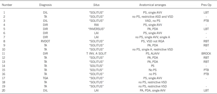
Table I – List of the pulmonary arteriovenous fistulae comprising the number of each case, the diagnosis, the situs, the anatomical arranges, and the previous operations (Prev Op)
Number Diagnosis Situs Anatomical arranges Prev Op
01 DIL “SOLITUS” PS, single AVV LBT
02 TA “SOLITUS” no PS, restrictive ASD and VSD
-03 DIL “SOLITUS” VAD, no PS PTB
04 DIR RAI PS, single AVV
-05 DIR “INVERSUS” PA, PDA LBT
06 DIR LAI PS, single AVV
-07 DIR LAI no PS, single AVV, single A
-08 RVDOT “SOLITUS” PS, VSD not RGA RBT
09 TA “SOLITUS” PA, PDA RBT
10 TA “SOLITUS” no PS, single A, restrictive VSD
-11 DIR T INV, A SOLIT PS, ALAVV BROCK
12 TA “SOLITUS” PA, PDA RBT
13 TA “SOLITUS” PA, PDA RBT
14 TA ’SOLITUS’’ PS
-15 TA ’SOLITUS’’ No PS PTB
16 TA “SOLITUS’’ no PS PTB
17 TGA ‘’SOLITUS’’ PS, single AVV
-18 TA ‘’SOLITUS’’ no PS, restrictive VSD
19 TA ‘’SOLITUS’’ no PS, restrictive VSD
-20 DIL LAI PA, PDA, single AVV LBT
DIL - double inlet to the left univentricular cavity; DIR - double inlet to the right univentricular cavity; TA - tricuspid atresia; RVDOT - right ventricular double outflow tract; TGA - complete transposition of the great arteries; RAI - right atrial isomerism; LAI - left atrial isomerism; T INV - thoracic “inversus”; A SOLIT - abdominal “solitus”; PS - pulmonary stenosis; PA - pulmonary atresia; AVV - atrioventricular valve; ASD - atrial septal defect; PDA - patent ductus arteriosus; VSD - ventricular septal defect; single A - single atrium; VSD not RGA - ventricular septal defect not related to the great arteries; RBT - right Blalock-Taussig; LBT - left Blalock-Taussig; PTB - pulmonary trunk banding; VAD - ventriculoarterial discordance; ALAVV - atresic left atrioventricular valve.
Fig. 2 - Case # 2 – Angiographic sequence in posteroanterior projection. A) contrast material injection into the right superior vena cava (RSVC), with opacification of both pulmonary arteries. Note the vascular dilations in the inferior lobe of the right lung, compatible with PAVFs; B) magnification of the right pulmonary artery (RPA), highlighting the angiomatoid lesions.
RSVC
RPA RPA
34
with continuation and drainage through the azygos system in one patient and through the hemiazygos system in 2 others.
The pulmonary biopsy revealed pulmonary parenchyma with ectatic and congestive arterial and venous vascular network com-patible with pulmonary arteriovenous fistulae (fig. 5 and 6).
In 13 (65.0%) of 20 patients with pulmonary arteriovenous fistulae, the right superior vena cava was used for connecting with the right pulmonary artery; in 2 (10.0%), the left superior vena cava was anastomosed to the left pulmonary artery; and in 5 (25.0%), both the left and right venae cavae were anastomosed to the respective left and right pulmonary arteries.
Blood flow from the ventricular cavity to the pulmonary trunk was maintained in 10 (50.0%) patients. In 5 (25.0%), blood flow was abolished by surgical ligature of the pulmonary trunk, and in the other 5, it never occurred due to the presence of pulmonary valve atresia. The comparative study considering the location of the operation and maintenance of the anterograde flow between the groups with and without pulmonary arteriovenous fistulae showed no statistical significance (P=0.31 and 0.19, respectively).
The duration of the follow-up of patients in whom pulmonary arteriovenous fistulae were detected after the bidirectional Glenn operation ranged from 4 to 84 months (mean, 32.4±21.65; me-dian, 25.0). Six patients had progressive dyspnea and stable cya-nosis on the occasion of the detection of the fistulae; 8 had progressive cyanosis; and 6 had progressive dyspnea and cyanosis. On echocardiographic study, the duration between injection of the contrasted material and the appearance of the microbubbles in the pulmonary veins ranged from 3 to 8 seconds, which corres-ponded to the mean of 5 cardiac cycles (range, 4 to 7). As no contrast material was observed in the inferior vena cava and in the systemic return atrium, the possibility of systemic venous
collaterals was excluded in 19 (95.0%) patients. In one (5.0%), the presence of microbubbles was observed in the inferior vena cava, which also allowed the diagnosis of systemic venovenous connection to that vein. In 9 (45.0%) patients, pulmonary arterio-venous fistulae were detected in the right lung; in 3 (15.0%) patients, in the left lung; and in 8 (40.0%) patients, in both lungs (tab. II). Of the 34 patients who developed no pulmonary arterio-venous fistulae, systemic arterio-venous collaterals were observed in 14 (41.1%) patients.
Of the 20 patients with pulmonary arteriovenous fistulae diag-nosed on microbubble contrast echocardiography, pulmonary angio-graphy detected fistulae in 16 (80.0%) due to the presence of one or more of the following factors: 1) reticular appearance of the lung parenchyma; 2) angiomatoid dilations of the lobar pul-monary arteries (figs. 2 and 4); 3) loss of the capillary phase; and 4) rapid arteriovenous transit. Four patients (cases 7, 13, 16, and 19) had no angiographic images of fistulae. Table III shows the echocardiographic and angiographic findings and the time interval between the bidirectional Glenn operation and the detection of the fistulae in 20 patients.
Angiography had a sensitivity of 80.0%, specificity of 100%, and positive and negative predictive values of 100% and 89.5%. Of the 20 patients with pulmonary arteriovenous fistulae, 10 underwent total cavopulmonary operation; 2 (10.0%) died in the immediate phase due to hypoxemia and low cardiac output syn-drome; 5 are awaiting surgical correction; and 4 are not indicated for surgery, because of the presence of bilateral diffuse fistulae in 2 patients, and hypoplasia of the pulmonary arteries, with a Nakata index 14 lower than 200 mm2/m2, in the other 2. One patient died after the hemodynamic restudy (case 8), which was performed for assessing the possibility of definitive correction (total
cavopul-Fig. 3 -Case # 20 - A) transesophageal study with visualization of the right
superior and inferior pulmonary veins (RSPV and RIPV) in a longitudinal view prior to contrast material injection; B) return of the contrast material through the right superior and inferior pulmonary veins, characterizing PAVFs in the entire right lung.
Fig. 4 - Case # 20 - A) injection of contrast material through the bidirectional Glenn operation to the right, with selective opacification of the right pulmonary artery, which shows diffuse angiomatoid dilations in the entire right lung. B) selective injection of contrast material into the left pulmonary artery through the bidirectional Glenn operation to the left, showing a normal angiographic appea-rance. RPA- right pulmonary artery; LPA- left pulmonary artery; RSVC- right superior vena cava; LSVC- left superior vena cava.
Fig. 5 –Microscopic view of the right lung showing ectatic * vessels in the
pulmonary parenchyma and in fibrous septa (HE, 40x).
35
monary operation). Of the 8 patients surviving the total cavopul-monary operation, disappearance of the pulcavopul-monary arteriovenous fistulae was detected in 3 (27.0%) patients (cases 2, 3, and 10) at 6, 26, and 36 months, respectively (figs. 7 and 8), according to contrast transesophageal echocardiography and hemodynamic study. On contrast transesophageal echocardiography performed 5 and 18 months after the operation, 2 patients (cases 11 and 15, respectively) had reversal of the left pulmonary fistulae, but not of the right pulmonary fistulae. In 3 patients (cases 4, 9, and 16), the pulmonary arteriovenous fistulae remained unaltered on contrast transesophageal echocardiography performed 20, 29, and 18 months after the operation, respectively.
The control group showed no return of microbubbles through the pulmonary veins, confirming the importance of contrast echo-cardiography for diagnosing the presence or absence of pulmonary arteriovenous fistulae.
Discussion
Pulmonary arteriovenous fistulae have been postulated as rem-nants of minute arteriovenous communications present in the fetus and neonatal infant, which, by dilation, become fistulous, causing precapillary shunts and arterial insaturation 15. Injections of gela-tinous calcium carbonate into the pulmonary arteries of stillborns
showed the presence of vascular channels that go beyond the capillary bed and directly communicate the arterial and venous systems 16. These channels may be responsible for the development of the pulmonary circulation, before the alveolar capillary network establishes. The lungs of neonates are more sensitive and the embryonic arteriovenous connections may persist, explaining the greater prevalence of pulmonary arteriovenous fistulae, which are clinically more significant in young infants 16.
In our study, in the 20 patients who developed pulmonary arteriovenous fistulae, statistical significance was observed in regard to neither the intensity of the event nor the age on the occasion of the bidirectional Glenn operation. In 2 patients, pulmonary ar-teriovenous fistulae were detected earlier (cases 12 and 18), 6 and 4 months after surgery, respectively, and their ages on the occasion of surgery were 39 and 17 months (tab. III). In regard to the patients with diffuse pulmonary arteriovenous fistulae (cases 1, 6, and 20), their ages on the occasion of operation were 31, 53, and 46 months, and the pulmonary arteriovenous fistulae were detected at 26, 40, and 84 months of follow-up, respectively (tab. III). Therefore, their follow-up was longer than 2 years, confirming the impression obtained in our study and in the literature that, the longer that time, the greater the possibility of occurrence of pulmonary arteriovenous fistulae 9,17,18. On the other hand, the 2 younger patients (cases 2 and 16), operated upon at the ages of 2 and 5 months, respectively, pulmonary arteriovenous fistulae were detected 23 and 24 months (tab. III) after surgery, without any relation between the precocity of the operation and the ap-pearance of pulmonary arteriovenous fistulae.
Therefore, pulmonary arteriovenous fistulae seem to be related to the time of evolution after the bidirectional Glenn operation. Cloutier et al 9 reported a 25.0% incidence of the event in 20 patients studied during an 8.8-year follow-up. Kopf et al 17, studying 62 cases, reported the presence of pulmonary arteriovenous fistulae in 19 (31.0%), and the only predictive variable was the interval between the operation and the detection of fistulae (P<0.05). Trusler et al 18, studying 61 patients, detected the development of pulmonary arteriovenous fistulae in 13 (21.0%) patients. The mean follow-up of patients with fistulae after the bidirectional Glenn operation was 125.6±28.5 months; the mean follow-up of those with no angiographic evidence of fistulae was 86.05±37.0 months (P=0.027).
The pathogenesis of the pulmonary arteriovenous fistulae has not yet been clarified. It has been suggested 8,19 that patients with left atrial isomerism have a predisposition for the occurrence of pulmonary arteriovenous fistulae as part of the spectrum of incom-plete development of organs affected by the syndrome, or that the fistulae are related to the biliary disease sometimes found in those patients. Studies carried out by Srivastava et al 20 have
Table II – Distribution of the PAVFs according to the site of the bidirectional Glenn operation
Site of operation PAVFs
Right lung Left lung Both lungs
N º % N º % N º %
Right 8 40.0 1 5.0 4 20.0
Left - - 1 5.0 1 05.0
Right and left 1 05.0 1 5.0 3 15.0
Fig. 7 – Case # 3 - A and B) Angiographic study of the bidirectional Glenn operation to the right showing angiomatoid dilations of the pulmonary vessels in the middle and inferior lobes of the right lung and in the hilum and superior lobe of the left lung (arrows). RPA- right pulmonary artery; LPA- left pulmonary artery.
Fig. 8 -Case # 3 – Angiographic restudy 2 years after total cavopulmonary
operation. A and B) Injection of contrast material into the tunnel that connects the inferior vena cava to the pulmonary arteries, showing a normal angiographic aspect, with complete disappearance of the vascular dilations in both lungs. RPA- right pulmonary artery; LPA- left pulmonary artery.
RPA LPA
RPA
36
reported a high incidence of pulmonary arteriovenous fistulae after the bidirectional Glenn operation in 6 of 28 (21.0%) patients with left atrial isomerism, with a mean follow-up of 4 years.
The bad distribution of the pulmonary flow may be another cause of appearance of pulmonary arteriovenous fistulae 21, because the passive pulmonary filling, without the propelling force of the right ventricle after the operation, results in an increase in the perfusion of the inferior lobe instead of the superior lobe. Although this bad distribution is present in some patients after the total cavopulmonary operation 9, pulmonary arteriovenous fistulae are not frequent complications.
Another cause could be the absence of pulsatile flow, but this possibility does not explain the appearance of pulmonary arterio-venous fistulae in patients with biliary atresia 22.
These considerations strongly suggest that the hepatic venous blood plays a role in preventing the pulmonary arteriovenous fistulae. Patients with hepatic cirrhosis 23,24 develop vascular dilations similar to those found after the bidirectional Glenn operation, impairing the precapillary and capillary regions of the pulmonary vessels. Although abnormal vasoactive agents were found in the hepatic venous blood of patients with cirrhosis 25,26, most patients with pulmonary arteriovenous fistulae consequent to the bidirectional Glenn operation have normal liver function 20.
This finding leads to the supposition that it is the absence of a normal hepatic factor, rather than the presence of an abnormal factor in the hepatic venous blood that produces pulmonary arte-riovenous fistulae 20. Lamberg et al 22 have reported that 2 patients with biliary atresia developed pulmonary arteriovenous fistulae, which reversed 3 months after orthotopic liver transplantation, suggesting that the return of the normal liver flow to the lungs was responsible for involution of the fistulae. Other studies 27,28 have also confirmed this hypothesis in adults with hepatic cirrhosis and children with biliary atresia.
On the other hand, the evidence of pulmonary arteriovenous fistulae in patients with left atrial isomerism with no cardiac ano-malies 19,29, the close relation between biliary atresia and pulmonary arteriovenous fistulae 30-32, and worsening or appearance of fistulae
Table III – Echocardiographic and angiographic findings and interval (Dt) in moths between the bidirectional Glenn operation and the detection of PAVFs in 20 patients.
N Echocardiography Angiography ∆t
01 Right lung Diffuse: right lung 26
02 Right lung Inferior lobe: right lung 23
03 Right and left lungs Middle and inferior lobes: right lungSuperior lobe: left lung 48
04 Right and left lungs Inferior lobe: right and left lungs 38
05 Right and left lungs Inferior lobe: left lung 16
06 Right and left lungs Diffuse: right lung. Hilum and superior lobe of the left lung 40
07 Left lung No images of PAVFs 48
08 Right lung Inferior lobe: right lung 24
09 Right and left lungs Base of both lungs 24
10 Right lung Base: right lung 18
11 Right and left lungs Inferior lobe: right and left lungs 20
12 Right lung Inferior lobe: right lung 06
13 Right lung No images of PAVFs 26
14 Right and left lungs Base of both lungs 29
15 Right and left lungs Hilum and inferior lobe: left lung Base: right lung 17
16 Right lung No images of PAVFs 24
17 Left lung Superior lobe: left lobe 48
18 Right lung Base: right lung 04
19 Left lung No images of PAVFs 84
20 Right lung Diffuse: right lung 84
after a total cavopulmonary operation 9 suggest that hepatic venous blood is partially responsible for, but does not play an exclusive role in the genesis of the pulmonary arteriovenous fistulae.
The studies by Moore et al 33, Knight and Mee 34, and Shah et al 35 confirm the hypothesis of the role played by the hepatic factor in the formation of the pulmonary arteriovenous fistulae by showing the appearance of fistulae after the Kawashima operation 32, and their partial 34 or complete 35 disappearance after the incorporation of the supra-hepatic veins into the pulmonary circulation. In our study, of the 8 patients with pulmonary arteriovenous fistulae un-dergoing total cavopulmonary operation, complete regression occurred in 3 and partial in 2, the latter with bilateral fistulae. A new inves-tigation with a longer follow-up may have more favorable results.
Despite the evidence showing that the hepatic venous blood plays a role in the genesis of pulmonary arteriovenous fistulae, their causal agent has not been detected. Vasodilators originating in the mesenteric venous blood, such as glucagon, have been proposed as the major agents involved in the appearance of the fistulae 25. Kawata et al 36 reported the appearance of pulmonary arteriovenous fistulae in 3 of 16 patients with left atrial isomerism and cyanogen congenital heart diseases and no previous operation, 2 of whom also had systemic arteriovenous fistulae. These results were compared with those of 50 patients with right atrial isome-rism, but none of them developed fistulae, either pulmonary or systemic. One patient with left atrial isomerism and systemic and pulmonary arteriovenous fistulae had an elevation in somatostatin, an antagonist of glucagon, with values around 92.8 pg/mL, when the normal level is 28 pg/mL. The increase in somatostatin may reflect a homeostatic mechanism to prevent the excessive vaso-dilation caused by an unknown mediator, which may account for the appearance of the fistulae 36. In addition, the unknown vaso-dilator agent may reach the systemic circulation without being metabolized by the liver, and, therefore, may similarly produce systemic arteriovenous fistulae.
37
in patients with interruption of the hepatic segment of the inferior vena cava, with continuation and drainage through the azygos or hemiazygos system.
Those considerations again shed light onto the liver, because, as already cited, the pulmonary arteriovenous fistulae consequent to biliary atresia 22 or hepatic cirrhosis 25,26 reverse after orthotopic liver transplantation.
Although the unknown vasodilator agent may be responsible for the genesis of the pulmonary and systemic arteriovenous fistulae in patients with left atrial isomerism, other mechanisms may be implicated. The following hypothesis may be formulated: in these cases, vasodilator agents, such as glucagon and vasoactive intes-tinal peptides originating from the mesenteric circulation, are not metabolized by the liver and pass directly to the lungs, heart, and other organs, stimulating the formation of the fistulae. Hypersen-sitivity of the pulmonary endothelium or a reduction in the sensi-tivity of the pulmonary vasculature to an endogenous vasoconstrictor should be considered alternative mechanisms 36.
Recognizing the importance of the hepatic venous blood return to the lungs for preventing fistulae, Macé et al 37 and the group of this study 13 modified the procedure of the bidirectional Glenn operation to allow a first mandatory passage of hepatic blood through the lungs. Aiming at this, the right pulmonary artery was connected to the inferior vena cava, a procedure called the inverted bidirectional Glenn operation, whose technical details have already been described in a previous publication 13. After surgery, blood of the inferior vena cava has to pass through the lungs, supplying them with the hypothetical hepatic factor. This technique was used in 2 patients in this series, who developed no pulmonary arteriovenous fistulae during 2 years of follow-up, when a total cavopulmonary operation was performed.
In regard to the prevention of the development of pulmonary arteriovenous fistulae, the literature has emphasized the possible benefits of pulsatile flow, both for the decrease in the incidence of the fistulae and for their reversal after the inclusion of hepatic venous blood 30,38,39.
In our study, anterograde pulmonary flow was maintained in 21 (39.0%) patients and was absent in 33 (61.0%) patients (P=0.19), nonsignificant for pulmonary arteriovenous fistulae.
Historically, the assessment of the presence of pulmonary ar-teriovenous fistulae has been difficult due to the lack of an appro-priate investigation medium for the supervision of patients at risk for developing fistulae. Although pulmonary angiography and pul-monary perfusion study with technetium-99m macroaggregated albumin have been used for demonstrating pulmonary arteriovenous fistulae 9,18, these techniques are very uncomfortable and seem to be less reliable than contrast echocardiography 40, 41-43. According to Bernstein et al 40, pulmonary angiography has low sensitivity, mainly in cases of localized and small fistulae. Those authors reported that pulmonary angiography allowed the diagnosis in only 2 of the 9 cases of fistulae. Chang et al 42 reported the detection of pulmonary arteriovenous fistulae by use of contrast echocardiography in 10 of 14 (71.0%) patients after the bidirec-tional Glenn operation, while pulmonary angiography provided the diagnosis in only 3 (21.0%).
In our study, the angiographic study of the pulmonary arteries
was completely normal in 4 (20.0%) patients (cases 7, 13, 16, and 19) (tab. III), although the contrast echocardiographic studies repeated every 3 months were clearly positive. The sensitivity of angiography was 80.0%, and specificity was 100%. Our findings established that contrast echocardiography is the most sensitive method for detecting pulmonary arteriovenous fistulae, and should be performed as part of the echocardiographic routine for assessing all patients undergoing the bidirectional Glenn operation.
Special attention should be given to the contrast technique and the correct interpretation of the images to reduce the possi-bilities of false-positive or false-negative results. An inadequate formation of microbubbles may result in a false-negative. The contrast material should only be injected if the mixture is opaque, when it has a grayish color. The injection should be rapid. The quality of the mixture is more important than the volume injected. The injections may and should be repeated as many times as necessary. No type of complication was recorded in 207 exami-nations performed.
The false-positive diagnosis may occur in the presence of systemic venous collaterals, which develop between the superior vena cava and the systemic return atrium, or between the superior vena cava or the innominate vein and the inferior vena cava, or between the innominate vein and the pulmonary veins. Therefore, it is recom-mended that the diagnosis of pulmonary arteriovenous fistulae on contrast echocardiography should be considered positive when the microbubbles are detected returning through the pulmonary veins. To rule out the possibility of connections between the innominate vein and the pulmonary veins, a rare situation (less than 4%), occlusion of the innominate vein with a balloon and contrast injection are mandatory through a cardiac catheter 44.
One of the most intriguing aspects of pulmonary arteriovenous fistulae is the scarcity of therapeutic options available. If not treated, they result in significant morbidity and mortality. In those cases, the options are: to undo the Glenn anastomosis 45; perform heart and lung transplantation 31; create an ipsilateral axillary arteriovenousfistula 17; and convert to a total cavopulmonary opera-tion. In patients with left atrial isomerism and the bidirectional Glenn operation, the inclusion of the suprahepatic veins into pulmonary circulation 34, 35 or into the azygos system 46 has led to resolution of the fistulae.
In our experience, the reversal of the fistulae occurred after surgical complementation to total cavopulmonary, which was com-plete in 3 and partial in 2, as reported.
38
children undergoing the bidirectional Glenn operation have a signi-ficant risk of developing pulmonary arteriovenous fistulae.
The results of the present study allowed the following conclu-sions: 1) the incidence of pulmonary arteriovenous fistulae after the bidirectional Glenn operation was high, with values around 37.0% (95% CI: 24.1 - 49.9%) in our case series; 2) the only independent variable that significantly correlated with the appea-rance of pulmonary arteriovenous fistulae was the time interval elapsed after the bidirectional Glenn operation, with an increased risk for those with an evolution time longer than 2 years (P=0.04); 3) contrast echocardiography was the standard method for diag-nosing pulmonary arteriovenous fistulae, suggesting its use every 3 months for all patients undergoing the bidirectional Glenn
ope-ration; 4) pulmonary angiography proved to be a method with sensitivity of 80% and specificity of 100%, and positive and negative predictive values of 100% and 89.5%, respectively, for detecting pulmonary arteriovenous fistulae. The exclusion of fistulae was not considered a finding of apparent normality of the pulmonary vascular parenchyma; 5) in our study, total cavopulmonary operation was an appropriate treatment for pulmonary arteriovenous fistulae because of the inclusion of hepatic blood into the lungs.
Further investigation is mandatory for determining other risk factors involved in the pathogenesis of pulmonary arteriovenous fistulae, for confirming the still hypothetic hepatic factor, and also for identifying the substance(s) responsible for dilation of pre-capillary and pre-capillary pulmonary vessels.
1. Carlon CA, Mondini PG, DemarchI R. Surgical treatment of some cardiovascular
diseases: a new vascular anastomosis. J Int Coll Surg 1951; 16: 1-3.
2. Meshalkin EN. Anastomosis of the superior vena cava with the Pulmonary artery
in patients with congenital heart disease with blood flow insufficiency in the lesser circulation experiment. Eksp Khir 1956; 1: 3-10.
3. Bakulev AN, Kolesnikov SA. Anastomosis of superior vena cava and pulmonary
artery in the surgical treatment of certain congenital defects of the heart. J Thorac Surg 1959; 37: 693-702.
4. Glenn WWL. Circulatory bypass of the right side of the heart. IV - Shunt between
superior vena cava and distal right pulmonary artery. Report of clinical
applica-tion. N Engl J Méd 1958;259:117-20.
5. Haller JA.Jr, Adkins JC, Rauenhorst J. Total bypass of the superior vena cava into
both lungs. Surg Fórum 1964;15:264-5.
6. Azzolina G, Eufrate S, Pensa P. Tricuspid atresia: experience in surgical management
with a modified cavopulmonary anastomosis. Thorax 1972; 27: 111-15.
7. Mathur M, Glenn WWL. Long-term evaluation of cava-pulmonary artery
anasto-mosis. Surgery 1998;74: 899-91.
8. McFaul RC, Tajik AJ, Mair DD, et al. Development of pulmonary arteriovenous
shunt after superior vena cava-right pulmonary artery (Glenn) anastomosis. Circu-lation 1997; 55: 212-6.
9. Cloutier A, Ash JM, Smallhorn J, et al. Abnormal distribution of pulmonary blood
flow after the Glenn shunt or Fontan procedure: risk of development of arteriove-nous fistulae. Circulation 1985; 72: 471-9.
10. Bridges ND, Jonas R A, Mayer JE, et al. Bidirecional Cavopulmonary anastomosis as interim palliation for high-risk Fontan candidates: early results. Circulation 1990 Supplement 4; 82: 170-6.
11. Van Hare GF, Silverman NH. Contrast two-dimensional echocardiography in con-genital heart disease: techniques, indication and clinical utility. J AM Coll. Cardiol 1989; 13: 673-86.
12. Huhta JC, Smallhorn JF, MaCartney FJ. Two dimensional echocardiographic
diag-nosis of situs. Br Heart J 1982:48:97-108.
13. Paulista PP, Santana MVT, Henriques Neto ATM, et al. New approach in partial cavopulmonary connection. Cardio. Young 1998; 8: 364-367.
14. Nakata S, Imai Y, Takanashi Y, et al. A new meted for the quantitative standardization of croo-section areas of the pulmonary arteries in congenital heart diseases with de-creased pulmonary blood flow. J Thorac cardiovasc. Surg 1984; 88: 610-19 15. Anabtawi IN, Ellison RG, Ellison LT. Pulmonary arteriovenous aneurysms and
fistu-las: anatomical variations, embryology and classification. Ann Thorac Surg 1965; 1: 277-85.
16. Groniowski J. Morphological investigation on pulmonary circulation in the neona-tal period. Am Dis Child 1960; 99: 516-23.
17. Kopf GS, LAaks H, Stansel HC, et al. Thirty-year follow up of superior vena
cava-pulmonary artery (Glenn) shunts. J Thorac Cardiovasc Surg 1990;100: 662-71.
18. Trusler GA., Willliams WG, Cohen AJ, et al. The cavo-pulmonary shunt: evolution of a concept. Circulation 1990 Supplement3; 82: 131-38.
19. Amodeo A, Di Donato R, Carotti A, et al. Pulmonary arteriovenous fistulas and
po-lysplenia syndrome. [Letter]. J Thorac Cardiovasc Surg 1994;107: 1378-9
20. Srivastava D, Preminger T, Lock JE, et al. Hepatic venous blood and the develop-ment of pulmonary arteriovenous malformation in congenital heart disease. Circu-lation 1995; 92: 1217-22.
21. Chang AC, Hanley FL., Wernovsky G, et al. Early bidirectional cavopulmonary shunt in young infants: postoperative course and early results. Circulation 1993 Part2; 88: 149-58.
22. Lamberg JM, Brandt ML, Lebecque P, et al. Reversal of cirrhosis-related pulmonary shunting in two children by orthotopic liver transplantation. Transplantation 1992; 53: 1135-8.
23. Berthelot P, Walker JG, Sherlok S, et al. Arterial changes in the lungs in cirrhosis of the liver: lung spider nevi. N Engl J Méd 1966; 274: 291-8.
24. Hind CR, Wong CM. Detection of pulmonary arteriovenous fistulae in patient with
cirrhosis by contrast 2D echocardiography. Gut 1981;22:1042-5.
25. Krowka MJ, Cortese DA. Hepatopulmonary syndrome: an evolving perspective in the era of liver transplantation. Hepatology 1990; 11:138-42.
26. Snell AM. The effects of chronic disease of the liver on the composition and phy-siochemical properties of blood changes in the serum proteins; reduction in the oxygen saturation of the arterial blood. Ann Intern Med 1935; 9: 690-711. 27. Fewtrell MS, Noble-Jamielson G, Revell S, et al. Intrapulmonary shunting in the
bi-liary atresia/polysplenia syndrome: reversal after liver transplantation. Arch Dis Child 1994; 70:501-4.
28. Stoller JK, Moodie D, Schiavone WA, et al. Reduction of intrapulmonary shunt and resolution of digital clubbing associated with primary biliary cirrhosis after li-ver transplantation. Hepatology 1990; 11: 54-8.
29. Papagiannis J, Kanter RJ, Effman EL, et al. Polysplenia with pulmonary arteriove-nous malformations. Pediatr Cardiol 1993; 14: 127-9.
30. Abramson SJ, Berdon WE, Altman RP, et a. Biliary atresia and non cardiac
polys-plenia syndrome: US and surgical considerations.Radiology 1987; 163: 377-9.
31. Fann JI, Wilson MK, Theodore J, et al. Combined heart and single-lung
transplan-tation in complex congenital heart disease. Ann ThoracSurg 1998; 65:823-5.
32. Kawashima Y. Cavopulmonary shunt and pulmonary arteriovenous malforma-tions. Ann Thorac Surg 1997; 63: 930-2.
33. Moore JW, Kirby WC, Madden WA., et al. Development of pulmonary arteriove-nous malformation after modified Fontan operation. J Thorac Cardiovasc Surg
1989;98:1045-50.
34. Knight W.B, Mee RBB. A cure for pulmonary arteriovenous fistulas? AnnThorac Surg
1995; 59: 999-1001.
35. Shah MJ, Rychik J, Fogel MA., et al.Pulmonary AV malformations after superior cavopulmonary connection: resolution after inclusion of hepatic vein in the pul-monary circulation. Ann Thorac Surg, 1997; 63: 960-3.
36. Kawata H, Kishimoto H, Ikawa S, et al. Pulmonary and systemic arteriovenous
fistulas in patients with left isomerism. Cardiol Young 1998;8: 290-4.
37. Macé L, Dervannian P, Losay J, et al. Bidirectional inferior vena cava-pulmonary
artery shunt. Ann Thorac Surg 1997; 63: 1321-5.
38. Shemin RJ, Merrill WH, Pfeifer JS, et al. Evaluation of right atrial-pulmonary artery conduits for tricuspid atresia: experimental study. J Thorac Cardiovasc Surg 1979; 77: 685-90.
39. Van Arsdell S, Williams WG, Maser CM, et al. Superior vena cava to pulmonary
artery anastomosis: an adjunct to biventricular repair. JThorac Cardiovasc Surg
1996; 112: 1143-49.
40. Bernstein HS, Brook MM, Silverman NH, et al. Development of pulmonary arterio-venous fistulae in children after cavopulmonary shunt. Circulation 1995 Supple-ment 2; 92: 309-14.
41. Bernstein HS, UrselPC, Brook MM, et al. Fulminant development of pulmonary arterivenous fistulas in infant after total cavopulmonary shunt. Pediatr. Cardiol 1996, 17: 46-50.
42. Chang RK., Alejos JC, Atkinson D, et al. Bubble contrast echocardiography in de-tecting pulmonary arteriovenous shunting in children with univentricular heart after
cavopulmonary anastomosis. J Am Coll Cardiol 1999; 33: 2052-8.
43. Hernandez A, Strauss AW, McKnight R, et al. Diagnosis of pulmonary arteriove-nous fistula by contrast echocardiography. J Pediatr 1978; 93:.258-61. 44. Ovaert C, Filippini LHPM, Benson LM, et al. ‘’You didn’t see them, but now you do!’’:
use of balloon occlusion angiography in the identification of systemic venous anoma-lies before and after cavopulmonary procedures. Cardiol Young 1999; 9:357-63. 45. Van Den Bogaert-Van Heesveld AM, Derom F, Kunnen M, et al. Surgery for
arterio-venous fistulas and dilated vessels in the right lung after the Glenn procedure. J Thorac Cardiovasc Surg 1978; 76: 195-7.