AR
TIGO ORIGINAL/ ORIGINAL AR
TICLE
THE VALUE OF HIGH-RESOLUTION
ANOSCOPY IN THE DIAGNOSIS OF
ANAL CANCER PRECURSOR LESIONS IN
HIV-POSITIVE PATIENTS
Felicidad
GIMENEZ
1, Ivan Tramujas da
COSTA-e-SILVA
2,
Adriana
DAUMAS
2, José de
ARAÚJO
3, Sara Grigna
MEDEIROS
4and Luiz
FERREIRA
5ABSTRACT – Context – Anal cancer, although a still rare disease, is being observed in ascending rates among some population segments known to be at risk for the development of the disease. Human papillomavirus (HPV) infection, immunodepression and anal intercourse are some factors associated with the development of the malignancy. Its similarities to cervical cancer have led to many studies aiming to establish guidelines for detecting and treating precursor lesions of anal cancer, with the goal of prevention. High-resolution anoscopy is routinely used for the diagnosis of anal cancer precursor lesions in many centers but the medical literature is still deicient concerning the role of this diagnostic modality. Objectives - To evaluate diagnostic validation and precision measures of high-resolution anoscopy in comparison to histopathological results of anal biopsies performed in HIV-positive patients treated at the Tropical Medicine Foundation of Amazonas, AM, Brazil. To observe any possible association between some risk factors for the development of anal cancer and the presence of anal squamous intraepithelial lesions. Methods – A hundred and twenty-eight HIV-positive patients were submitted to anal canal cytological sampling for the detection of HPV infection by a PCR based method. High-resolution anoscopy was then performed after topical application of acetic acid 3% in the anal canal for 2 minutes. Eventual acetowhite lesions that were detected were recorded in respect to location, and classiied by their tinctorial pattern, distribution aspect, relief, surface and vascular pattern. Biopsies of acetowhite lesions were performed under local anesthesia and the specimens sent to histopathological analysis. The patients were interviewed for the presence of anal cancer risk factors. Results - The prevalences of anal HPV infection and of anal squamous intraepithelial lesions in the studied population were, respectively, 79% and 39.1%. High-resolution anoscopy showed sensibility of 90%, speciicity of 19.23%, positive predictive value of 41.67%, negative predictive value of 75%, and a kappacoeficient of 0.076. From the analyzed lesions, high-grade squamous intraepithelial lesionswas more frequently observed in association to dense (68%), lat (61%), smooth (61%), non-papillary (83%) and normal vascular pattern (70%) acetowhite lesions, while low-grade squamous intraepithelial lesions tended to be associated to dense (66%), lat-raised or raised (68%), granular (59%), non-papillary (62%) and normal vascular pattern (53%) acetowhite lesions. No statistical signiicance was observed as to the association of epidemiological characteristics and of most of the investigated anal cancer risk factors and presence of acetowhite lesions or anal squamous intraepithelial lesions. However, anal receptive sex and anal HPV infection were signiicantly associated to anal squamous intraepithelial lesions (P = 0.0493 and P = 0.006, respectively). Conclusion - High-resolution anoscopy demonstrated to be a sensitive, but not speciic test for the detection of anal squamous intraepithelial lesions. Risk factors anal receptive sex and anal HPV infection were signiicantly associated to the presence of anal squamous intraepithelial lesions. Based on high-resolution anoscopy image data, acetowhite lesions relief and surface pattern were prone to distinguish between low-grade squamous intraepithelial lesions and high-grade squamous intraepithelial lesions.
HEADINGS –Anus neoplasms. HIV. Papillomavirus infection. Proctoscopy.
This research was developed at the Tropical Medicine Foundation of Amazonas, Manaus, AM, Brazil
This research was supported by the National Program of DST-AIDS, Ministry of Health of Brazil – UNESCO (grant 914BRA1101). Conflict of Interest: none
1 Getulio Vargas University Hospital, Federal University of Amazonas; 2 Division of Clinical Surgery, Federal University of the Amazonas; 3 Pathological Anatomy Department
Tropical Medicine Foundation of Amazonas; 5 Federal University of Amazonas; 6 Department of Pathology and Forensic Sciencies, Federal University of Amazonas,
Manaus, AM, Brasil
INTRODUCTION
Anal cancer has a low incidence corresponding to 1.5% of all tumors detected in the digestive tract in the United
States(42). In Brazil, according to the National Institute of
Cancer (INCA), the incidence of anal cancer is reported in association to the incidence of colorectal cancer. In 2009, 4.24 men/100,000 and 4.44 women/100,000 were reported to have colorectal cancer in the state of Amazonas. This type of cancer is ranked as the 4th most detected type in Brazil,
and 6th in the north of Brazil(4).
The number of new cases of cancer has signiicantly increased in distinct groups of population, deined as groups
at risk to develop the malignancy(28). They are composed
of either immune compromised individuals or individuals engaged in recognized risky activities, namely participation
in anal receptive intercourse (especially males)(9, 10, 33, 40),
immunosuppressed transplanted patients(36), individuals with a
history of sexually transmitted diseases (STD)(5, 24, 25,30), women
presenting a history of cervical cancer or of cervical, vulvar
or vaginal squamous intraepithelial lesions(10), individuals
with chronic anal inlammation due to istulas, issures and
hemorrhoids(40), tobacco smokers(16), and individuals with
anal cancer due to genetic factors(14).
Among STD, the human papillomavirus (HPV) is well known for causing epithelial proliferative lesions and, based on its oncogenic potential, to evolve into benign warts or high-grade lesions that can turn to skin and mucous malignant
tumors(31, 50).
HPV infection and development of precursor lesions of cervical cancer generally occur in the transformation zone (squamocolumnar junction) of the uterine cervix. Similarly, the anal transitional zone corresponds to the junction of the anal stratiied squamous epithelium to the glandular
epithelium of the rectum(27), an area where HPV infects cells
of the basal layer of the epithelium through micro lesions(12).
Cell and tissue atypical changes in the anogenital area are called anal squamous intraepithelial lesions (ASIL), which, according to the oncogenic potential of the resident HPV
type, may or may not evolve into anal cancer(45).
The incidence in the number of cases of cervical cancer has been reduced from 40/100,000 to 8/100,000 in countries where
cervical cancer precursor lesions are routinely monitored(41).
Based on similarity between ASIL and cervical precursor lesions, a similar routine has been proposed for diagnosing
pre-cancerous anal lesions(29). Consequently, a cytology test
equivalent to the Pap smear to diagnose cervical cancer is also used to diagnose anal lesions. However, even though
this test is eficient, inexpensive(44) and highly sensitive (98%)
to detect cellular alterations, it has low speciicity (50%)(37).
So in cases of anal cell changes with signs of dysplasia of any magnitude, tend to indicate the use of anuscopy with
magniied a procedure similar to cervical(8, 23).
In this example, it is observed, with special attention, the squamocolumnar transition zone after topical application of acetic acid 3%, lesions suspected to be produced by the cytopathic effect of HPV will become whitish and underwent
biopsy(26). The histopathological result of a high resolution
anoscopy (HRA) monitored biopsy is considered the gold-standard test for conirming the presence of an anal squamous
intraepithelial lesion(35, 43).
However, even though HRA is a tool routinely used in the diagnosis of anal lesions, there is little information in the medical literature regarding the success of this medical diagnostic technique regarding the detection of anal cancer precursor lesions.
This study was designed to investigate the importance of high-resolution anoscopy as a diagnostic tool to recognize anal lesions associated to ASIL, in order to validate its utilization in the continuous monitoring program of HIV-positive patients treated at Tropical Medicine Foundation of Amazonas (FMT-AM), Manaus, AM, Brazil, a population sub-group that has a well-known high potential to develop anal cancer precursor lesions. The research will also evaluate in the population sample treated at the institution: 1) the association among the presence of risk factors for the development of anal cancer and the indings of acetowhite lesions (AWL) at HRA and of ASIL at histopathological analyses; 2) the association between the histopathological results that followed HRA monitored biopsies and the presence of HPV anal infection, according to PCR analysis; and 3) the prevalence of ASIL and HPV infection in the studied population.
METHODS
After approval at the Ethics in Research Committee of FMT-AM, a primary transversal descriptive diagnostic study was performed in 128 HIV-positive patients of both genders seen in the coloproctology outpatient clinic of the institution. All the patients signed an informed consent agreement.
After anal canal cytological sampling for the performance of a PCR based HPV detection test according to the
method described by Bauer and Manos(2), patients were
submitted to HRA, by three colorectal surgeons (FSG, ITCS, AGDPG), after topical application of 3% acetic acid, in the anal canal, for 2 minutes. Observed AWL had their location, tinctorial characteristics (acetowhite negative, tenuous or dense), aspect (focal or coalescent), relief (lat, slightly elevated, or elevated lesions), surface (smooth or granular; papillary or nonpapillary), as well as vascular proile (normal or atypical; warty vessels or no warty vessels, punctation or no punctation, mosaicism or no mosaicism) documented according to a modiied version
of the Barcelona classiication (2002)(49). Biopsies of AWL
were performed for histopathological analysis under local anesthesia. When no AWL was observed, biopsies were performed in a standardized location (just above the pectinate line at the 7 o’clock position, considering 12 o’clock the anterior commissure).
The histopathological results of the anal biopsies were classiied as: negative (including benign inlammatory alterations), low-grade squamous intraepithelial lesions (LSIL, including anal condyloma) or high-grade squamous
The criteria investigated to deine groups at risk to develop anal cancer were as follows: report of anal receptive intercourse, number of sexual partners in the last 5 years, age at which individuals became sexually active, actual and past patient information for STD, HPV infection status, T CD4+
lymphocyte levels below 200 cell/μL, use of antiretroviral
therapy (HAART), presence of benign concomitant diseases, history of smoking and of drug addiction.
Data were evaluated in contingency tables in which values were statistically analyzed by Pearson’s chi-square test or Fisher’s exact test. Independent variables such as age were analyzed by the Mann-Whitney U test. Agreement between the presence of AWL at HRA and the histopathological diagnosis of ASIL was evaluated by kappa statistics interpreted according to Landis and Koch criteria (<0.00 = poor; 0.00 – 0.20 = weak; 0.21 – 0.40 = regular; 0.41 – 0.60 = moderate; 0.61 – 0.80 = strong; 0.81 – 1.00 = almost perfect).
Signiicance level values of 0.05 and conidence intervals of 95% were established for all analysis.
RESULTS
Table 1 shows the distribution of HIV-positive patients as to receptive anal sex, number of sexual partners in the last 5 years, onset of sexual activity, presence or history of STD and HPV infection in relation to the presence of ACW lesions and ASIL.
Table 2 correlates CD4 cells counts, use of highly-active antiretroviral therapy, presence of concomitant benign diseases,
smoking and illicit drug addiction with the presence of ACW lesions and ASIL in HIV-positive patients.
Table 3 depicts the diagnostic validation and precision measures of high-resolution anoscopy obtained in this study in comparison to the gold-standard histopathology.
Table 4 denotes the phenotypic characteristics of HRA of HIV-positive patients, relative to the results of the histopathological analyses of the anal biopsies performed after topical application of 3% acetic acid.
Table 5 correlates the results of PCR for HPV and histopathological results of anal biopsies performed in HIV-positive patients.
Figures 1 to 6 depict phenotypic HRA characteristics of AWL and indicate their correlation with corresponding PCR for HPV and histopathological indings.
DISCUSSION
Data analysis of factors involved in anal carcinogenesis
Acetowhite lesion
No statistical signiicance was observed in the correlation between risk factors for anal cancer and the occurrence of
AWL (P>0.05) (Tables 1 and 2).
Intraepithelial lesions
Nevertheless, of 89 patients participating in anal intercourse, 40 presented ASIL (44.94%) at anal biopsies, while only 10
Variable
Acetowhite lesions Total P-value* Anal intraepithelial lesion Total P-value*
Positive Negative Positive Negative
n % n % n % n %
Anal intercourse 0.6083* 0.0493*
Yes 76 85.39 13 14.61 89 40 44.94 49 55.06 89
No 32 82.05 7 17.95 39 10 25.64 29 74.36 39
Nr sexual partners
last 5 years 0.7632* 0.3806*
≤ 10 80 83.33 16 16.67 96 35 36.46 61 63.54 96
> 10 24 88.89 3 11.11 27 13 48.15 14 51.85 27
Sexual activity
initiation 0.128* 0.1688*
≤ 14 years-old 35 76.09 11 23.91 46 14 30.43 32 69.57 46
> 14 years-old 67 88.16 9 11.84 76 34 44.74 42 55.26 76
History of STD 0.647* 0.9547*
Yes 60 85.71 10 14.29 70 27 38.57 43 61.43 70
No 48 82.76 10 17.24 58 23 30.66 35 60.34 58
History of HPV 0.241* 0.006*
Yes 88 86.27 14 13.73 102 46 45.10 56 54.90 102
No 20 76.92 6 23.08 26 4 15.38 22 84.62 26
TABLE 1. Distribution of HIV-positive patients, individuals participating in anal receptive intercourse, number of partners in the last 5 years, age at which individuals became sexually active, actual and past patient information for STD, HPV infection information regarding presence of squamous intraepithelial lesions
Value in bold indicates P-value with statistical difference at the 5% level
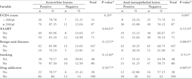
TABLE 2. Distribution of HIV-positive patients according to T-CD4 cell counts, use of highly active anti-retroviral therapy (HAART), presence of concomitant benign anal diseases, smoking, drug addiction in correlation to high-resolution anoscopy and histopathological indings.
Value in bold indicates P-value with statistical difference at the 5% level
* Pearson’s chi-square test or Fisher’s exact test Variable
Acetowhite lesions Total P-value* Anal intraepithelial lesion Total P-value*
Positive Negative Positive Negative
n % n % n % n %
T-CD4 count 0.26* 0.06*
< 200/μl 26 78.78 7 21.21 33 8 24.24 25 75.76 33
≥ 200/μl 76 87.35 11 12.64 87 38 43.68 49 56.32 87
HAART 0.8423* 0.3134*
Yes 49 85.96 8 14.04 57 19 33.33 38 66.67 57
No 59 83.10 12 16.90 71 31 43.66 40 56.34 71
Benign anal diseases 0.3215* 0.8845*
Yes 92 85.98 15 14.02 107 42 39.25 65 60.75 107
No 16 76.19 5 23.81 21 8 38.10 13 61.90 21
Smoking 0.3146* 0.513*
Yes 38 79.17 10 20.83 48 17 35.42 31 64.58 48
No 70 87.50 10 12.50 80 33 41.25 47 58.75 80
Drug addiction 0.5077* 0.8053*
Yes 22 78.57 6 21.43 28 12 42.86 16 57.15 28
No 86 86 14 14 100 38 38 62 62 100
TABLE 3. Diagnostic eficiency of high resolution anoscopy
HRA/Pathology ASIL+* ASIL-* Total
AWL+* 45 63 108
AWL-* 5 15 20
Total 50 78 128
*AWL+ = positive acetowhite lesions; Positive predictive value = 41.67% AWL- = negative acetowhite lesions. Negative predictive value = 75%
ASIL+ = positive histopathology; False positive = 89.76%
ASIL- = negative histopathology. False negative = 10%
Accuracy = 46.87% ASIL prevalence = 39.1%
Sensitivity = 90% Chi-square test P = 0.2142
Speciicity = 19.23% Kappa test = 0.076
(25.64%) of those who did not mention anal intercourse
activity had the same diagnosis (P = 0.04930) (Table 1).
Palefsky et al.(29) observed a relationship between ASIL and
anal receptive sex in HIV-positive patients. They reported that 50% of HIV-positive men-who-have-sex-with-men
(MSM) in their study presented ASIL. Fox et al.(13) reported
ASIL was found in HIV-positive MSM in a prevalence of 26% and 36% in two cohort studies undertaken at the USA
mainly before the advent of HAART. Gimenez et al.(18) have
previously reported a prevalence of ASIL in HIV-positive
MSM of 42% (P = 0.03).
Regarding the number of sexual partners in the last 5 years, there was no statistical signiicance in the correlation with histopathological diagnoses of ASIL of any grade. Thirty-ive (36.46%) individuals who reported having less than 10 partners presented ASIL, while 13 (48.15%) of those that had more than 10 partners also presented the same histopathological inding (Table 1). These results are inconsistent with a study
by Frisch et al.(15) in which it was observed that more than
10 sexual partners increased the risk of cancer by 5 times in
women and 2.8 times in men. Klencke and Palefsky(22) observed
that, in this case, the exposure to various HPV types is the real risk factor to the increasing incidence of ASIL and of anal and cervical cancer observed in these patients.
The average age for irst engaging in sexual activity was 14.65 years in the patients studied herein. There was no statistical signiicance in the correlation between age at which individuals began sexual activity and ASIL. Of 46 patients who began having sex as early as 14 years old, 14 (30.43%) were ASIL positive. Seventy-six individuals started having sex after the age of 14; among those, 34 (44.74%) were ASIL positive (Table 1). In comparison, Frisch et
al.(15) observed moderately elevated risk for anal cancer in
females who started having sex earlier than 16 years old in comparison to women who irst had sexual intercourse after the age of 20 years.
TABLE 4. Correlation between image characteristics of high-resolution anoscopy and histopathological results
Neg: ASIL negative, including inlammatory alterations, LSIL: low-grade squamous intraepithelial lesion, HSIL: high-grade squamous intraepithelial lesion, AWL: acetowhite lesion Histopathology
Anoscopy Neg % LSIL % HSIL % Total P*
Tinctorial characteristics 0.005
Negative AWL 15 75.00 4 20% 1 5% 20
Dense AWL 28 44.44 22 34.92 13 20.63 63
Tenuous AWL 35 77.78 6 13.33 4 8.89 45
Lesion distribution aspect 0.921
Focal 30 56.60 14 26.42 9 16.98 53
Coalescent 33 60.00 14 25.45 8 14.55 55
Relief 6 <0.001
Flat 49 74.24 6 9.09 11 16.67 66
Slightly elevated 9 37.50 10 41.67 5 20.83 24
Elevated 5 27.78 12 66.67 1 5.56 18
Surface 0.002
Smooth 44 68.75 9 14.06 11 17.19 64
Granular 18 41.86 19 44.19 6 13.95 43
Papillary 4 21.05 12 63.16 3 15.79 19 <0.001
Nonpapillary 74 67.89 20 18.35 15 13.76 109
Vascular Proile
Normal 52 63.41 17 20.73 13 15.85 82 0.3631
atypical 6 46.15 5 38.46 2 15.38 13
warty vessels 3 42.86 3 42.86 1 14.29 7 0.5691
no warty vessels 59 59.00 25 25 16 16 100
punctation 1 33.33 2 66.67 0 0 3 0.2248
no punctation 62 59.61 26 25 16 15.38 104
mosaicism 1 50.00 1 50 0 0 2 0.666
no mosaicism 61 58.09 27 25.71 17 16.19 105
two groups (Table 1). Regarding anal cancer, Frisch et al.(15)
observed a relationship between STD and anal neoplastic
development. In 2002, Frisch(17) reported that most STD
related to anal cancer were also associated with HPV infection. Statistical signiicance was observed between the presence of anal papillomavirus infection, according to PCR results,
and ASIL (P = 0.006). Forty-six HPV positive patients
(45.10%) presented ASIL, while only four (15.38%)
non-infected patients presented lesions (Table 1). Palefsky et al.(32)
observed, back in 1998, that nearly all HIV-positive male
patients with lymphocytes T-CD4 counts less than 500/mm³ they were following in the city of San Francisco presented PCR detected anal HPV infection. They also reported that 72% of HIV-positive males with CD4 counts less than 200/mm³ had abnormal anal cytology, so that, taken together, both indings pointed to a probable high prevalence of ASIL in these patients. Indeed, commenting about a work performed by his group in the San Francisco bay to investigate his earlier assumptions and published in 2008, Palefsky states that the prevalence of ASIL of any grade in HIV-positive MSM was
TABLE 5. Correlation between presence of HPV anal infection and anal cancer precursor lesions
HPV+: presence of HPV, HPV-: absence of HPV.
ASIL+: presence of anal cancer precursor lesion; ASIL-: absence of anal cancer precursor lesion PCR/
histopathology ASIL+ % ASIL- % Total P-value
HPV+ 46 45.10 56 54.90 102 0.006
HPV- 4 15.38 22 84.62 26
57% (43% HSIL) and that HPV infection was detected in
88% of these patients, 72% of whom were oncogenic HPV(34).
Thirty-three of the patients studied herein presented T-CD4 levels below 200 cells/µL, eight (24.24%) of those were ASIL positive. Among those patients presenting T-CD4 cells counts above 200/µL, 38 (43.68%) were ASIL positive, demonstrating no correlation between T-CD4 levels and ASIL (Table 2). The same was observed in patients who were under HAART. Nineteen patients (33.33%) that received the medication were ASIL positive, while 31 (43.66%) patients who did not use the medication were also positive for ASIL
(Table 2). Abramowitz et al.(1) also observed no correlation
in the association of levels of T-CD4 lymphocytes, use of
HAART and ASIL. On the other hand, Piketty et al.(38)
reported a high prevalence of ASIL (64%) in patients using
HAART when their immunity was restored and Palefsky(34)
described an increase in number of cases of anal and cervical cancer despite the use of HAART.
There was also no statistical signiicance in the association between ASIL and the presence of anal benign diseases (hemorrhoids, hypertrophied papilla, anal istula, anal issure, mucosal prolapse, anal pruritus, and proctitis) in the 107 individuals in whom these characteristics were studied. Among these patients, 42 (39.25%) were positive for ASIL, while 8 of 27 patients (38.10%) who did not present any listed disease were also positive (Table 2). These results are not
consistent with the indings of Frisch et al.(14) and Tseng et
al.(47) who noticed a signiicant relation between benign anal
lesions and anal cancer, although no case of anal cancer was observed among our patients.
Our data did not ind any association of smoking with the presence of ASIL. We observed that among 48 smokers, 17 (35.42%) were positive for ASIL, while 70 (87.50%) non-smokers also presented ASIL (Table 2). Contrarily (considering
ASIL a stage of anal cancer development), Tseng at al.(47)
reported an association between smoking and anal cancer
and Daling et al.(11) showed that smoking was associated with
an odds ratio of 3.9 to develop anal cancer.
There was no statistical signiicance between the presence of ASIL and addiction to hallucinogen drugs. Among drug users, 42.86% presented intraephitelial lesions, while 38% of non-drug users also presented ASIL (Table 2). In contrast,
Ching-hong et al.(6) reported statistical signiicance for this
factor (P = 0.03), while Piketty et al.(37) reported 34% of cases
of ASIL in HPV positive heterosexual individuals using drugs.
Diagnostic accuracy of HRA
HRA was associated with high sensitivity (90%) but low speciicity (19.23%) for the diagnosis of ASIL, with a negative predictive value of 75% and a low positive predictive value of 41.67% (Table 3). False positive and false negative rates were, respectively, 89.76% and 10%. Sensitivity and speciicity rates of HRA vary greatly in the literature, since this is still a very subjective, as well as user, equipment and
other-comorbidity-dependent test(7, 19, 26, 46). Values vary from
59%(26) to 100%(46) for sensitivity and from 66%(46) to 74%(24)
for speciicity. However, more concordantly with our results,
Tuon et al.(48) reported for cervical colposcopy, a much more
widely employed and studied method, a speciicity rate as low as 19%.
Correlating the presence of AWL with the histopathological diagnosis of ASIL, there was no statistical signiicance between the variables according to the chi-square test (0.2142), whereas the kappa test (0.076) showed low evidence of agreement between the variables. This is possibly a consequence of the fact that lesions such as hypertrophied papillae, hemorrhoids and inlammation in anal canal used to stain positively by acetic acid, a relection of the 63 acetowhite-positive lesions that were negative for ASIL, as well as due to HRA interpretative differences among three individual observers.
Diagnostic agreement between HRA images and histopathology
Regarding tinctorial quality of HRA a signiicant relationship was observed relative to the presence of
histopathologically conirmed ASIL (P = 0.005), for of 20
patients with HRA negative for AWL, 15 (75%) had negative histopathological results, 4 (20%) presented LSIL and 1 (5%) HSIL. Likewise, regarding dense AWL, 28 (44.44%) of 63 patients had negative histopathological results, 22 (34.92%) presented LSIL and 13 (20.63%) HSIL. On the other hand, of 45 individuals with tenuous AWL, 35 (77.78%) had negative histopathological results, 6 (13.33%) presented LSIL and 4 (8.89%) HSIL. A proportion of HSIL (72%) and LSIL (69%) lesions were described as dense AWL. Based on the odds ratio, dense AWL were 2.3 times more likely to be an HSIL than a tenuous lesion (21% of dense AWL lesions were HSIL compared to 9% of tenuous AWL lesions). But, accordingly, a dense AWL was 2.7 times more likely to be an LSIL than a tenuous lesion.
Regarding the relief of lesions observed at HRA, lat lesions tended to be mainly negative for ASIL, but HSIL lesions were more frequently lat at HRA than LSIL. While slightly elevated lesions were more often either negative for ASIL or LSIL, elevated lesions tended to be LSIL. These
results showed statistical signiicance (P<0.001). HSIL lesions
tended to be signiicantly more associated to lat AWL than
LSIL (P<0.001). A high-grade lesion was 3 times more prone
to be lat than to be elevated (16% of lat lesions were HSIL compared to 5% of elevated lesions). Comparatively, Jay et
al.(21) observed that lat lesions were 4 times more frequently
associated to HSIL than elevated lesions (39% of lat lesions were HSIL compared to 9% of elevated lesions).
When surface aspect was analyzed, smooth lesions were more frequently observed in relation to negative histopathological results, whereas a granular aspect was not prone to be HSIL and a papillary aspect was more
frequently associated to low-grade lesions (P = 0.002).
Among lesions described as smooth, the probability of them being HSIL were 1.2 times higher than being LSIL (17% of HSIL lesions were smoothv compared to 14% of
granular lesions). Jay et al.(21) found that smooth lesions
FIGURE 5. Acetowhite lesion dense, coalescent, elevated, granular, punctation, HPV positive. Histopathology: condiloma
FIGURE 6. Acetowhite lesion dense, slightly elevated, granular, HPV positive. Histopathology: LSIL
FIGURE 1. Positive acetowhile lesion, dense, focal, lat, smooth, non-papillary, HPV positive. Histopathology: HSIL
FIGURE 2. Acetowhite lesion dense, coalescent, slightly elevated, papillary, HPV positive. Histopathology: HSIL
FIGURE 3. Acetowhite lesion, dense, coalescent, slightly elevated, papillary, HPV positive. Histopathology: LSIL
(31% of smooth lesions were HSIL compared to 13% of granular lesions).
Regarding the description of lesions being either papillary or non-papillary, a higher proportion of HSIL (83%) than
LSIL (62%) was described as non-papillary (P = 0.001).
Non-papillary lesions had a 0.87 times greater chance to be
associated to HSIL than LSIL. Jay et al.(21) reported that
non-papillary lesions were twice as likely to be HSIL than papillary lesions (34% of non-papillary lesions were HSIL compared to 14% of papillary lesions).
There was no statistical signiicance for the remaining studied aspects of the lesions and their vascular pattern (Table
4), which is not consistent with the indings of Jay et al.(21), in
which warty vessels, punctation and mosaicism all presented a statistical signiicance at the level of 0.001.
Our data indicate that of the HSIL lesions analyzed 68% were dense AWL, 61% were lat, 61% were smooth, 83% were non-papillary and 70% presented a normal vascular pattern. Regarding LSIL lesions, 66% of them were dense AWL, 68% were slightly elevated or elevated, 59% were granular, 62% were non-papillary and 53% presented normal vascular pattern.
ASIL prevalence
Fifty patients were diagnosed as having ASIL (39.1%) at histopathology: 32 (25%) presented low grade lesions and 18 (14.1%) presented high-grade lesions. These results are similar to other indings in the literature for the prevalence
of ASIL, where LSIL ranges from 35.7%(20) to 42%(38), while
HSIL from 7.1%(18) to 26%(21).
HPV prevalence
The observed HPV prevalence in the HIV-positive patients studied was of 79%. Other similar indings in the literature
present prevalence from 80%(38) to 98%(39).
Correlation between anal cancer precursor lesions and HPV infection
Among 50 patients presenting ASIL, 46 were HPV positive, consistent with a 92% prevalence. Among 32 patients who had LSIL, 30 (93.75%) were HPV positive, while of 18 patients with HSIL, 16 (88.88%) had HPV infection. In a systematic review, 1,824 patients were studied, including 472 and 360 presenting HSIL and LSIL, respectively. The prevalence of HPV in patients presenting HSIL and LSIL was 71.91% and
88%, respectively(20). Based on the data presented in Table
5, a prevalence of 45% of ASIL was observed HIV-positive patients co-infected with HPV.
CONCLUSIONS
HRA was a highly sensitive method for the detection of ASIL considering the tinctorial quality, relief and surface aspect of AWL which enhanced the capability of distinction between high- and low-grade anal squamous lesions. Nevertheless, due to its poor observed speciicity a complementary histopathological study is mandatory in order not to miss unsuspected atypical lesions. The prevalence of anal HPV infection and of ASIL in the studied HIV-positive patients was of, respectively, 79% and 39.1% (25% LSIL and 14.1% HSIL). Anal intercourse and anal HPV infection were highly correlated with the presence of ASIL.
ACKNOWLEDGMENTS
Gimenez F, Costa-e-Silva IT, Daumas A, Araújo J, Medeiros SG, Ferreira L. Anuscopia de alta resolução: valor diagnóstico em lesões precursoras de câncer anal em pacientes soropositivos.. Arq Gastroenterol. 2011;48(2):136-45.
RESUMO – Contexto– O câncer anal, muito embora ainda seja uma doença rara, vem sendo observado com frequência ascendente em alguns grupos populacionais considerados sob risco para o desenvolvimento da doença. Infecção pelo vírus do papiloma humano (HPV), imunossupressão e o sexo anoreceptivo são alguns dos fatores associados ao desenvolvimento da neoplasia. Suas semelhanças com o câncer do colo do útero levaram muitos estudos voltados para o estabelecimento de regras para a detecção e tratamento de lesões precursoras do câncer anal, tudo com o objetivo de prevenir a doença. A anuscopia com magniicação de imagem é rotineiramente utilizada para o diagnóstico de lesões precursoras do câncer anal em muitos centros, mas a literatura médica ainda é escassa a respeito do papel a ser desempenhado por essa modalidade diagnóstica.Objetivos– Avaliar as medidas de validação e precisão diagnósticas da anuscopia com magniicação de imagem em comparação com resultados histopatológicos de biopsias anais realizadas em pacientes HIV-positivos tratados na Fundação de Medicina Tropical do Amazonas, Manaus, AM, Brasil. Observar qualquer possível associação entre alguns fatores de risco para o desenvolvimento do câncer anal e a presença de lesões intraepiteliais escamosas anais.
Métodos– Cento e vinte e oito pacientes HIV-positivos foram submetidos a coleta de material celular anal para a realização da detecção da presença de HPV pela reação em cadeia da polimerase. Anuscopias com magniicação de imagem foram realizadas após a aplicação tópica de ácido acético a 3% no canal anal por 2 minutos. As lesões acetobrancas eventualmente observadas foram registradas com respeito a sua localização e classiicadas quanto ao seu padrão tintorial, aspecto de distribuição, relevo, características de sua superfície e vascularidade. Foram realizadas biopsias das lesões acetobrancas sob anestesia local e os espécimes foram remetidos para estudo histopatológico. Os pacientes foram entrevistados em relação à presença de fatores de risco para o câncer anal. Resultados– As prevalências de infecção anal pelo HPV e de lesões intraepiteliais escamosas anais na amostra populacional estudada foram de 79% e 39,1%, respectivamente. A sensibilidade e a especiicidade da anuscopia com magniicação de imagem foram, respectivamente, de 90% e 19,23%, enquanto que o valor preditivo positivo foi de 41,67%, o valor preditivo negativo foi de 75% e o coeiciente kappa de 0,076. Com respeito às lesões analisadas de alto grau foram mais frequentemente observadas em associação com lesões acetobrancas densas (68%), planas (61%), lisas (61%), não-papilíferas (83%) e com padrão vascular normal (70%), enquanto que lesões de baixo-grau tenderam a se associar a lesões aetobrancas densas (66%), plano-elevadas ou elevadas (68%), granulares (59%), não-papilíferas (62%) e de padrão vascular normal (53%). Não se observou signiicância estatística na associação entre características epidemiológicas e a maioria dos fatores de risco para o câncer anal e a presença de lesão acetobrancas ou de lesões intraepiteliais escamosas anais. Entretanto, o sexo anorreceptivo e a detecção de infecção anal por HPV, segundo a técnica da reação da cadeia de polimerase, associaram-se signiicantemente com lesões intraepiteliais escamosas anais (P = 0,0493 e P =0,006, respectivamente). Conclusões – A anuscopia com magniicação de imagem demonstrou ser um método diagnostico sensível, mas inespecíico para a detecção de lesões intraepiteliais escamosas anais. Os fatores de risco sexo anorreceptivo e infecção anal pelo HPV associaram-se signiicantemente à presença de lesões intraepiteliais escamosas anais. Com base nos achados da anuscopia com magniicação de imagem, o relevo e o aspecto morfológico da distribuição das lesões acetobrancas na superfície do canal anal tenderam a permitir a distinção entre lesões de baixo e alto grau.
DESCRITORES – Neoplasias do ânus. HIV. Infecções por papilomavírus. Proctoscopia.
REFERENCES
1. Abramowitz L, Benabderrahmane D, Ravaud P, Walker F, Rioux C, Jestin C, Bouvet E, Soulé JC, Leport C, Duval X. Anal squamous intraepithelial lesions and condyloma in HIV-infected heterosexual men, homosexual men and women: prevalence and associated factors. AIDS. 2007;21:1457-65.
2. Bauer HM, Manos MM. PCR Detection of genital human papillomavirus. In: Persing DH, Smith TF, Tenove FC, White TJ, editors. Diagnostic molecular microbiology principles and applications. Washington D.C.: American Society for Microbiology; 1993. p.407-13.
3. Bethesda System 2001 Terminology. [online]. Bethesda (MD): National Cancer Institute [cited 2005 Sep 29]. Available from: <http://bethesda2001.cancer.gov/ terminology.html>
4. Brasil. Ministério da Saúde. Incidência de câncer no Brasil. Estimativa 2010. Rio de Janeiro: Instituto Nacional do Câncer; 2010.
5. Chin-Hong PV, Palefsky JM. Natural history and clinical management of anal human papillomavirus disease in men and women infected with human immunodeiciency virus. Clin Infect Dis. 2002;35:1127-34.
6. Chin-Hong PV, Vittinghoff E, Cranston RD, Browne L, Buchbinder S, Colfax G, Da Costa M, Darragh T, Benet DJ, Judson F, Koblin B, Mayer KH, Palefsky JM. Age-related prevalence of anal cancer precursors in homosexual men: the explore study. J Natl Cancer Inst. 2005;97:896-905.
7. Costa-e-Silva IT, Gimenez FS, Fujimoto LBM, Ferreira LCL. Valor da anuscopia com magniicação de imagem no diagnóstico de lesões associadas ao câncer anal [resumo]. In: 55º Congresso Brasileiro de Coloproctologia. 2006, Rio de Janeiro. Rev Bras Coloproctol. 2006;26:98-9.
8. Coutinho JRH. Rastreamento de lesões pré-neoplasicas do ânus: citologia anal e anuscopia de alta resolução, novas armas para prevenção. Rev Col Bras Cir. 2006;33:311-7.
9. Daling JR, Weiss NS, Klopfenstein LL, Cochran LE, Chow WH, Daifuku R. Correlates of homosexual behavior and the incidence of anal cancer. JAMA. 1982;247:1988-90.
10. Daling JR, Weiss NS, Hislop TG, Maden C, Coates RJ, Sherman KJ, Ashley RL, Beagrie M, Ryan JA, Corey L. Sexual practices, sexually transmitted diseases and the incidence of anal cancer. N Engl J Med. 1987;317:973-7.
11. Daling JR, Madeleine MM, Johnson LG, Schwartz SM, Shera KA, Wurscher MA, Carter JJ, Porter PL, Galloway DA, McDougall JK. Human papillomavirus, smoking and sexual practices in the etiology of anal cancer. Cancer. 2004;101:270-80. 12. Fenger C. The anal transitional zone. Location and extent. Acta Pathol Microbiol
Scand A. 1979;87A:379-86.
13. Fox PA. Human papillomavirus and anal intraepithelial neoplasia. Curr Opin Infect Dis. 2006;19:62-6.
14. Frisch M, Olsen JH, Bautz A, Melbey M. Benign anal lesions and the risk of anal cancer. N Engl J Med. 1994;331:300-2.
15. Frisch M, Glimelius B, van den Brule AJ, Wohlfahrt J, Meijer CJ, Walboomers JM, Goldman S, Svensson C, Adami HO, Melbye M. Sexually transmitted infection as a cause of anal cancer. N Engl J Med. 1997;337:1350-8.
16. Frisch M, Fenger C, van den Brule AJ, Sorensen P, Meijer CJ, Wallboomers JM, Adami HO, Melbye M, Glimelius B. Variants of squamous cell carcinoma of the anal canal and perianal skin and their relation to human papillomaviruses. Cancer Res. 1999;59:753-7.
18. Gimenez FS, Costa e Silva IT, Guimarães ADP, Ferreira LCL, Araújo JR, Rocha RP, Atala LS, Avi SV, Talhari S. Prevalência de lesões precursoras do câncer anal em indivíduos HIV positivos atendidos na Fundação de Medicina Tropical do Amazonas, experiência inicial em Manaus. Rev Bras Coloproctol. 2008;28:72-6. 19. Hammes LS. Correlação entre achados colposcópicos e diagnóstico histológico
segundo a classiicação colposcópica da federação internacional de patologia cervical e colposcopia de 2002 [dissertação]. Porto Alegre: Universidade Federal do Rio Grande do Sul; 2004.
20. Hoots BE, Palefsky JM, Pimenta JM, Smith JS. Human papillomavirus type distribution in anal cancer and anal intraepithelial lesions. Int J Cancer. 2009;124:2375-83.
21. Jay N, Berry JM, Hogeboom CJ, Holly EA, Darragh TM, Palefsky JM. Colposcopic appearance of anal squamous intraepithelial lesions: relationship to histopathology. Dis Colon Rectum. 1997;40:919-28.
22. Klencke BJ, Palefsky JM. Anal cancer: an HIV-associated cancer. Hematol Oncol Clin North Am. 2003;17:859-72.
23. Kuppers V. Signiicance of colposcopy in cancer prevention. Gynakol Prax. 2005;29:69-86.
24. Martin F, Bower M. Anal intraepithelial neoplasia in HIV positive people. Sex Transm Infect. 2001;77:327-31.
25. Martins CR. HPV-induced anal dysplasia: what do we know and what can we do about it? Hopkins HIV Rep. 2001;133-5.
26. Mathews WC, Sitapati A, Caperna JC, Barber RE, Tugent A, Go U. Measurement characteristics of anal citology, histopathology and high-resolution anoscopic visual impression in an anal dysplasia screening program. J Acquir Immune Deic Syndr. 2004;37:1610-5.
27. Melbye M, Sprogel P. Aetiological parallel between anal cancer and cervical cancer. Lancet. 1991;338:657-9.
28. Nadal RS, Manzione CR. Citologia como método para detecção de lesões precursoras do carcinoma anal. Rev Bras Coloproctol. 2005;25:72-4. 29. Palefsky JM, Holly EA, Hogeboom CJ, Berry JM, Jay N, Darragh TM. Anal
cytology as a screening tool for anal squamous intraepithelial lesions. J Acquir Immune Deic Syndr Hum Retrovirol. 1997;14:415-22.
30. Palefsky JM. Human papillomavirus infection and anogenital neoplasia in human immunodeiciency virus-positive men and women. J Natl Cancer Inst Monogr. 1998;(23):15-20.
31. Palefsky JM, Holly EA, Hogeboom CJ, Ralston ML, DaCosta MM, Botts R, Berry JM, Jay N, Darragh TM. Virologic, immunologic, and clinical parameters in the incidence and progression of anal squamous intraephitelial lesions in HIV-positive and HIV-negative homosexual men. J Acquir Immune Deic-Syndr Hum Retrovirol. 1998;17:314-9.
32. Palesky JM, Holly EA, Ralstson ML, Jay N. Prevalence and risk factors for human papillomavirus infection of the anal canal in human immunodeiciency virus (HIV)-positive and high-risk HIV-negative homosexual men. J Infect Dis. 1998;177:361-7. 33. Palefsky JM, Holly EA, Ralston ML, Jay N, Berry JM, Darragh TM. High
incidence of anal high-grade squamous intraepithelial lesions among HIV-positive and HIV-negative homosexual and bisexual men. AIDS. 1998;12:495-503. 34. Palefsky J. Human papillomavirus–related disease in people with HIV. Curr Opin
HIV AIDS. 2009;4:52-6.
35. Panther LA, Wagner K, Proper J, Fugelso DK, Chatis PA, Weeden W, Nasser IA, Doweiko JP, Dezube BJ. High resolution anoscopy indings for men who have sex with men: inaccuracy of anal cytology as a predictor of histologic high-grade anal intraepithelial neoplasia and the impact of HIV serostatus. Clin Infect Dis.
2004;38:1490-2.
36. Penn I. Occurrence of cancers in immunosuppressed organ transplant recipients. Clin Transpl. 1998;147-58.
37. Piketty C, Darragh TM, Da Costa M, Bruneval P, Heard I, Kazatchkine MD, Palefsky JM. High prevalence of anal human papillomavirus infection and anal cancer precursors among HIV-infected persons in the absence of anal intercourse. Ann Intern Med. 2003;138:453-9.
38. Piketty C, Darragh TM, Heard I, Da Costa M, Bruneval P, Kazatchkine MD, Palefsky JM. High prevalence of anal squamous intaepithelial lesions in HIV– positive men despite the use of highly active antiretroviral therapy. Sex Transm Dis. 2004;31:96-9.
39. Pokomandy A, Rouleau D, Ghattas, G, Vézina S, Coté P, Macleod J, Allaire G, Franco EL, Coutlée F. Prevalence, clearance and incidence of anal human papillomavirus infection in HIV-infected men: the HIPVIRG cohort study. J Infect Dis. 2009;199:965-973.
40. Prieto Reyes M, Vázquez Márquez L. [Anal epidermoid carcinoma: a rare incidence or a rare diagnosis?] Rev Esp Enferm Dig. 1997;2:128-32.
41. Qualters JR, Lee LNC, Smith RA, Aubert RE. Breast and cervical cancer surveillance, United State, 1973–1987. MMWR CDC Surveill Summ. 1992;41(2):1-7.
42. Ryan DP, Compton CC, Mayer RJ. Carcinoma of the anal canal. N Engl J Med. 2000;342:792-800.
43. Scholeield JH, Johnson J, Hitchcock A, Kocjan G, Smith JH, Smith PA, Ferryman S, Byass P. Guidelines for anal cytology-to-make cytological diagnosis and follow up much more reliable. Cytopathology. 1998;9:15-22.
44. Silva ITC, Gimenez FS, Guimarães RAS, Camelo RT, Melo MND, Barros FS, Guimarães ADP, CabralCRB, Guimarães EL. Citologia anal como método de rastreamento para detecção precoce do câncer anal: esfregaços com algodão hidróilo são mesmos insatisfatórios? Acta Cir Bras. 2005;20:109-114. 45. Spence AR, Franco EL, Ferenczy A. The role of human papillomavirus in cancer:
evidence to date. Am J Cancer. 2005;4:49-64.
46. Tramujas da Costa e Silva I, de Lima Ferreira LC, Santos Gimenez F, Gonçalves Guimarães RA, Botinelly Fujimoto L, Barbosa Cabral CR, Venturim Mozzer R, de Souza Atala L. High-resolution anoscopy in the diagnosis of anal cancer precursor lesions in renal graft recipients. Ann Surg Oncol. 2008;15:1470-5. 47. Tseng HF, Morgenstern H, Mack TM, Peters RK. Risk factors for anal cancer:
results of a population-based case-control study. Cancer Causes Control. 2003;14:837-46.
48. Tuon FFB, Bittencourt MS, Panichi MA, Pinto AP. Avaliação da sensibilidade e especiicidade dos exames citopatológicos e colposcópico em relação ao exame histológico na identiicação de lesões intra-epiteliais cervicais. Rev Assoc Med Bras. 2002;48:140-4.
49. Walker P, Dexeus S, De Palo G, Barrasso R, Campion M, Girardi F, Jakob C, Roy M. International terminology of colposcopy: an update report from the International Federation for Cervical Pathology and Colposcopy. Obstet Gynecol. 2003;101:175-77.
50. Zaki SR, Judd R, Cofield LM, Greer P, Rolston F, Evatt BL. Human papillomavirus infeccion and anal carcinoma. Retrospective analysis by in situ hybridization and the polymerase chain reaction. Am J Pathol. 1992;140:1345-55.