Correlação entre genótipo-fenótipo em famílias brasileiras com glaucoma primário de
ângulo aberto determinada por mutações no exon 3 do gene TIGR/MYOC
Trabalho realizado na Clínica Oftalmológica da Faculda-de Faculda-de Medicina, UniversidaFaculda-de Faculda-de São Paulo - USP - São Paulo (SP), Brasil e no Laboratório de Investigação Médica (LIM25) da Faculdade de Medicina da USP -São Paulo (SP) - Brasil.
1Doutora em Oftalmologia pela Faculdade de Medicina da Universidade de São Paulo USP São Paulo (SP) -Brasil.
2Professor Livre Docente da Clínica Oftalmológica do Hospital das Clínicas da USP - São Paulo (SP) - Brasil. 3Biomédica estagiária do Laboratório de Investigação Médica (LIM-25), Faculdade de Medicina da USP - São Paulo (SP) - Brasil.
4Doutora em Endocrinologia ela Faculdade de Medici-nada - USP - São Paulo (SP) - Brasil.
5Professor Livre Docente pela Faculdade de Medicina da USP - São Paulo (SP) - Brasil.
Endereço para correspondência: Rua 38, número 296, Setor Marista - Goiânia (GO) CEP 74823-350 E-mail: [email protected]
[email protected] Recebido para publicação em 01.06.2005 Aprovação em 13.09.2005
Nota Editorial: Depois de concluída a análise do artigo sob sigilo editorial e com a anuência dos Drs. Augusto Paranhos Jr e Juliana Maria Ferraz Sallum sobre a divulgação de seu nome como revisor, agradece-mos sua participação neste processo.
Cristine Araújo Povoa1
Roberto Freire Santiago Malta2 Mariana de Moraes Rezende3 Karla Fabiana Santana de Melo4 Daniel Giannella-Neto5
Keywords: Glaucoma, open-angle; Genes, Phenotype; Genotype; Mutation; Genetic
screening; Brazil
INTRODUCTION
Glaucoma is a potentially severe ocular disease and the second leading worldwide cause of blindness, after cataract(1). Primary open-angle
glauco-ma (POAG) represents more than half of all glaucoglauco-ma cases in North Ameri-can Caucasian populations and affects nearly 2% of subjects older than 45 years(1-2).
The Brazilian prevalence of glaucoma is unknown. The results of scree-ning studies suggest that the disease is frequent, the majority of the subjects is affected by POAG and the disease represents an important social problem(3-4).
primary open angle glaucoma of Brazilian families
with mutations in exon 3 of the TIGR/MYOC gene
Purpose: To investigate the phenotype of primary open-angle glaucoma (POAG) in Brazilian families with mutation in exon 3 of TIGR/MYOC.
Methods: Seventy-eight POAG patients with a positive family history and eighteen unrelated patients with POAG were screened by automated DNA sequencing for mutations in exon 3 of the TIGR/MYOC gene. The pedigrees of POAG patients with mutations that lead to amino acid change were built. All available relatives of the index cases were also examined and genotyped by sequencing. Results: Four sequence variants were identified in exon 3 of the TIGR/MYOC gene (Tyr347Tyr, Pro370Pro, Lys398Arg and Cys433Arg) from the 96 initially screened patients. The Lys398Arg mutation was previously described as a polymorphism and in our study did not segregate with POAG. The most prevalent mutation was Cys433Arg, affecting 3 index cases (3.1% or 3/96). In two different families, 8/56 subjects presented Cys433Arg mutation and had POAG, 5/56 had ocular hyperten-sion and 8/56 had no disease manifestation. POAG patients had a median age at diagnosis of 43.25 yr (17-58 yr) and intraocular pressure (IOP) with a mean of 36.3 ± 3.8mmHg for the right eye and 37.6 ± 9.75 mmHg for the left eye. The group of patients with Cys433Arg mutation had significantly higher IOP (p<0.0007) and vertical cup/disc ratio when compared to the patients without mutation (p<0.023). Conclusions: Cys433Arg mutation in exon 3 of the TIGR/MYOC gene is related to juvenile-onset POAG (J-POAG) in Brazilian families and autosomal dominant inheritance. The phenotype of this mutation is characterized by varied ages at diagnosis, causing J-POAG and late-onset POAG, associated with high IOP.
The first locus responsible for POAG, named GLC1-A, was identified in 1993(5) by genetic linkage analysis in 5
genera-tions of a large family with juvenile glaucoma, on the long arm of chromosome 1.
Stone et al.(6) found mutations in exon 3 of the TIGR/
MYOC gene in 2.9% of patients with unselected glaucoma and in 4.4% of the patients with glaucoma family history. The most frequent mutation in this study was the Gln368STOP.
The subsequent studies, analyzing the prevalence of the mutation on GLC1-A, repeated the initial results and identified additional mutations, almost all located in exon 3 of the TIGR/ MYOC. Most of these mutations were missense, located in the
exon 3 that encodes the olfatomedin homology domain(7-9).
In Brazil, studies of patients with juvenile glaucoma identi-fied a new mutation (Cys433Arg), not found in other popula-tions, and that may be the most common TIGR/MYOC gene mutation in the Brazilian population (10).
Considering that POAG is relatively prevalent, that our population shows specific racial characteristics resulting from strong miscegenation and that presence of mutations in the TIGR/MYOC gene influences the variation of phenotypic expression, we aimed to identify mutations in exon 3 of the TIGR/MYOC gene of POAG patients and observe the pheno-typic expression of the families with the identified mutations.
METHODS
The initial cases consisted of 96 index glaucomatous pa-tients, admitted at the Ophthalmologic Department of the “Hospital das Clínicas da Universidade de São Paulo” (HC-FMUSP). The patients were selected according to the follo-wing criteria: 1) patients with POAG either juvenile-onset or adult, with or without family history of the disease; 2) ocular hypertensive patients (OH) with positive family history of glaucoma; 3) patients with optic disc with suspicion of glau-coma (SOD), and a family history of glauglau-coma and 4) patients with no manifestation of disease (NDM) who had glaucoma-tous relatives.
The patients with glaucoma had an open angle on gonios-copy, glaucomatous optic disc features and alteration of automated visual field. The patients with open-angle gonios-copy that had intraocular pressure (IOP) of 22 mmHg or higher, (in the absence of IOP lowering therapy), and no optic disc or visual field features suggestive of glaucoma were considered ocular hypertensives.
The patients with SOD had open angle on gonioscopy, cup/disc ratio 0.7 or greater; asymmetry of cup/disc ratio greater than 0.2, in the absence of other alterations of the neural rim that suggest glaucoma; absence of visual field alteration and IOP levels lower than 22 mmHg.
The patients without disease manifestation (NDM) were characterized by the presence of open angle, IOP lower than 22 mmHg, optic cup/disc ratio lower than 0.7, regular neural rim and normal visual field.
The visual field was considered altered according to the
Anderson criteria for minimal abnormality in glaucoma(11). The
visual field was not performed in children under 6 years. Informed parental consent, patient consent, and approval by the Hospital Ethics Committee were obtained before initiating the study, according to the Declaration of Helsinki.
Family studies
Seventy-seven available relatives of 2 of the 3 index cases with mutation Cys433Arg in exon 3 of the TIGR/MYOC gene, were submitted to ophthalmologic examination, performed at the “Hospital das Clínicas da Universidade de Sao Paulo”, and were screened to identify the family’s mutation.
The following information was collected from the relatives: IOP, use of IOP lowering therapy or other medications, history of laser treatment and surgery for glaucoma.
The ophthalmologic examination consisted of evaluation of visual acuity with optical corrections, biomicroscopy, Gold-mann applanation tonometry, gonioscopy with Sussman lens
(Ocular® Instruments Bellevue, WA USA), biomicroscopy of
the optic disc with 90DP lens (Ocular® Instruments Bellevue,
WA USA), standard automated visual field test (program C24-2,
SITA fast strategy from Humphrey Field Analyzer (Humphrey®
Instruments, CA) and stereophotography of the disc.
Study of exon 3 of the TIGR/MYOC gene
Genomic DNA of the patients and their available relatives was obtained from peripheral blood leukocytes, according to
the protocol of Genomic PrepTM Blood DNA Isolation Kit
(Amersham Pharmacia Biotech, Inc.). A fragment of 487 bp corresponding to exon 3 of the TIGR/MYOC gene was ampli-fied by PCR. The forward primer CGGGTGCTGTGGTGTACTC and reverse primer AAGAGCTTCTTCTCCAGGGG were used.
Amplification conditionsconsisted of an initial denaturing
step of 95ºC for 3 min, 30cycles of 94ºC for 1 min; 55ºC for 1 min; 72ºC for 1 min; followed by a final extension step at 72ºC for 10 min. For sequencing 5 ng of amplified DNA fragment were purified in a column using the supplier’s instructions
(GibcoBRL® Lifetechnologies, Gasthersburg/EUA) and
direc-tly sequenced with the ABI PRISM Genetic Analyser 377
automatic DNA sequencer(PE Applied Biosystems, Foster
City, CA). The sequencing was made according to the
sup-plier’s protocol (ABI Prism® Big Dye Terminator Cycle
Se-quencing ready Reaction).
Statistical analysis was performed using the non-parame-tric of Mann-Whitney test. The used software was “Statisti-ca” (version 5.5). Value of p<0.05 was considered significant.
RESULTS
Initially, 96 individuals were screened to identify index ca-ses: 75 (78.1%) were diagnosed as POAG, 10 (10.4%) were OH, 2 (2.0%) presented SOD and 9 (9.3%) did not have manifestation of the disease (NDM). Eighteen patients (18.7%) with POAG had no family history of glaucoma (Table 1 and Figure 1).
Fifty-eight patients (60.4%) were female and 38 (39.5%) were male, of which 56 (58.3%) were Caucasians, 39 (40.4%) Negroid and 1 (1%) Mongol.
Mutational analysis of exon 3 of the TIGR/MYOC gene
Four heterozygous variants (Tyr347Tyr, Pro370Pro, Lys398Arg and Cys433Arg) were detected in 6/96 subjects (6.2%) (Table 2). Three of them (Tyr347Tyr, Pro370Pro and Lys398Arg) are not considered disease-causing mutation.
The C→T transition at nucleotide 1361 (Cys433Arg) was
the most prevalent mutation, affecting 3 of 96 studied patients
(3.1%). The index cases with this disease-causing mutation belonged to the same diagnosis group of POAG with a family history. Considering only this group, the frequency of the mutation Cys433Arg was 5.2% (3/57). The Lys398Arg muta-tion was found in a female patient with SOD and positive family history, being present in 1% of the total sample (1/96). Pro370Pro mutation was identified in 1 patient and Tyr347Tyr mutation in another patient. Both were found in patients with POAG and a positive family history, affecting 3.5% (2/57) of the patients of this group (Table 2).
No alterations were identified in exon 3 of the TIGR/MYOC gene in the group of OH patients and NDM patients (Table 2).
Study of the families
The segregation study of Cys433Arg mutation and its phenotypic expression was made in 2 of the 3 families with this mutation (LFN and MAF families). The third index case did not authorize her family to participate in this study (Figure 1).
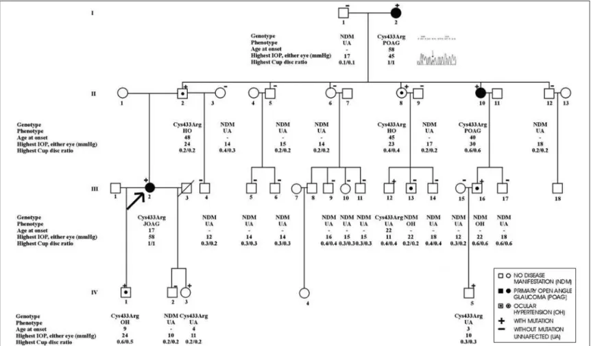
The pedigree of LFN and MAF families is shown in Figures 2 and 3, respectively.
The LFN family index case had juvenile-onset POAG. This family had 27 members submitted to the study of exon 3 of the TIGR/MYOC gene and ophthalmologic evaluation. Ten members of this family were heterozygous for the mutation Cys433Arg (Figure 1). Two had a diagnosis of adult POAG, one of J-POAG, three were considered OH and four had NMD (Figure 2).
The MAF family index case had adult POAG. Fifty members of this family were submitted to analysis of exon 3 of the TIGR/ MYOC gene; but only 29 were available for ophthalmologic evaluation. Eleven members of this family were heterozygous for the Cys433Arg mutation (Figure 3): 4 had a diagnosis of adult POAG, 1 of juvenile-onset POAG; 2 were OH, and 4 NDM. One patient from each family was considered OH and did not present mutations in exon 3 of the TIGR/MYOC gene.
Phenotypic characteristics of patients with POAG and Cys433Arg
The Cys433Arg mutation was present in all available relati-ves of the families LFN and MAF with a diagnosis of POAG (Figures 2 and 3). Five of 8 patients (62.5%) were female and 3/8 (37.5%) were male. The median age at diagnosis of POAG
Table 1. Diagnosis and family data of the 96 index cases
Diagnosis
FH POAG OH SOD NDM N
FH + 57 (59.3%) 10 (10.4%) 2 (2.0%) 9 (9.3%) 78 (081.2%) FH - 18 (18.7%) 0 0 0 18 (018.7%) N 75 (78.1%) 10 (10.4%) 2 (2.0%) 9 (9.3%) 96 (100.0%)
FH+: family history positive; FH-: family history negative; N: total patients; POAG: primary open-angle glaucoma patients; OH: ocular hipertensive patients; SOD: patients with optic disc with suspicion of glaucoma; NDM: patients with no manifestation of disease
Figure 1 - Summary of the studied families, after the identification of the mutations in the index cases
INITIAL SAMPLE:
96 representatives of families
LFN Caucasian Juvenile POAG Age: 17 yr IOP: 50mmHg Cys433Arg (Family 1) CA Negroid POAG Age: 53 yr IOP: 45mmHg Cys433Arg Did not authorize
MAF Negroid POAG Age: 49 yr IOP: 33mmHg Cys433Arg (Family 2) LMS Caucasian SOD Age: 42 yr IOP: 14mmHg Lys398Arg
(Family 3)
Family 1 Study of exon 3 and Ophthalmologic examination 27 members 10 members with Cys433Arg
02 Adult POAG
01 Juvenile POAG
03 OH
04 NDM
Family 2 Study of exon 3
050 members Study of exon 3 and Ophthalmologic examination
029 members 11 members with Cys433Arg
04 Adult POAG
01 Juvenile POAG
02 O H
04 NDM
Family 3 Study of exon 3 and Ophthalmologic examination
014 members
5 members with Lys398Arg 2 SOD
3 NDM
Table 2. Mutations in exon 3 of the TIGR/MYOC gene
Diagnosis groups
Variations on exon 3 POAG OH SOD NDM N
FH + FH - FH+ FH+ FH+
Cys433Arg 03 (03.1%) 0 0 0 0 03 (03.1%) Lys398Arg 0 0 0 1 0 01 (01.0%)
Pro370Pro 01 (01.0%) 0 0 0 0 01 (01.0%) 06 (06.2%) Tyr347Tyr 01 (01.0%) 0 0 0 0 01 (01.0%)
No alterations on exon 52 (54.1%) 18 (18.7%) 10 (10.4%) 1 (1.0%) 9 (9.3%) 90 (93.7%) 90 (93.7%) N 57 (59.3%) 18 (18.7%) 10 (10.4%) 2 (2.1%) 9 (9.3%) 96(100.0%)
Figure 2 - Heredogram of family LFN. All the patients with POAG presented the mutation in one of the alleles. Of the ocular hypertensives (OH) only patient LFN III-13 did not present the mutation.
Figure 3 - Heredogram of family MAF. All the patients with POAG, and with examined genotype presented the mutation Cys433Arg. Of the OH patients, only patient MAF III-17 did not present the mutation. All the patients NDM with mutation had a mean age less than the lowest limit of the median
was 45 yr, ranging from 17 to 58 yr. Two patients, one from each family, presented juvenile-onset POAG (Table 3).
The age at diagnosis of juvenile-onset POAG of the LFN family’s index case III-2 was 17 yr and that of the index case of MAF family was 49 (Table 3). This group of patients presen-ted a mean of the maximal untreapresen-ted IOP of 36.3±3.8 mmHg (median of 37 mmHg, ranging from 30 to 40 mmHg) in the right eye and 37.6±9.75mmHg (median 35 mmHg, ranging from 28 to 58 mmHg) in the left eye (Table 3).
The median vertical cup/disc ratio in the right and left eyes was 0.9 and 1.0, respectively. It ranged from 0.4 to 1.0 in the right eye and 0.6 to 1.0 in the left eye.
Only 2 of 8 patients with POAG (LFN and MAF families) were not submitted to surgery to treat glaucoma and were controlled on medical therapy. The remaining six patients un-derwent trabeculectomy and in two of them a surgery to im-plant a glaucoma drainage device was also necessary.
Five of these 8 patients (62.5%) presented visual acuity lower than 20/400 secondary to POAG in at least one of the eyes (Table 3).
Phenotypic characteristics of OH patients
Seven patients, five of LFN family and two of MAF family, were considered OH (Table 4). Two OH subjects, one female patient of LFN family (LFN III–13) and a male of MAF family (MAF III–17) did not present the mutation Cys433Arg in the
TIGR/MYOC gene. The patient LFN III-13 had a diagnosis of OH when she was 21 yr old with IOP of 22 mmHg in the right eye and 21 mmHg in the left eye, vertical cup/disc ratio in the right and left eyes of 0.2 and visual acuity of 20/20 in both eyes. The patient MAF III-17 had a diagnosis of OH when he was 40 yr old. He presented initial IOP of 30 in both eyes, vertical cup/disc ratio of 0.5 in the right eye and 0.6 in the left eye and visual acuity of 20/20 in both eyes. Among the five OH patients with mutation, one (20%) was female.
The median age at diagnosis in the OH patients with muta-tion was 39 yr, ranging from 9 to 48 yr.
The OH patients presented median IOP for the right and left eye of 22 mmHg. The median vertical cup/disc ratio was 0.4 in both eyes.
All OH patients, with and without mutation, presented visual acuity of 20/20 with best optical correction.
Phenotypic characteristics of NDM patients
The Cys433Arg mutation was identified in 8 of 40 subjects with no disease manifestation of both families. Among these 8 patients the median age was 14.5 yr, ranging from 3 to 43 yr and four were female (Table 5).
The mean IOP of the NDM patients with Cys433Arg muta-tion was 12.5 mmHg in the right eye and 14 mmHg in the left eye. The IOP ranged from 10 to 19 mmHg in the right eye and
Table 4. Genotype and clinical data of the OH patients from LFN and MAF families
Patients Mutation Age Gender Visual acuity Max. IOP C/D
with OH Cys433Arg RE LE RE LE RE LE
V H V H
LFN IV-1 Present 9 M 20/20 20/20 24 22 0.6 0.5 0.5 0.5 LFN II-2 Present 48 M 20/20 20/20 22 24 0.2 0.2 0.2 0.2 LFN III-13 Absent 21 F 20/20 20/20 22 21 0.2 0.2 0.2 0.2 LFN II-8 Present 45 F 20/20 20/20 21 23 0.4 0.4 0.4 0.4 LFN III-16 Present 20 M 20/20 20/20 22 19 0.6 0.6 0.6 0.6 MAF III-8 Present 39 M 20/20 20/20 25 21 0.3 0.2 0.2 0.2 MAF III-17 Absent 40 M 20/20 20/20 30 30 0.5 0.5 0.6 0.6
OH: ocular hipertensive patient; Max. IOP: max intraocular pressure recorded without medication; C/D: Cup/disc ratio; F: female; M: male; RE: right eye; LE: left eye; V: vertical; H: horizontal; NLP: no light perception
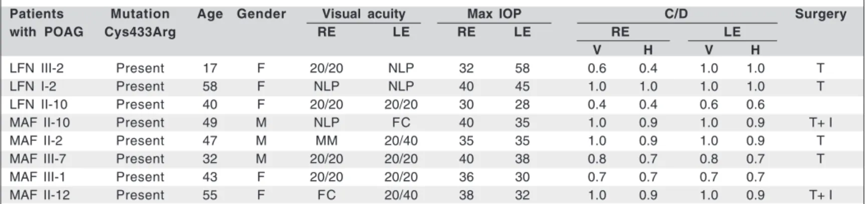
Table 3. Phenotypic characteristics of the 8 patients with POAG and Cys433Arg mutation from LFN and MAF families
Patients Mutation Age Gender Visual acuity Max IOP C/D Surgery
with POAG Cys433Arg RE LE RE LE RE LE
V H V H
LFN III-2 Present 17 F 20/20 NLP 32 58 0.6 0.4 1.0 1.0 T LFN I-2 Present 58 F NLP NLP 40 45 1.0 1.0 1.0 1.0 T LFN II-10 Present 40 F 20/20 20/20 30 28 0.4 0.4 0.6 0.6
MAF II-10 Present 49 M NLP FC 40 35 1.0 0.9 1.0 0.9 T+ I MAF II-2 Present 47 M MM 20/40 35 35 1.0 0.9 1.0 0.9 T MAF III-7 Present 32 M 20/20 20/20 40 38 0.8 0.7 0.8 0.7 T MAF III-1 Present 43 F 20/20 20/20 36 30 0.7 0.7 0.7 0.7
MAF II-12 Present 55 F FC 20/40 38 32 1.0 0.9 1.0 0.9 T+ I
from 10 to 18mmHg in the left eye. The median cup/disc ratio in the right eye of NDM patients was 0.2 and in the left eye it was 0.25. The cup/disc ratio ranged from 0.1 to 0.4 in both eyes.
Thirty-two NDM subjects had no mutation in exon 3 of the TIGR/MYOC gene, 72% (23/32) were male and the median age of this group was 26 yr, ranging 4 to 79 yr.
The IOP median of NDM patients without mutation was 14 mmHg in the left eye. The median vertical cup/disc ratio was 0.3 in the right and left eye, ranging from 0.1 to 0.6 in both eyes.
The patients LFN I-1 and MAF III-22 presented deficient visual acuity due to the presence of bilateral senile cataract and refractive amblyopic respectively.
Phenotypic characteristics of patients with and without mutation
The IOP and cup/disc ratio of the subjects with or without mutation showed statistical differences (Tables 3, 4, 5). The IOP in the group of patients with mutation was higher (p<0.001) than Table 5. Genotype and clinical data of NDM subjects from LFN e MAF families
Patients Mutation Age Gender Visual acuity Max IOP C/D
with NDM Cys433Arg RE LE RE LE RE LE
V H V H
LFN II-3 Absent 53 F 20/20 20/20 14 14 0.4 0.3 0.4 0.3 LFN IV-2 Absent 6 M 20/20 20/20 10 10 0.2 0.2 0.2 0.2 LFN IV-3 Present 4 F 20/20 20/20 11 10 0.2 0.2 0.2 0.2 LFN III-4 Absent 4 M 20/20 20/20 12 12 0.3 0.2 0.3 0.2 LFN I-1 Absent 79 M 20/100 20/40 17 17 0.1 0.1 0.1 0.1 LFN III-17 Absent 11 M 20/20 20/20 18 18 0.3 0.3 0.6 0.6 LFN III-9 Absent 31 M 20/20 20/20 16 16 0.4 0.4 0.4 0.4 LFN II-9 Absent 46 M 20/20 20/20 17 15 0.2 0.2 0.2 0.2 LFN III-14 Absent 18 M 20/20 20/20 16 18 0.4 0.4 0.4 0.3 LFN III-12 Present 22 M 20/20 20/20 11 11 0.3 0.3 0.4 0.4 LFN III-6 Absent 8 M 20/20 20/20 14 14 0.3 0.3 0.3 0.3 LFN II-6 Absent 49 F 20/20 20/20 13 14 0.2 0.2 0.2 0.2 LFN III-5 Absent 13 M 20/20 20/20 13 14 0.3 0.3 0.3 0.3 LFN II-12 Absent 48 M 20/20 20/20 16 18 0.2 0.2 0.2 0.2 LFN II-5 Absent 39 M 20/20 20/20 15 15 0.2 0.2 0.2 0.2 LFN III-10 Absent 19 F 20/20 20/20 15 15 0.3 0.3 0.3 0.3 LFN III-11 Absent 11 M 20/20 20/20 10 13 0.7 0.6 0.6 0.5 LFN IV-5 Present 3 M 20/25 20/25 10 10 0.3 0.3 0.3 0.3 LFN III-15 Absent 19 F 20/20 20/20 12 11 0.3 0.2 0.3 0.2 MAF III-19 Absent 35 F 20/20 20/20 12 12 0.3 0.3 0.2 0.2 MAF III-20 Absent 26 M 20/20 20/20 20 18 0.1 0.1 0.1 0.1 MAF III-16 Absent 45 F 20/20 20/20 12 12 0.3 0.3 0.3 0.3 MAF IV-15 Absent 14 M 20/20 20/20 10 12 0.1 0.1 0.1 0.1 MAF II-9 Absent 68 F 20/60 20/30 12 12 0.3 0.3 0.3 0.3 MAF IV-19 Absent 13 M 20/20 20/20 10 10 0.1 0.1 0.1 0.1 MAF II-11 Absent 59 M 20/20 20/20 12 13 0.3 0.3 0.3 0.3 MAF III-24 Absent 44 M 20/20 20/20 14 15 0.3 0.3 0.3 0.3 MAF III-26 Absent 35 M 20/20 20/20 14 14 0.2 0.2 0.2 0.2 MAF IV-16 Absent 16 F 20/20 20/20 12 12 0.1 0.1 0.1 0.1 MAF IV-22 Absent 11 M 20/20 20/20 10 11 0.2 0.2 0.2 0.2 MAF IV-23 Absent 18 F 20/20 20/20 15 15 0.6 0.5 0.5 0.5 MAF III-27 Present 43 F 20/20 20/20 17 18 0.2 0.2 0.3 0.2 MAF III-22 Present 28 F 20/30 20/80 19 17 0.3 0.3 0.4 0.4 MAF IV-20 Absent 12 M 20/20 20/20 18 19 0.4 0.4 0.3 0.3 MAF IV-18 Absent 15 M 20/20 20/20 10 10 0.2 0.2 0.2 0.2 MAF III-23 Absent 35 F 20/20 20/20 10 10 0.2 0.2 0.2 0.2 MAF III-25 Present 39 F 20/20 20/20 11 10 0.2 0.2 0.2 0.2 MAF III-28 Absent 41 M 20/20 20/20 18 16 0.5 0.5 0.4 0.4 MAF IV-24 Absent 12 M 20/20 20/20 11 11 0.2 0.2 0.2 0.2 MAF IV-21 Present 7 M 20/20 20/20 14 17 0.1 0.1 0.1 0.1 MAF IV-25 Present 6 M 20/20 20/20 14 17 0.1 0.1 0.1 0.1
IOP of the patients without mutation. The NDM patients with Cys433Arg mutation had a greater cup/disc ratio when compa-red with those without mutation (p<0.026).
Penetrance of mutation Cys433Arg by age groups
The Cys433Arg mutation was identified in 5 subjects with ages between 0 and 10 yr, in 4 between 10 and 30 yr, in 4 between 30 and 40 yr and in 8 with up to 40 yr. POAG was present in one (LFN IV-1), 2 (LFN III-2 and LFN III-16), 3 (LFN II-10, MAF III-7 and MAF III-8) and 7 patients (LFN I-2, LFN II-2, LFN II-8, MAF II-2, MAF II-10, MAF II-12, MAF III-1) with Cys433Arg mutation corresponding to 20%, 50%, 75% and 87,5%, respectively, according to the age groups (Tables 3, 4, 5).
DISCUSSION
POAG is a complex genetic disease that presents a range of ocular signs that are poorly understood in regard to their pathophysiology(12).
One of the main benefits obtained by genetic study in this type of disease is the identification of subjects who are at risk of developing it(12). Considering that the mutation in the TIGR/
MYOC gene is responsible for the disease, the observation of the phenotype caused by different mutations may result in characterization of specific clinical situations determined by
mutations(12-13). Therefore, clinical and molecular analyses
were performed in 96 Brazilian unrelated subjects.
The Cys433Arg residue is located in the region of the more preserved amino acids in the olfatomedin domain of myocilin. Thus, any alteration in this region is expected to alter the structure of the protein causing disease(17). This residue might
play an important role in the establishment of intermolecular connections of the “myocilin” protein. Amino acid substitu-tion in this region could prevent myocilin olygomeric comple-xes formation. Therefore, endangering myocilin intermolecular relations may imply in increased resistance of aqueous humor drainage, through the trabecular meshwork(18).
The non-conservative Cys433Arg mutation was first des-cribed by Vasconcellos et al (10) in patients with juvenile and
adult POAG. These authors analyzed the haplotypes of the patients with Cys433Arg mutation and observed that these patients had a common ancestor, suggesting that this muta-tion, described only in the Brazilian populamuta-tion, should have founder effect(19).
Specific mutations are described in different population
groups(8,9). The Cys433Arg mutation was the most prevalent
amino acid substitution in a series of 28 Brazilian patients with J-POAG and in our series of 96 patients with POAG. Therefore, Cys433Arg probably is the most frequent POAG causing mutation in the Brazilian population(10,19).
In all but Eastern population, the most frequent identified mutation in the TIGR/MYOC gene is Gln368STOP. This muta-tion has been associated with juvenile-onset and adult POAG.
The average age at diagnosis of POAG in subjects with this mutation varied according to the different authors: Shimizu et al.(20) found an average age of 37 yr, ranging from 27 to 49 yr,
whereas Allingham et al(21) reported an average age of 62 yr,
ranging from 41 to 75 yr and Alward et al(14) reported an average
age of 59 yr, ranging from 36 to 77 yr. The mutation Gln368STOP has variable relevance and can be found in normal patients, ocular hypertensive patients and those with POAG. Neverthe-less, both population and family studies suggest that there is a greater risk of development of the disease in subjects with Gln368STOP mutation.
The Cys433Arg mutation seems to present phenotypic heterogeneity similar to Gln368STOP. It was found in patients with juvenile-onset and adult POAG. In both families evaluated in this study the median age at diagnosis of POAG was 45 yr, ranging from 17 to 63 yr. The average age at diagnosis found by Vasconcellos(10), evaluating patients with J-POAG, was 27.6 yr,
ranging from 20 to 35 years.
The study of the families LFN and MAF showed that Cys433Arg mutation co-segregates with the disease. All sub-jects with diagnosis of POAG had this mutation. These data suggest that the Cys433Arg mutation, in exon 3 of the TIGR/ MYOC gene is associated with the development of POAG.
The patients with mutation presented greater median IOP without medication, and also greater median of vertical cup/ disc ratio when compared to patients without mutation.
In practice, the analysis of these results allowed the iden-tification of patients likely at high risk of disease development Two OH patients (LFN III-13 and MAF III-17) did not have mutation in exon 3 of the TIGR/MYOC gene. Patient LFN III-13, 21 years old, may be part of a group of OH patients that shall never develop glaucoma. Patient MAF III-17, 40 years old, presented maximal IOP without medication of 30 mmHg and had his ocular hypertensive pressure treated by profes-sionals from another institution. As patient MAF III-17 and LFN III-13 had no mutation in exon 3 of the TIGR/MYOC gene we may suggest that another region of this gene or another
gene can determine this phenotype(23-27). Environmental
ef-fects could also be implied, making this patient a phenocopy. What turns the interpretation of these data difficult is the fact that the patient comes from a family with a mutation of high penetrance that possibly causes this disease. Nevertheless, the evaluation of central corneal thickness could explain the ocular hypertension(28).
Although some studies show relationship between the TIGR/MYOC gene and POAG, the role of myocilin in the glau-coma pathophysiology is not completely understood (20,29). In
this context, other genes and proteins maybe interact in the genesis of POAG. The presence of sequence variants in the consensus region of the promoter region could interfere alte-ring the MYOC/TIGR expression(30).
Unless another translation initiation codon (methionine) oc-curs downstream the mutated codon, this gene or this region of the gene plays no key role in the genesis of this disease. This observation could weaken the role of myocilin in glauco-ma pathophysiology.
In respect to Cys433Arg mutation, only the evaluation of other families and, mainly, the follow-up of patients with this mutation without manifestation of the disease and ocular hy-pertension will provide reliable conclusions about penetrance. The screening of mutations for POAG is not a factual method of diagnosis and there are many and different invol-ved loci, with countless identified mutations. The pathophy-siology of POAG remains little understood and functional studies of the Cys433Arg mutation could improve the know-ledge about the structure and function of myocilin. Greater accuracy and availability of the techniques of genetic scree-ning are necessary before these tools can be incorporated into clinical practice.
RESUMO
Objetivo: Identificar nos representantes de famílias com glauco-ma primário de ângulo aberto (GPAA) mutações no exon 3 do gene TIGR/MYOC e avaliar a expressão fenotípica associada às mutações encontradas em seus respectivos núcleos familiares.
Métodos: Setenta e oito pacientes (81,2%), com pelo menos um representante na família com GPAA, e dezoito pacientes (18,7%) com glaucoma esporádico tiveram o exon 3, do gene TIGR/MYOC, submetido a seqüenciamento automático para identificação de mutações. Os pacientes, com mutação não silenciosa identificadas nesta triagem inicial, tiveram os here-dogramas de suas famílias construídos. Todos os seus familia-res disponíveis foram submetidos a exame oftalmológico e se-qüenciamento automático do exon 3, do gene TIGR/MYOC.
Resultados: Foram identificados quatro tipos de variações na seqüência do exon 3 do TIGR/MYOC (Cys433Arg, Pro370Pro, Lys398Arg e Tyr347Tyr) nos 96 pacientes inicialmente estuda-dos. A mutação Lys398Arg previamente descrita como polimor-fismo não segregou com a doença na família estudada. A muta-ção Cys433Arg foi a mais prevalente afetando 3,1% da amostra inicial (3/96). Em duas diferentes famílias (56 integrantes dispo-níveis para exame), 8/56 carregavam a mutação Cys433Arg e tinham GPAA, 5/56 com mutação eram hipertensos oculares e 8/56 com mutação não apresentavam manifestações da doença. Pacientes com GPAA apresentaram mediana de idade de diag-nóstico de 43,25 anos, variando entre 17-58, e média de pressão intra-ocular (PIO) de 36,3±3,8 mmHg para olho direito e 37,6±9,75 mmHg para olho esquerdo. O grupo com a mutação Cys433Arg apresentou PIO significantemente mais elevada (p<0,0007) e relação escavação/disco vertical mais
comprometi-da (p<0,023) que o grupo de pacientes sem mutação.
Conclu-são: A mutação no exon 3 do gene TIGR/MYOC associa-se com famílias brasileiras portadoras de GPAA de início precoce. O
fenótipo desta mutação é caracterizado por variável idade de diagnóstico, causando GPAA-juvenil e GPAA do adulto, PIO bastante elevada, de difícil controle, freqüentemente levando a grave comprometimento visual.
Descritores: Glaucoma de ângulo aberto; Genes; Fenótipo; Genótipo; Mutação; Triagem genética; Brasil
REFERENCES
1. Quigley HA. Number of people with glaucoma worldwide. Br J Ophthalmol. 1996;80(5):389-93. Review.
2. Thylefors B, Negrel AD. The global impact of glaucoma. Bull World Health Organ. 1994;72(3):323-6.
3. Ghanem CC. Levantamento de casos de glaucoma em Joinville - Santa Cata-rina, 1984. Arq Bras Oftalmol. 1989;52(2):40-3.
4. Povoa CA, Nicolela MT, Valle ALSL, Gomes LES, Neustein I. Prevalência de glaucoma identificada em campanha de detecção em São Paulo. Arq Bras Oftalmol. 2001;64(4):303-7.
5. Sheffield VC, Stone EM, Alward WL, Drack AV, Johnson AT, Streb LM, Nichols BE. Genetic linkage of familial open angle glaucoma to chromosome 1q21-q31. Nat Genet. 1993;4(1):47-50.
6. Stone EM, Fingert JH, Alward WL, Nguyen TD, Polansky JR, Sunden SL, et al. Identification of a gene that causes primary open angle glaucoma. Science. 1997;275(5300):668-70.
7. Rozsa FW, Shimizu S, Lichter PR, Johnson AT, Othman MI, Scott K, et al. GLC1A mutations point to regions of potential functional importance on the TIGR/MYOC protein. Mol Vis. 1998;4:20.
8. Fingert JH, Heon E, Liebmann JM, Yamamoto T, Craig JE, Rait J, et al. Analysis of myocilin mutations in 1703 glaucoma patients from five different populations. Hum Mol Genet. 1999;8(5):899-905.
9. Lam DS, Leung YF, Chua JK, Baum L, Fan DS, Choy KW, Pang CP. Truncations in the TIGR gene in individuals with and without primary open-angle glaucoma. Invest Ophthalmol Vis Sci. 2000;41(6):1386-91. 10. Vasconcellos JP, Melo MB, Costa VP, Tsukumo DM, Basseres DS, Bordin
S, et al. Novel mutation in the MYOC gene in primary open glaucoma patients. J Med Genet. 2000;37(4):301-3.
11. Anderson, DR. Automated static perimetry. St Louis: Mosby Year Book; c1992. 12. Quigley HA. The search for glaucoma genes-implications for pathogenesis and
disease detection. N Engl J Med. 1998;338(15):1063-4.
13. Mathew C. Science, medicine, and the future: Postgenomic technologies: hun-ting the genes for common disorders. BMJ. 2001;322(7293):1031-4. Review. 14. Alward WL, Fingert JH, Coote MA, Johnson AT, Lemer SF, Junqua D, et al. Clinical features associated with mutations in the chromosome 1 open-angle glaucoma gene (GLC1A). N Engl J Med. 1998;338(15):1022-7. 15. Richards JE, Ritch R, Lichter PR, Rozsa FW, Stringham HM, Caronia RM,
et al. Novel trabecular meshwork inducible glucocorticoid response mutation in an eight-generation juvenile-onset primary open-angle glaucoma pedigree. Ophthalmology. 1998;105(9):1698-707.
16. Craig JE, Baird PN, Healey DL, McNaught AI, McCartney PJ, Rait JL, et al. Evidence for genetic heterogeneity within eight glaucoma families, with the GLC1A Gln368STOP mutation being an important phenotypic modifier. Ophthalmology. 2001;108(9):1607-20.
17. Nguyen TD, Chen P, Huang WD, Chen H, Johnson D, Polansky JR. Gene structure and properties of TIGR, an olfactomedin-related glycoprotein cloned from glucocorticoid-induced trabecular meshwork cells. J Biol Chem. 1998; 273(11):6341-50.
18. Fingert JH, Stone EM, Sheffield VC, Alward WL. Myocilin glaucoma. Surv Ophthalmol. 2002;47(6):547-61. Review.
19. de Vasconcellos JP, de Melo MB, Schimiti R, Costa FF, Costa VP. Penetran-ce and phenotype of the Cys433Arg myocilin mutation in a family pedigree with primary open-angle glaucoma. J Glaucoma. 2003;12(2):104-7. 20. Shimizu S, Lichter PR, Johnson AT, Zhou Z, Higashi M, Gottfredsdottir M,
et al. Age-dependent prevalence of mutations at the GLC1A locus in primary open-angle glaucoma. Am J Ophthalmol. 2000;130(2):165-77.
B, et al. Gln368STOP myocilin mutation in families with late-onset primary open-angle glaucoma. Invest Ophthalmol Vis Sci. 1998;39(12):2288-95. 22. Trifan OC, Traboulsi EI, Stoilova D, Alozie I, Nguyen R, Raja S, Sarfarazi
M. A third locus (GLC1D) for adult-onset primary open-angle glaucoma maps to the 8q23 region. Am J Ophthalmol. 1998;126(1):17-28.
23. Wirtz MK, Samples JR, Rust K, Lie J, Nordling L, Schilling K, et al. GLC1F, a new primary open-angle glaucoma locus, maps to 7q35-q36. Arch Ophthalmol. 1999;117(2):237-41.
24. Stoilova D, Child A, Trifan OC, Crick RP, Coakes RL, Sarfarazi M. Locali-zation of a locus (GLC1B) for adult-onset primary open angle glaucoma to the 2cen-q13 region. Genomics. 1996;36(1):142-50.
25. Wirtz MK, Samples JR, Kramer PL, Rust K, Topinka JR, Yount J, et al. Mapping a gene for adult-onset primary open-angle glaucoma to chromosome 3q. Am J Hum Genet. 1997;60(2):296-304.
26. Sarfarazi M, Child A, Stoilova D, Brice G, Desai T, Trifan OC, et al.
Localization of the fourth locus (GLC1E) for adult-onset primary open-angle glaucoma to the 10p15-p14 region. Am J Hum Genet. 1998;62(3):641-52. 27. Rezaie T, Child A, Hitchings R, Brice G, Miller L, Coca-Prados M, et al.
Adult-onset primary open-angle glaucoma caused by mutations in optineurin. Science. 2002;295(5557):1077-9.
28. Copt RP, Thomas R, Mermoud A.Corneal thickness in ocular hypertension, primary open-angle glaucoma, and normal tension glaucoma. Arch Ophthal-mol. 1999;117(1):14-6.
29. Gould DB, Miceli-Libby L, Savinova OV, Torrado M, Tomarev SI, Smith RS, John SW. Genetically increasing Myoc expression supports a necessary patholo-gic role of abnormal proteins in glaucoma. Mol Cell Biol. 2004;24(20):9019-25. 30. Saura M, Cabana M, Ayuso C, Valverde D. Mutations including the promo-ter region of myocilin/TIGR gene. Eur J Human Genet. 2005;13(3):384-7. 31. Cohen CS, Allingham RR. The dawn of genetic testing for glaucoma. Curr
Opin Ophthalmol. 2004;15(2):75-9. Review.