tropicalis
Resistant to Fluconazole
Cecília Rocha da Silva,a,bJoão Batista de Andrade Neto,aJosé Júlio Costa Sidrim,bMaria Rozzelê Ferreira Ângelo,c
Hemerson Iury Ferreira Magalhães,a,dBruno Coêlho Cavalcanti,dRaimunda Sâmia Nogueira Brilhante,bDanielle Silveira Macedo,d Manoel Odorico de Moraes,dMarina Duarte Pinto Lobo,eThalles Barbosa Grangeiro,eHélio Vitoriano Nobre Júniora,b,d
Department of Clinical and Toxicological Analysis, School of Pharmacy, Laboratory of Bioprospection and Experiments in Yeast (LABEL), Federal University of Ceará, Fortaleza, CE, Brazila; Department of Pathology and Legal Medicine, School of Medicine, Specialized Medical Mycology Center, Federal University of Ceará, Fortaleza, CE, Brazilb; Central Public Health Laboratory (LACEN-CE), Fortaleza, CE, Brazilc; Department of Physiology and Pharmacology, Federal University of Ceará, Fortaleza, CE, Brazild; Molecular Genetics Laboratory, Department of Biology, Center of Sciences, Federal University of Ceará, Fortaleza, CE, Brazile
There have recently been significant increases in the prevalence of systemic invasive fungal infections. However, the number of
antifungal drugs on the market is limited in comparison to the number of available antibacterial drugs. This fact, coupled with
the increased frequency of cross-resistance, makes it necessary to develop new therapeutic strategies. Combination drug
thera-pies have become one of the most widely used and effective strategies to alleviate this problem. Amiodarone (AMD) is classically
used for the treatment of atrial fibrillation and is the drug of choice for patients with arrhythmia. Recent studies have shown
broad antifungal activity of the drug when administered in combination with fluconazole (FLC). In the present study, we
in-duced resistance to fluconazole in six strains of
Candida tropicalis
and evaluated potential synergism between fluconazole and
amiodarone. The evaluation of drug interaction was determined by calculating the fractional inhibitory concentration and by
performing flow cytometry. We conclude that amiodarone, when administered in combination with fluconazole, exhibits
activ-ity against strains of
C. tropicalis
that are resistant to fluconazole, which most likely occurs via changes in the integrity of the
yeast cell membrane and the generation of oxidative stress, mitochondrial dysfunction, and DNA damage that could lead to cell
death by apoptosis.
I
n recent decades, there has been an increased incidence of
inva-sive fungal infections (IFIs) in immunocompromised
hospital-ized patients. These infections have been associated with
signifi-cant levels of morbidity and mortality and have caused a serious
public health problem (1–5). The epidemiology of IFIs is
chang-ing, although
Candida albicans
remains the most important
fun-gal agent. However, a notable increase in infections caused by
non-
albicans
species (
C. tropicalis
,
C. glabrata
,
C. parapsilosis
, and
C. krusei
) has been reported, and infections by these species
ac-count for 36 to 63% of all cases (2,
6–9). These changes in IFI
epidemiology can be explained by the high level of resistance of
non-
albicans
species to certain antifungal drugs (10).
Fluconazole is one of the most commonly used antifungal
agents, and it is used both for prophylaxis and for therapy to
combat candidemia. Various mechanisms of resistance to azoles
in
Candida
spp. have been described (11,
12). Far fewer antifungal
drugs than antibacterial drugs are available on the market, and
most of them exhibit fungistatic effects. This fact, together with
the increased frequency of cross-resistance, creates greater
ur-gency in the search for new therapeutic strategies (13–15).
Amiodarone (AMD) represents a promising new class of
anti-fungal drug, the class III antiarrhythmics (7), and the drug is
viewed as a potential alternative to currently available antifungal
therapies. Various studies have demonstrated the fungicidal
activ-ity of amiodarone against several species, including a variety of
clinically important fungi, such as
C. albicans
,
Cryptococcus
neo-formans
,
Fusarium oxysporum
, and
Aspergillus nidulans
(17,
18).
Additionally, several studies have also demonstrated that the
com-bination of low doses of amiodarone with azoles
in vitro
exhibited
a synergistic effect against strains of
C. neoformans
,
C. albicans
,
and
A. fumigatus
(7,
19–21).
The aim of the current study was to evaluate and compare the
synergistic effect of amiodarone and fluconazole in fluconazole
(FLC)-sensitive (clinically isolated in Ceará State, Northeastern
Brazil) and FLC-resistant (resistance induced secondarily in the
present study) strains of
C. tropicalis
using different standard
tech-niques, such as broth microdilution (BMD) susceptibility
meth-ods, flow cytometry procedures, and single-cell gel electrophoresis
(comet assay).
MATERIALS AND METHODS
Isolates.We used six strains ofC. tropicalisthat had been isolated from blood samples between 2009 and 2010 at the Central Public Health Lab-oratory (LACEN-CE) and that were part of the Collection of Yeasts of the Laboratory of Bioprospection and Experiments in Yeast affiliated with the School of Pharmacy at the Federal University of Ceará (LABEL/FF/UFC). The strains were inoculated on Sabouraud dextrose agar (Himedia, Mum-bai, India) and incubated at 35°C for 24 h. They were then plated on CHROMagar Candida (Himedia) to assess purity and submitted for mo-lecular identification.
Molecular identification.Genomic DNA was purified using a cetyltri-methylammonium bromide (CTAB)-based protocol, as described previ-ously (18). The nuclear DNA region comprising internal transcribed
Received10 May 2012Returned for modification27 July 2012
Accepted8 January 2013
Published ahead of print28 January 2013
Address correspondence to Hélio Vitoriano Nobre Júnior, label_ufc@yahoo.com.br.
Copyright © 2013, American Society for Microbiology. All Rights Reserved.
spacers 1 and 2 (ITS1 and ITS2) and the 5.8S rRNA gene was amplified by PCR using primers ITS4 (5=-TCCTCCGCTTATTGATATGC-3=) and ITS5 (5=-GCAAGTAAAAGTCGTAACAAGA-3=), as suggested by White et al. (16). Once the specificity of the amplifications was confirmed, the PCR products from the remaining reactions were purified using the GFX PCR DNA and Gel Band Purification kit (GE Healthcare Life Sciences, Piscataway, NJ) (23). The concentrations of the purified PCR products were determined by measuring the absorbance of a 10-fold dilution at 260 nm. DNA sequencing was performed using the DYEnamic ET terminator cycle-sequencing kit (GE Healthcare Life Sciences) according to the pro-tocol supplied by the manufacturer, and both strands were sequenced using primers ITS4 and ITS5. The sequencing reactions were then ana-lyzed in a MegaBACE 1000 automatic sequencer (GE Healthcare Bio-Sciences). The parameters for the sequencing runs included injection at 3 kV for 50 s and electrophoresis at 6 kV for 180 min. Automated base calling was performed using Cimarron 3.12 software, and the electro-pherograms were visualized with Sequence Analyzer v4.0 (Amersham Biosciences, Sunnyvale, CA). The base sequences were then deduced by inspection of each processed data trace, and the complete sequences were assembled using Cap3 software (24). The sequences determined were compared to those previously deposited in the GenBank database using the program BLAST (25).
Development of FLC resistance.A single randomly selected colony from eachC. tropicalisstrain was inoculated into 1 ml of RPMI 1640 (Cultilab, São Paulo, Brazil) and incubated overnight in a rotating drum at 35°C. A 200-l aliquot of this culture was transferred into 1 ml of fresh culture medium (RPMI 1640) with fluconazole and was further incubated overnight as described above. The following day, an aliquot from each culture was again transferred into fresh medium containing fluconazole and incubated as described. Each day, a 200-l aliquot from each subcul-ture was mixed with 500l of 50% glycerol and frozen at⫺20°C until testing. In this experiment, the strains were incubated in the presence of fluconazole at a concentration four times the MIC for the compound (26,27).
Antifungal susceptibility test and evaluation of drug interaction.
The BMD susceptibility test was performed according to the document M27-A3 using RPMI broth (pH 7.0) buffered with 0.165 M MOPS (mor-pholinepropanesulfonic acid) (Sigma Chemical, St. Louis, MO). Flucona-zole (Merck Sharp & Dohme, São Paulo, Brazil) and amiodarone were dissolved in distilled water and dimethyl sulfoxide (DMSO) (Sigma Chemical), respectively. Fluconazole was tested in the range of 0.125 to 64
g/ml and amiodarone in the range of 0.125 to 64M. The 96-well cul-ture plates were incubated at 35°C for 48 h, and the results were read visually, as recommended by the CLSI (28). The MIC50was considered to
be the concentration that inhibited 50% of fungal growth. The strains were classified as susceptible (S) or resistant (R) to fluconazole according to the following cutoff points: S, MICⱕ2g/ml; R, MICⱖ8g/ml (29). The strainsC. parapsilosisATCC 22019 andC. kruseiATCC 6258 were used as controls (28).
After determining the MIC of each drug, the checkerboard technique was performed. In this technique, each well of the culture plate contained a unique combination of concentrations of the drugs being tested. The percent inhibition of cell growth in the presence of the various drug com-binations was determined in relation to the control well containing cells only (30,31). Thus, the cells were exposed to various concentrations (0.125 to 64g/ml) of fluconazole in combination with 0.20M amio-darone, and the interaction between fluconazole and amiodarone was determined by calculating the fractional inhibitory concentration index (FICI) as follows: FICI⫽[FC]/[CFS]⫹[AC]/[CAS], where [FC] and [AC] represent the MICs of fluconazole and amiodarone acting in com-bination, whereas [CFS] and [CAS] are the MICs of the same drugs, re-spectively, acting alone. The interaction between the drugs was classified as synergistic (SYN) (FICI⬍0.5), indifferent (IND) (0.5⬍FICIⱕ4.0), or antagonistic (ANT) (FICI⬎4.0).
Cell treatments.For the determination of cell density, membrane in-tegrity, mitochondrial transmembrane potential (⌬m), and caspase ac-tivation, resistant strains were treated with fluconazole (64g/ml), ami-odarone (64M), or fluconazole (16g/ml) plus amiodarone (0.20M), and susceptible strains were treated with fluconazole (64g/ml) for 4, 6, or 24 h at 35°C. For the evaluation of oxidative stress, DNA damage, and the induction of apoptosis following treatment, the resistant and sensitive strains ofC. tropicaliswere exposed to fluconazole (64g/ml), amioda-rone (64M), or fluconazole (16g/ml) plus amiodarone (0.20M) for 24 h (11,32,33). All tests were performed in triplicate in three indepen-dent experiments.
Preparation of yeast suspensions.Cell suspensions were prepared from cultures in the exponential phase of growth. Cells were harvested by centrifugation (1,600⫻gfor 10 min at 4°C), washed twice with 0.85% saline solution (1,200⫻gfor 5 min at 4°C), and then resuspended (⬃106
cells/ml) in HEPES buffer (Sigma Chemical) supplemented with 2% glu-cose at pH 7.2 (32).
Determination of cell density and membrane integrity.The cell den-sity and membrane integrity of the fungal strains were evaluated by the exclusion of 2g/ml propidium iodide (PI). Aliquots removed after 4, 6, and 24 h of incubation with drugs were analyzed by flow cytometry. For each experiment, 10,000 events were evaluated, and cell debris was omit-ted from the analysis. Cell fluorescence was then determined by flow cy-tometry using a Guava EasyCyte Mini System cytometer (Guava Technol-ogies, Inc., Hayward, CA) and CytoSoft 4.1 software (34,35).
Measurement of mitochondrial transmembrane potential.⌬m was determined according to retention of the dye rhodamine 123 (Rho 123) by the fungal strains following 24 h of drug exposure. The cells were washed with phosphate-buffered saline (PBS), incubated with Rho 123 (1
g/ml) at 35°C for 15 min in the dark, and then washed twice with PBS. Next, the cells were incubated again in PBS at 35°C in the dark for 30 min, and the fluorescence was measured using flow cytometry (Guava EasyCyte Mini System). For each experiment, 10,000 events were evalu-ated, and cell debris was omitted from the analysis (35,36).
Efflux of rhodamine 6G.The activity of efflux pumps was determined according to retention of the dye rhodamine 6G (Rho 6G) by the fungal strains following 24 h of drug exposure. Cells were washed with PBS, incubated with Rho 6G (10M) and deoxy-D-glucose (5M) at 35°C for
2 h, and then washed twice with PBS. The cells were incubated again in PBS at 35°C in the dark for 30 min, and the fluorescence was measured using a multiplate reader (Spectra Count; Packard, Ontario, Canada) at 529 nm for excitation and 553 nm for emission (12).
Detection of ROS.For the detection of reactive oxygen species (ROS) produced over a 24-h culture period, cells were incubated with 20M CM-H2DCFDA [5-(and-6)-chloromethyl-2=,7=
-dichlorodihydrofluores-cein diacetate acetyl ester] for 30 min in the dark at 35°C. Then, the cells were harvested, washed, resuspended in PBS, and immediately analyzed by flow cytometry (Guava EasyCyte Mini System). CM-H2DCFDA
read-ily diffuses through the cell membrane and is hydrolyzed by intracellular esterases to nonfluorescent dichlorofluorescein (DCFH), which is then rapidly oxidized to highly fluorescent DCF (2=,7=-dichlorofluorescein) as a result of a broad range of intracellular oxidative stresses other than H2O2
(37). The fluorescence intensity of DCF is proportional to the intracellular amount of ROS formed (38).
Yeast comet assay.The alkaline comet assay was performed essentially as described by Miloshev et al. (39). Up to 200l of 0.5% agarose was spread on each slide, and this supportive agarose layer was air dried prior to the application of the cell suspension. Yeast cells were collected by centrifugation in an Eppendorf microcentrifuge for 5 min, washed with distilled water, and resuspended in S buffer (1 M sorbitol, 25 mM KH2PO4, pH 6.5). Aliquots of approximately 5⫻104cells were mixed
performed in a cold room at 8°C to 10°C. The coverslips were removed, and the slides were incubated in 30 mM NaOH, 1 M NaCl, 0.1% lauryl-sarcosine, 50 mM EDTA (pH 12.3) for 1 h to lyse the spheroplasts. The slides were rinsed three times for 20 min each time in 30 mM NaOH, 10 mM EDTA (pH 12.4) to unwind the DNA, and the slides were then sub-jected to electrophoresis in the same buffer. Electrophoresis was carried out for 20 min at 0.5 V/cm and 24 mA. After electrophoresis, the gels were neutralized by submerging the slides in 10 mM Tris-HCl, pH 7.5, for 10 min, followed by consecutive 10-min incubations in 76% and 96% etha-nol, respectively. Finally, the slides were left to air dry and were stained with ethidium bromide (1 mg/ml) and visualized by fluorescence micros-copy (35).
All of the above-mentioned steps were conducted in the dark to pre-vent additional DNA damage. Images of 100 randomly selected cells (50 cells from each of 2 replicate slides) were analyzed for each experimental group. The cells were scored visually and assigned to one of five classes according to tail size (from undamaged [class 0] to maximally damaged [class 4]), and a damage index value was calculated for each sample of cells. The damage index values, therefore, ranged from 0 (completely un-damaged: 100 cells⫻0) to 400 (maximum damage: 100 cells⫻4) (40). The frequency of tailed cells, which were taken as an indicator of DNA damage, was calculated based on the numbers of cells with tails (DNA strand breaks) and without them.
Analysis of oxidized purine bases in yeast DNA.The levels of oxi-dized purine bases were estimated using the alkaline comet assay, as
de-scribed above. Briefly, the slides were removed from the lysis solution and washed three times in enzyme buffer (40 mM HEPES, 100 mM KCl, 0.5 mM EDTA, 0.2 mg/ml bovine serum albumin [BSA], pH 8.0), drained, and incubated with 70l formamidopyrimidine DNA glycosylase (FPG) for 30 min at 35°C. Images of 100 randomly selected cells (50 cells from each of two replicate slides) were visually analyzed per group. The amount of oxidized purines (FPG-sensitive sites) was then determined by sub-tracting the number of strand breaks (samples incubated with buffer alone) from the total number of breaks obtained after incubation with FPG, according to the methods described previously (9).
Caspase 3/7 activation.Treated and untreatedC. tropicaliscells were harvested by centrifugation and digested with 2 mg/ml zymolyase 20T (Seikagaku Corp.) in potassium phosphate buffer (PPB) (1 M sorbitol, pH 6.0) for 2 h at 30°C. Caspase 3/7-like activity was determined by flow cytometry using a caspase 3/7 FAM kit from EMD Millipore (Darmstadt, Germany). Yeast cells were incubated with fluorescence-labeled inhibitor of caspases (FLICA) and PI (a necrosis indicator) for 1 h at 35°C in a CO2
incubator. After incubation, 80l of wash buffer was added, and the cells were centrifuged at 5,000 rpm for 5 min. The resulting pellet was resus-pended in 200l of wash buffer and centrifuged again. The cells were then resuspended in the working solution (PI and wash buffer) and analyzed immediately by flow cytometry (35).
Statistical analysis.Thein vitrosusceptibility experiments and the profiles of synergism and expression were repeated at least three times on different days. Arithmetic means and standard deviations were used to statistically analyze continuous variables (FICI), whereas geo-metric means were used to compare the MIC results. The data ob-tained from the flow cytometry experiments and the alkaline comet assay were compared using one-way analysis of variance (ANOVA), followed by the Newman-Keuls test (P⬍0.05).
RESULTS
Molecular identification.
To confirm the identities of the species
used in the present work, the complete ITS/5.8S regions (ITS1, 5.8S,
and ITS2) of the nuclear ribosomal DNAs from all yeast strains were
amplified, sequenced, and compared to sequences deposited in the
GenBank database. BLAST searches revealed that the ITS sequences
from the six isolates studied here were identical to the ITS/5.8S
se-quences from different isolates and strains of
C. tropicalis
, and all
matching sequences were also from this species (data not shown).
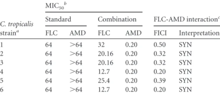
Synergistic effect of amiodarone and fluconazole.
Resistance
to fluconazole was experimentally induced in the six clinical
strains of
C
.
tropicalis
(Table 1). The development of fluconazole
resistance was achieved in approximately 100 days, and the strains
were also resistant to amiodarone. However, when these
experi-TABLE 1Synergistic effect of FLC and AMD against FLC-resistantstrains ofC. tropicalisisolated in Ceará (MIC50at 48 h)
C. tropicalis straina
MIC50b
FLC-AMD interactionc
Standard Combination
FLC AMD FLC AMD FICI Interpretation
1 64 ⬎64 32 0.20 0.50 SYN
2 64 ⬎64 20.16 0.20 0.32 SYN
3 64 ⬎64 20.16 0.20 0.32 SYN
4 64 ⬎64 12.7 0.20 0.20 SYN
5 64 ⬎64 25.4 0.20 0.39 SYN
6 64 ⬎64 12.7 0.20 0.20 SYN
aFLC-resistant strains ofC. tropicalisisolated from biological samples. b
The MIC50was defined as the lowest concentration that produced a 50% reduction in
growth of fungal cells after 48 h of incubation. The procedure was performed according to CLSI protocol M27-A3. The values are expressed ing/ml for FLC and inM for AMD. The MICs represent geometric means of at least three MICs determined on different days.
cThe synergistic effect of FLC and AMD was calculated based on the FICI (see the text).
FIG 1Effect of FLC (64g/ml) on an FLC-sensitive strain ofC. tropicalisafter 24 h. (Left) Analysis of changes in cell size/granularity (forward scatter [FSC]⫻
mentally induced resistant strains were exposed to various
con-centrations of fluconazole plus 0.20
M amiodarone, a synergistic
effect of the drugs on cell growth was observed (FICI
ⱕ
0.50)
(Table 1). Based on these findings, experiments were devised
aimed at elucidating the mechanisms involved in the cytotoxic
action of amiodarone and fluconazole against
C. tropicalis
.
Changes in cell size/granularity are synergistically induced
by amiodarone and fluconazole in fluconazole-resistant strains.
The flow cytometry analysis (side scatter
⫻
forward light scatter)
of the fluconazole-susceptible strains treated with fluconazole
re-vealed cell shrinkage and nuclear condensation, as evidenced by a
decrease in forward light scattering and a transient increase in side
scattering (Fig. 1, left). Both of these changes in cell
size/granular-ity observed in fluconazole-sensitive strains are compatible with
the presence of dying or dead cells. Regarding
fluconazole-resis-tant
C. tropicalis
, changes in cell size/granularity were not detected
in cells subjected to shorter treatments (4 and 6 h) with either
drug, alone or in combination (data not shown). On the other
hand, all of the fluconazole-resistant
C. tropicalis
strains showed
changes in cell size/granularity, which were observed only after 24
h of exposure to both fluconazole and amiodarone (see
Fig. 3A).
Loss of cell viability and damage to the plasma membrane are
observed after cotreatment of fluconazole-resistant
C
.
tropicalis
strains with amiodarone and fluconazole.
Treatment of
flucona-zole-sensitive
C. tropicalis
strains with fluconazole caused
dis-ruption of the yeast cell membrane (Fig. 1, middle), and this
effect was stronger than that found in the fluconazole-resistant
strains (see
Fig. 3B), as demonstrated by the propidium iodide
exclusion assay. As depicted in
Fig. 2, the exposure of
flucona-zole-resistant strains to the azole did not cause reductions in
the number of viable cells compared to the control. However,
cells treated with fluconazole and amiodarone for 24 h showed
a significant decrease in cell viability (
P
⬍
0.05). Moreover, the
plasma membrane stability of fluconazole-resistant cells was
not affected after exposure to fluconazole or amiodarone
(sin-gle treatment), respectively (Fig. 3B). On the other hand,
co-treatment (fluconazole plus amiodarone) of the
fluconazole-resistant strains induced a significant increase (
P
⬍
0.05) in the
population of cells with lesions in the plasma membrane
(Fig. 3B) after 24 h of exposure.
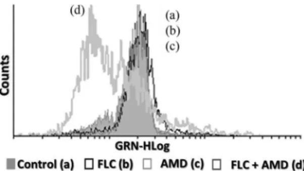
Amiodarone inhibits rhodamine 6G efflux in
fluconazole-resistant
C
.
tropicalis
strains.
As shown in
Fig. 4, the treatment of
fluconazole-resistant strains with fluconazole for 24 h did not
cause any significant change (
P
⬍
0.05) in the efflux of Rho 6G in
comparison to the control group (untreated cells). On the other
hand, when these fluconazole-resistant strains were treated with
amiodarone or amiodarone and fluconazole for 24 h, significant
inhibition (
P
⬍
0.05) in the efflux of Rho 6G was observed,
sug-gesting that amiodarone interferes with the operation of the efflux
pump, as described previously (7,
21).
Amiodarone and fluconazole synergistically increase the
in-tracellular production of ROS in fluconazole-resistant
C.
tropi-calis
strains.
A significant increase (
P
⬍
0.05) in ROS production
was observed when fluconazole-sensitive
C. tropicalis
strains were
exposed to fluconazole or fluconazole plus amiodarone compared
FIG 2Effects of the different treatments on the viability of FLC-resistant cellsofC. tropicalisas evaluated by flow cytometry after 24 h. Cells were treated with RPMI (negative control), AMD (64M), FLC (FLUCO; 64g/ml), and FLC (16g/ml) plus AMD (0.20M). The data are presented as mean values⫾
standard errors of the means (SEM) from experiments performed in triplicate.
with the negative-control group (Fig. 5A
and
B). On the other
hand, a significant increase (
P
⬍
0.05) in ROS production by the
fluconazole-resistant strains was observed only in cells treated
with both compounds (Fig. 5C
and
D).
Depolarization of the mitochondrial membrane (
⌬
m).
After
24-h exposure to fluconazole, alterations in the
⌬
m were observed
in the fluconazole-sensitive strains of
C. tropicalis
(Fig. 1, right). In
contrast, mitochondrial dysfunctions in fluconazole-resistant strains
of
C. tropicalis
were observed only in cells treated with amiodarone
plus fluconazole, suggesting that the cotreatment induces a reduction
in the mitochondrial transmembrane potential (Fig. 6).
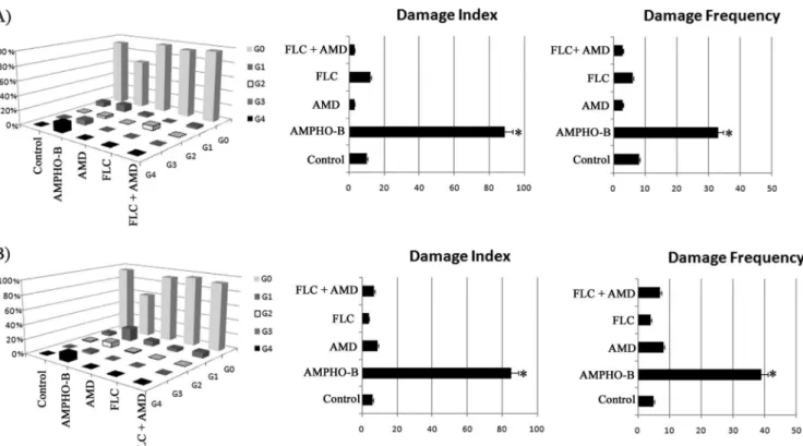
Cotreatment with fluconazole and amiodarone did not
in-duce strand breaks in the genomic DNA of
C. tropicalis
.
Strand
breaks in genomic DNA were evaluated by a standard alkaline
FIG 4Analysis of Rho 6G uptake and glucose-induced Rho 6G efflux onFLC-resistant strains ofC. tropicalisexposed to FLC (64g/ml), AMD (64
M), and FLC (16g/ml) plus AMD (0.20M) for 24 h. Untreated cells were used as a control. *,P⬍0.05 compared to the control (ANOVA and Newman-Keuls test). The error bars indicate SEM.
FIG 5Histograms obtained by flow cytometry analysis of yellow fluorescence (YLW-HLog) of FLC-sensitive (A) and FLC-resistant (C) strains ofC. tropicalisand percentages of DCF fluorescence-positive cells (ROS production) in FLC-sensitive (B) and FLC-resistant (D) strains exposed to RPMI (a), FLC (64g/ml) (b), AMD (64M) (c), and FLC (16g/ml) plus AMD (0.20M) (d) for 24 h. *,P⬍0.05 compared to the control (lanes C) (ANOVA and Newman-Keuls test). The error bars indicate SEM.
version of the comet assay, and the results were expressed as the
damage index (DI) and damage frequency (DF). Thus, the
treat-ment of the
C
.
tropicalis
strains (sensitive and resistant to
flucona-zole) with the drugs alone or in combination (fluconazole plus
amiodarone) was not able to induce strand breaks in their
genomic DNA (Fig. 7A
and
B) after 24-h exposure compared to
untreated cells. On the other hand, the known antifungal agent
amphotericin B (4
g/ml), which was used as a positive control,
induced a significant increase in DI and DF compared to
un-treated cultures of both fluconazole-sensitive and
fluconazole-re-sistant strains of
C
.
tropicalis
(Fig. 7A
and
B).
Oxidative DNA damage induced by cotreatment of
C
.
tropi-calis
with amiodarone and fluconazole.
As we had observed a
pro-oxidative effect of fluconazole, we next decided to evaluate
the level of oxidative DNA damage exerted by fluconazole alone or
in combination with amiodarone. To this end, we performed the
alkaline version of the comet assay using formamidopyrimidine
DNA glycosylase (FPG), a base excision repair enzyme that
recog-nizes and removes a wide range of oxidized purines from damaged
DNA (41).
Figure 8
shows that, following incubation with FPG,
there were significant increases in DI and DF in cells from both
strains (sensitive and resistant to fluconazole) that were treated
with fluconazole alone and strains that were treated with
flucona-zole plus amiodarone compared to cells not incubated with FPG.
Furthermore, amiodarone did not influence the oxidative DNA
damage induced by fluconazole. Interestingly, in cultures treated
with fluconazole or exposed to fluconazole and amiodarone, the
lev-els of FPG-sensitive sites were more pronounced in the
fluconazole-FIG 7Effects of different treatments on the distribution of damage classes (grades [G]), DNA damage index, and frequency (percent) using the standard alkaline version of the comet assay inC. tropicalisstrains sensitive (A) and resistant (B) to fluconazole after 24-h exposure. The yeasts were exposed to RPMI, FLC (64g/ml), AMD (64M), FLC (16g/ml) plus AMD (0.20M), and amphotericin B (AMPHO-B) (4g/ml) as a control. *,P⬍0.05 compared to the control (ANOVA and Newman-Keuls test). The error bars indicate SEM.
sensitive strains than in the resistant strains. Moreover, amiodarone
did not cause significant differences (
P
⬍
0.05) in the levels of
FPG-sensitive sites that were induced by fluconazole in the genomic DNAs
of both strains (sensitive and resistant to fluconazole).
Caspase 3/7 is synergistically activated by amiodarone and
fluconazole in fluconazole-resistant
C. tropicalis
strains.
In the
fluconazole-sensitive strains of
C. tropicalis
(Fig. 9A), an expanded
population of caspase-positive cells was detected by flow
cytom-etry following exposure to fluconazole alone (67.39%
⫾
1.15%) or
fluconazole and amiodarone (54.28%
⫾
2.56%). On the other hand,
in the fluconazole-resistant strains, a significant population of
caspase-positive cells was detected only when the cells were treated
with fluconazole and amiodarone (61.55%
⫾
2.21%) (Fig. 9B).
DISCUSSION
bility of
C. tropicalis
to azoles can be reduced by exposure
in vitro
or
in vivo
to these drugs (26,
46).
In our model, clinical strains of
C. tropicalis
have acquired
resistance to fluconazole, but the development of resistance
(achieved in
⬃
100 days) was slower than the time frame reported
by Barchiesi et al. (26). This difference was likely due to the lower
drug concentration used in our protocol.
Moreover, the synergistic effect of amiodarone with azoles against
azole-resistant strains of
C. albicans
and
A. fumigatus
has been
dem-onstrated
in vitro
(7,
19,
21). Knorre et al. (47) have reported that the
treatment of azole-resistant strains of
Saccharomyces cerevisiae
with
amiodarone decreased multidrug resistance due to the inhibition of
cellular efflux pumps. In the present study, the synergistic effect of
amiodarone and fluconazole was demonstrated using
fluconazole-resistant strains of
C. tropicalis
(Table 1).
As shown in
Fig. 2, amiodarone plus fluconazole caused
cyto-toxic effects on fluconazole-resistant strains of
C
.
tropicalis
, and
these changes were not significant in cultures treated with
amio-darone or fluconazole alone. Therefore, by blocking efflux pumps,
amiodarone likely enhances the function of fluconazole to
de-crease cell size and inde-crease granularity, as shown in
Fig. 3A.
As revealed by the Rho 123 test (Fig. 6), amiodarone plus
flu-conazole produced mitochondrial dysfunction in fluflu-conazole-re-
fluconazole-re-sistant strains, suggesting that these drugs affect the
mitochon-drial respiratory function. In this case, the
⌬
m value would be
expected to collapse and Rho 123 would likely not accumulate in
the mitochondria (36). Studies have shown that amiodarone has
the ability to induce a decrease in the mitochondrial
transmem-brane potential (7,
48). Therefore, according to the results shown
in
Fig. 2,
3, and
6, treatment of the fluconazole-resistant strains of
C
.
tropicalis
with amiodarone plus fluconazole altered not only cell
size/granularity, but also the
⌬
m value. These changes were
likely due to the different sites of action of the two drugs (7).
According to Gamarra et al. (7), the synergism between
amio-darone and fluconazole can be explained by efflux pump blockage
caused by amiodarone. On the other hand, Zhang et al. (49)
pro-posed that fluconazole exacerbated the intracellular levels of Ca
2⫹and H
⫹ions, thus interfering with the ionic homeostasis.
Ergos-terol is the major lipid of fungal cells, and changes in its
biosyn-thetic pathway can alter cellular responses to stress (7,
49).
Our findings also indicate that the synergistic effect of
amio-darone and fluconazole involves an increase in the intracellular
levels of ROS in the fluconazole-resistant strains of
C. tropicalis
.
Other works have reported that the azoles are responsible for
en-dogenous production of ROS in
Candida
spp. (50,
51), and this
has been observed in sensitive strains treated with fluconazole.
As shown in
Fig. 3B, the cell membrane integrity of
C. tropicalis
strains treated with fluconazole plus amiodarone was clearly
al-tered, as evaluated by the incorporation of PI, and this finding was
likely due to the severe membrane damage caused by increased
levels of ROS.
Yeast cells exhibit a wide range of dose-dependent responses to
increasing ROS concentrations (52–54). At higher doses, cell
death occurs in a fraction of cells and results in the acquisition of
phenotypes characteristic of caspase-dependent apoptosis (55).
Extensive DNA fragmentation occurs frequently in the early
stages of apoptosis, and it is an irreversible step that leads to cell
death (52,
56). The alkaline version of the comet assay (standard
protocol) is a sensitive procedure used to quantify the different
types of DNA damage in cells, including alkali-labile sites,
single-stranded breaks, and double-single-stranded breaks (57). Our data
re-vealed that exposure of
C. tropicalis
strains to fluconazole plus
amiodarone (Fig. 7) did not induce any significant increase in the
DNA migration pattern (DNA fragments). However, the standard
comet assay did reveal a significant increase in DNA breaks in
C.
tropicalis
strains treated with amphotericin B (4
g/ml), which
was used as a positive control.
Due to these results, the alkaline version of the comet assay was
conducted in the presence of FPG to verify oxidative damage to
the DNA (9,
58,
59). The results shown in
Fig. 8
indicate that
significant DNA damage was caused by the
fluconazole-plus-ami-odarone treatment, and these results are expressed as the index of
DNA damage after treatment with the DNA repair enzyme (FPG).
Moreover, the results obtained after incubation with FPG clearly
show that DNA migration was increased in the treated strains of
C.
tropicalis
, and the extent of oxidative DNA damage caused by the
synergism between fluconazole and amiodarone was significantly
higher. This increase was likely due to the ability of FPG to
recog-nize purines (adenine and guanine) within DNA that were
oxi-dized by the ROS produced following
fluconazole-plus-amioda-rone treatment (Fig. 5). Based on such results, we suggest that the
synergistic effect of fluconazole plus amiodarone facilitates
oxida-tive damage to DNA via ROS formation.
These results prompted us to perform further study on the
physiological state of the cells, particularly with regard to the
oc-currence of apoptosis and/or necrosis induced by fluconazole plus
amiodarone via caspase 3/7. As shown in
Fig. 9, an increase in the
number of apoptotic cells was observed in the
fluconazole-resis-tant strains treated with fluconazole plus amiodarone (61.55%
⫾
2.21%).
Conclusion.
In conclusion, treatment with fluconazole plus
amiodarone caused moderate
in vitro
cytotoxicity in
fluconazole-resistant strains of
C. tropicalis.
Although this treatment altered
the integrity of the plasma and mitochondrial membranes, as
de-scribed above, the synergism between fluconazole and
amioda-rone also seemed to cause DNA damage, leading to apoptotic cell
death.
ACKNOWLEDGMENTS
This research was supported by grants and fellowships from CNPq, CAPES/Brazil, and FUNCAP/Ceará.
We declare no conflicts of interest concerning this article.
REFERENCES
1.Horn D, Fishman J, Steinbach W, Anaissie E, Marr K, Olyaei A, Pfaller M, Weiss M, Webster K, Neofytos D.2007. Presentation of the PATH Alliance registry for prospective data collection and analysis of the epide-miology, therapy, and outcomes of invasive fungal infections. Diagn. Mi-crobiol. Infect. Dis.59:407– 414.
2.Horn DL, Neofytos D, Anaissie EJ, Fishman JA, Steinbach WJ, Olyaei AJ, Marr KA, Pfaller MA, Chang CH, Webster KM.2009. Epidemiology and outcomes of candidemia in 2019 patients: data from the prospective antifungal therapy alliance registry. Clin. Infect. Dis.48:1695–1703. 3.Pfaller MA, Castanheira M, Diekema DJ, Messer SA, Jones RN.2011.
Wild-type MIC distributions and epidemiologic cutoff values for flucona-zole, posaconaflucona-zole, and voriconazole when testingCryptococcus neofor-mansas determined by the CLSI broth microdilution method. Diagn. Microbiol. Infect. Dis.71:252–259.
4.Pfaller MA, Diekema DJ.2007. Epidemiology of invasive candidiasis: a persistent public health problem. Clin. Microbiol. Rev.20:133–163. 5.Sellami A, Sellami H, Neji S, Makni F, Abbes S, Cheikhrouhou F, Chelly
6.Chi HW, Yang YS, Shang ST, Chen KH, Yeh KM, Chang FY, Lin JC.
2011.Candida albicansversus non-albicansbloodstream infections: the comparison of risk factors and outcome. J. Microbiol. Immunol. Infect.
44:369 –375.
7.Gamarra S, Rocha EM, Zhang YQ, Park S, Rao R, Perlin DS.2010. Mechanism of the synergistic effect of amiodarone and fluconazole in Candida albicans. Antimicrob. Agents Chemother.54:1753–1761. 8.Graybill JR, Najvar LK, Holmberg JD, Luther MF.1995. Fluconazole,
D0870, and flucytosine treatment of disseminatedCandida tropicalis in-fections in mice. Antimicrob. Agents Chemother.39:924 –929. 9.da Silva EN, Jr, Cavalcanti BC, Guimaraes TT, Pinto Mdo C, Cabral IO,
Pessoa C, Costa-Lotufo LV, de Moraes MO, de Andrade CK, Dos Santos MR, de Simone CA, Goulart MO, Pinto AV.2011. Synthesis and evaluation of quinonoid compounds against tumor cell lines. Eur. J. Med. Chem.46:399 – 410.
10. Gonzalez GM, Elizondo M, Ayala J.2008. Trends in species distribution and susceptibility of bloodstream isolates ofCandidacollected in Monter-rey, Mexico, to seven antifungal agents: results of a 3-year (2004 to 2007) surveillance study. J. Clin. Microbiol.46:2902–2905.
11. Pina-Vaz C, Rodrigues AG, Costa-de-Oliveira S, Ricardo E, Mardh PA.
2005. Potent synergic effect between ibuprofen and azoles onCandida resulting from blockade of efflux pumps as determined by FUN-1 staining and flow cytometry. J. Antimicrob. Chemother.56:678 – 685.
12. Vandeputte P, Larcher G, Bergès T, Renier G, Dominique C, Bouchara JP.2005. Mechanisms of azole resistance in a clinical isolate ofCandida tropicalis. Antimicrob. Agents Chemother.49:4608 – 4615.
13. Pina-Vaz C, Sansonetty F, Rodrigues AG, Costa-de-Oliveira S, Marti-nez-de-Oliveira J, Fonseca AF. 2001. Susceptibility to fluconazole of Candidaclinical isolates determined by FUN-1 staining with flow cytom-etry and epifluorescence microscopy. J. Med. Microbiol.50:375–382. 14. Tobudic S, Kratzer C, Presterl E.2012. Azole-resistantCandidaspp.—
emerging pathogens? Mycoses55:24 –32.
15. Vanden Bossche H, Warnock DW, Dupont B, Kerridge D, Sen Gupta S, Improvisi L, Marichal P, Odds FC, Provost F, Ronin O. 1994. Mechanisms and clinical impact of antifungal drug resistance. J. Med. Vet. Mycol.32(Suppl. 1):189 –202.
16. White T, Bruns T, Lee S, Taylor J. 1990. Amplification and direct sequencing of fungal ribosomal RNA genes for phylogenetics, p 315–322. InInnis MA, Gefland DH, Sninsky JJ, White TJ (ed), PCR protocols: a guide to methods and applications. Academic Press, San Diego, CA. 17. Courchesne WE.2002. Characterization of a novel, broad-based
fungi-cidal activity for the antiarrhythmic drug amiodarone. J. Pharmacol. Exp. Ther.300:195–199.
18. Wakiec R, Prasad R, Morschhauser J, Barchiesi F, Borowski E, Milewski S.2007. Voriconazole and multidrug resistance inCandida albicans. My-coses50:109 –115.
19. Afeltra J, Vitale RG, Mouton JW, Verweij PE.2004. Potent synergis-tic in vitro interaction between nonantimicrobial membrane-active compounds and itraconazole against clinical isolates ofAspergillus fu-migatusresistant to itraconazole. Antimicrob. Agents Chemother.48: 1335–1343.
20. Gupta SS, Ton VK, Beaudry V, Rulli S, Cunningham K, Rao R.2003. Antifungal activity of amiodarone is mediated by disruption of calcium homeostasis. J. Biol. Chem.278:28831–28839.
21. Guo Q, Sun S, Li Y, Yu J, Shi C.2008. Synergistic activity of azoles with amiodarone against clinically resistantCandida albicanstested by che-querboard and time-kill methods. J. Med. Microbiol.57:457– 462. 22. Reference deleted.
23. Sambrook J, Fritsch EF, Maniatis T.1989. The condensed protocols from Molecular Cloning: a Laboratory Manual. Cold Spring Harbor Lab-oratory Press, Cold Spring Harbor, NY.
24. Huang X, Madan A.1999. CAP3: a DNA sequence assembly program. Genome Res.9:868 – 877.
25. Altschul SF, Gish W, Miller W, Myers EW, Lipman DJ.1990. Basic local alignment search tool. J. Mol. Biol.215:403– 410.
26. Barchiesi F, Calabrese D, Sanglard D, Falconi Di Francesco L, Caselli F, Giannini D, Giacometti A, Gavaudan S, Scalise G.2000. Experimental induction of fluconazole resistance inCandida tropicalisATCC 750. An-timicrob. Agents Chemother.44:1578 –1584.
27. Pinto e Silva AT, Costa-de-Oliveira S, Silva-Dias A, Pina-Vaz C, Ro-drigues AG.2009. Dynamics of in vitro acquisition of resistance by Can-dida parapsilosisto different azoles. FEMS Yeast Res.9:626 – 633.
28. Clinical and Laboratory Standards Institute.2008. Reference method for broth dilution antifungal susceptibility testing of yeasts. Approved standard M27-A3, 3rd ed. Clinical and Laboratory Standards Institute, Wayne, PA.
29. Pfaller MA, Diekema DJ.2012. Progress in antifungal susceptibility test-ing ofCandidaspp. by use of Clinical and Laboratory Standards Institute broth microdilution methods, 2010 to 2012. J. Clin. Microbiol.50:2846 – 2856.
30. Endo E.2007. Synergistic effect of the crude extract and fractions of Punica granatumagainstCandida albicansand synergy with fluconazole. Ph.D. dissertation. State University of Maringá, Maringá, Brazil. 31. Odds FC.2003. Synergy, antagonism, and what the chequerboard puts
between them. J. Antimicrob. Chemother.52:1.
32. Pina-Vaz C, Rodrigues AG.2010. Evaluation of antifungal susceptibility using flow cytometry. Methods Mol. Biol.638:281–289.
33. Rudensky B, Broide E, Berko N, Wiener-Well Y, Yinnon AM, Raveh D.
2008. Direct fluconazole susceptibility testing of positiveCandidablood cultures by flow cytometry. Mycoses51:200 –204.
34. Joung YH, Kim HR, Lee MK, Park AJ.2007. Fluconazole susceptibility testing ofCandidaspecies by flow cytometry. J. Infect.54:504 –508.
35. Pinkerton DM, Banwell MG, Garson MJ, Kumar N, Moraes MO,
Cavalcanti BC, Barros FWA, Pessoa C.2010. Antimicrobial and cyto-toxic activities of synthetically derived tambjamines C and E–J, BE-18591, and a related alkaloid from the marine bacteriumPseudoalteromonas tu-nicate. Chem. Biodivers.7:1311–1324.
36. Ludovico P, Sansonetty F, Corte-Real M.2001. Assessment of mitochon-drial membrane potential in yeast cell populations by flow cytometry. Microbiology147:3335–3343.
37. Hempel SL, Buettner GR, O’Malley YQ, Wessels DA, Flaherty DM.
1999. Dihydrofluorescein diacetate is superior for detecting intracellular oxidants: comparison with 2=,7=-dichlorodihydrofluorescein diacetate, 5(and 6)-carboxy-2=,7=-dichlorodihydrofluorescein diacetate, and dihy-drorhodamine 123. Free Radic. Biol. Med.27:146 –159.
38. LeBel CP, Ischiropoulos H, Bondy SC.1992. Evaluation of the probe 2=,7=-dichlorofluorescin as an indicator of reactive oxygen species forma-tion and oxidative stress. Chem. Res. Toxicol.5:227–231.
39. Miloshev G, Mihaylov I, Anachkova B.2002. Application of the single cell gel electrophoresis on yeast cells. Mutat. Res.513:69 –74.
40. Collins AR.2004. The comet assay for DNA damage and repair: princi-ples, applications, and limitations. Mol. Biotechnol.26:249 –261. 41. Serre L, Jesus KP, Boiteux S, Zelwer C, Castaing B. 2002. Crystal
structure of theLactococcus lactisformamidopyrimidine-DNA glycosylase bound to an abasic site analogue-containing DNA. EMBO J.21:2854 – 2865.
42. Almirante B, Rodriguez D, Park BJ, Cuenca-Estrella M, Planes AM, Almela M, Mensa J, Sanchez F, Ayats J, Gimenez M, Saballs P, Fridkin SK, Morgan J, Rodriguez-Tudela JL, Warnock DW, Pahissa A.2005. Epidemiology and predictors of mortality in cases ofCandida blood-stream infection: results from population-based surveillance, Barcelona, Spain, from 2002 to 2003. J. Clin. Microbiol.43:1829 –1835.
43. Kucukates E, Erturan Z, Susever S, Yegenoglu Y.2005. In vitro suscep-tibility of yeasts isolated from patients in intensive care units to flucona-zole and amphotericin B during a 3-year period. APMIS113:278 –283. 44. Wingard JR.1995. Importance ofCandidaspecies other thanC. albicans
as pathogens in oncology patients. Clin. Infect. Dis.20:115–125. 45. Wingard JR, Merz WG, Saral R. 1979.Candida tropicalis: a major
pathogen in immunocompromised patients. Ann. Intern. Med. 91: 539 –543.
46. Barchiesi F, Arzeni D, Del Prete MS, Sinicco A, Falconi Di Francesco L, Pasticci MB, Lamura L, Nuzzo MM, Burzacchini F, Coppola S, Chiodo F, Scalise G.1998. Fluconazole susceptibility and strain variation of Can-dida albicansisolates from HIV-infected patients with oropharyngeal can-didosis. J. Antimicrob. Chemother.41:541–548.
47. Knorre DA, Krivonosova TN, Markova OV, Severin FF.2009. Amio-darone inhibits multiple drug resistance in yeastSaccharomyces cerevisiae. Arch. Microbiol.191:675– 679.
48. Maresova L, Muend S, Zhang YQ, Sychrova H, Rao R.2009. Membrane hyperpolarization drives cation influx and fungicidal activity of amioda-rone. J. Biol. Chem.284:2795–2802.
49. Zhang Y-Q, Gamarra S, Garcia-Effron G, Park S, Perlin DS, Rao R.
50. Kobayashi D, Kondo K, Uehara N, Otokozawa S, Tsuji N, Yagihashi A, Watanabe N.2002. Endogenous reactive oxygen species is an important mediator of miconazole antifungal effect. Antimicrob. Agents Che-mother.46:3113–3117.
51. Rogers PD, Vermitsky JP, Edlind TD, Hilliard GM.2006. Proteomic analysis of experimentally induced azole resistance inCandida glabrata. J. Antimicrob. Chemother.58:434 – 438.
52. Guerreiro-Salvador V.2009. Assessment of Apoptosis and Necrosis in Saccharomyces cerevisiaein Fermentation Vinárias. Ph.D. dissertation. University of Minho, Braga, Portugal.
53. Perrone GG, Tan SX, Dawes IW.2008. Reactive oxygen species and yeast apoptosis. Biochim. Biophys. Acta1783:1354 –1368.
54. Temple MD, Perrone GG, Dawes IW.2005. Complex cellular responses to reactive oxygen species. Trends Cell Biol.15:319 –326.
55. Madeo F, Herker E, Maldener C, Wissing S, Lachelt S, Herlan M, Fehr M, Lauber K, Sigrist SJ, Wesselborg S, Frohlich KU.2002. A caspase-related protease regulates apoptosis in yeast. Mol. Cell9:911–917.
56. Ribeiro GF, Corte-Real M, Johansson B.2006. Characterization of DNA damage in yeast apoptosis induced by hydrogen peroxide, acetic acid, and hyperosmotic shock. Mol. Biol. Cell17:4584 – 4591.
57. Burlinson B, Tice RR, Speit G, Agurell E, Brendler-Schwaab SY, Collins AR, Escobar P, Honma M, Kumaravel TS, Nakajima M, Sasaki YF, Thybaud V, Uno Y, Vasquez M, Hartmann A.2007. Fourth Interna-tional Workgroup on Genotoxicity Testing: results of the in vivo Comet assay workgroup. Mutat. Res.627:31–35.
58. Cavalcanti BC, Barros FW, Cabral IO, Ferreira JR, Magalhaes HI, Junior HV, da Silva Junior EN, de Abreu FC, Costa CO, Goulart MO, Moraes MO, Pessoa C.2011. Preclinical genotoxicology of nor-beta-lapachone in human cultured lymphocytes and Chinese hamster lung fibroblasts. Chem. Res. Toxicol.24:1560 –1574.