Berberine Antifungal Activity in Fluconazole-Resistant Pathogenic
Yeasts: Action Mechanism Evaluated by Flow Cytometry and Biofilm
Growth Inhibition in
Candida
spp.
Anderson Ramos da Silva,a
João Batista de Andrade Neto,a
Cecília Rocha da Silva,a
Rosana de Sousa Campos,a Rose Anny Costa Silva,a
Daniel Domingues Freitas,a
Francisca Bruna Stefany Aires do Nascimento,a
Larissa Nara Dantas de Andrade,a Letícia Serpa Sampaio,a
Thalles Barbosa Grangeiro,b
Hemerson Iury Ferreira Magalhães,c
Bruno Coêlho Cavalcanti,d Manoel Odorico de Moraes,d
Hélio Vitoriano Nobre Júniora
School of Pharmacy, Laboratory of Bioprospection and Experiments in Yeast (LABEL), Federal University of Ceara, Fortaleza, CE, Brazila; Department of Biology, Science Center, Molecular Genetics Laboratory, Federal University of Ceara, Fortaleza, CE, Brazilb; School of Pharmacy, Federal University of Paraiba, João Pessoa, PB, Brazilc; Department of Physiology and Pharmacology, Federal University of Ceara, Fortaleza, CE, Brazild
The incidence of fungal infections and, in particular, the incidence of fungal antibiotic resistance, which is associated with
bio-film formation, have significantly increased, contributing to morbidity and mortality. Thus, new therapeutic strategies need to
be developed. In this context, natural products have emerged as a major source of possible antifungal agents. Berberine is a
pro-toberberine-type isoquinoline alkaloid isolated from the roots, rhizomes, and stem bark of natural herbs, such as
Berberis
aqui-folium
,
Berberis vulgaris
,
Berberis aristata
, and
Hydrastis canadensis
, and of
Phellodendron amurense
. Berberine has been
proven to have broad antibacterial and antifungal activity. In the present study, the potential antifungal effect of berberine
against fluconazole-resistant
Candida
and
Cryptococcus neoformans
strains, as well as against the biofilm form of
Candida
spp.,
was assessed. The antifungal effect of berberine was determined by a broth microdilution method (the M27-A3 method of the
Clinical and Laboratory Standards Institute) and flow cytometry techniques, in which the probable mechanism of action of the
compound was also assessed. For biofilm assessment, a colorimetric 3-(4,5-dimethyl-2-thiazolyl)-2,5-diphenyl-2H-tetrazolium
bromide (MTT) assay was used to determine the susceptibility of sessile cells. The isolates used in the study belonged to the
Lab-oratory of Bioprospection and Experiments in Yeast (LABEL) of the Federal University of Ceará. After 24 and 72 h,
fluconazole-resistant
Candida
and
Cryptococcus neoformans
strains showed berberine MICs equal to 8
g/ml and 16
g/ml, respectively.
Cytometric analysis showed that treatment with berberine caused alterations to the integrity of the plasma and mitochondrial
membranes and DNA damage, which led to cell death, probably by apoptosis. Assessment of biofilm-forming isolates after
treat-ment showed statistically significant reductions in biofilm cell activity (
P
<
0.001).
Y
easts are the most common agents in opportunistic fungal
infections, which mainly affect immunocompromised
pa-tients (
1
).
Candida
yeasts are pathogens that appear in nosocomial
infections.
Candida
spp. are the third most commonly isolated
agents of fungal infections acquired in hospitals (
2
) and are noted
for their significant prevalence in different medical centers and for
the complications that they cause. In addition, these infections are
difficult to diagnose, leading to mortality rates of 50% (
3
,
4
).
The opportunistic and pathogenic fungus
Cryptococcus
neofor-mans
can cause life-threatening meningitis. Each year, it is
esti-mated that about 1 million new cases of cryptococcal meningitis
occur throughout the world, making
C. neoformans
infection a
global public health concern (
5
,
6
). Although antifungal drugs for
the treatment cryptococcosis are available, they often fail for
sev-eral reasons, especially because of
C. neoformans
resistance to the
antifungal agents (
7
).
Another current problem are nosocomial infections associated
with biofilms. These infections are difficult to treat because these
microbial structures are resistant both to the activity of
conven-tional antimicrobial agents and to host defense mechanisms.
Can-dida
spp. are among the main organisms responsible for these
infections, which are mainly associated with implanted medical
devices (
2
).
In this context, herbal antifungal agents have gained
impor-tance due to their natural origin (
8
). Berberine is a
protober-berine-type isoquinoline alkaloid isolated from the roots,
rhi-zomes, and stem bark of natural herbs, such as
Berberis
aquifolium
,
Berberis vulgaris
,
Berberis aristat
a,
Hydrastis
canaden-sis
,
Phellodendron amurense
,
Coptis chinensis
, and
Tinospora
cordi-folia
(
9–11
). In some studies, it was shown that berberine extract
has significant antimicrobial activity against bacteria, viruses,
pro-tozoa, fungi, and yeasts (
12
).
Studies have also shown that berberine has a significant
anti-fungal effect against
Candida albicans
strains (
13–15
).
Received31 July 2015 Returned for modification14 November 2015
Accepted19 March 2016
Accepted manuscript posted online28 March 2016
Citationda Silva AR, de Andrade Neto JB, da Silva CR, Campos RDS, Costa Silva RA, Freitas DD, do Nascimento FBSA, de Andrade LND, Sampaio LS, Grangeiro TB, Magalhães HIF, Cavalcanti BC, de Moraes MO, Nobre Júnior HV. 2016. Berberine antifungal activity in fluconazole-resistant pathogenic yeasts: action mechanism evaluated by flow cytometry and biofilm growth inhibition inCandidaspp. Antimicrob Agents Chemother 60:3551–3557.
doi:10.1128/AAC.01846-15.
Address correspondence to Hélio Vitoriano Nobre Júnior, label_ufc@yahoo.com.br.
Copyright © 2016 da Silva et al. This is an open-access article distributed under the terms of theCreative Commons Attribution 4.0 International license.
on June 14, 2016 by UNIVERSIDADE FEDERAL DO CEARÕ
http://aac.asm.org/
The aim of the present study was to evaluate the antifungal
potential of berberine against different fluconazole-resistant
Can-dida
and
Cryptococcus neoformans
strains. In addition, the effect of
berberine against
C. albicans
and its activity against biofilms
pro-duced by
Candida tropicalis
were assessed through flow cytometry
procedures and a standard single cell gel electrophoresis (comet)
assay.
MATERIALS AND METHODS
Isolates.ElevenCandidastrains that were isolated from blood samples at the Central Public Health Laboratory (LACEN-CE) and that are part of the Yeast Collection from the Laboratory of Bioprospection and Experi-ments in Yeast, affiliated with the School of Pharmacy, Federal University of Ceara (LABEL/FF/UFC), and two ATCC strains (Candida parapsilosis
ATCC 22019 andCandida kruseiATCC 6258) were used. The strains were inoculated in Sabouraud dextrose agar (HiMedia, Mumbai, India) and incubated at 37°C for 24 h. Then, the strains were grown in CHROMagar
Candida(HiMedia), in order to evaluate their purity.
Molecular identification.Genomic DNA was purified using a proto-col based on the cetyltrimethylammonium bromide (CTAB) surfactant, as previously described (16). The nuclear DNA region comprising inter-nal transcribed spacer 1 (ITS1), ITS2, and the 5.8S rRNA gene was ampli-fied by PCR using primers ITS4 (5=-TCCTCCGCTTATTGATATGC-3=) and ITS5 (5=-GCAAGTAAAAGTCGTAACAAGA-3=), as suggested by White et al. (17). Amplification reactions were performed in a final vol-ume of 25l, which contained genomic DNA (300 to 400 ng), 1⫻GoTaq reaction buffer (Promega, Madison, WI, USA), 1.5 mM MgCl2, 200M each deoxynucleoside triphosphate (GE Healthcare Life Sciences, Piscat-away, NJ, USA), 0.5M each primer, and 1.25 U of GoTaq DNA poly-merase (Promega). Reactions were carried out in a Mastercycler gradient thermocycler (Eppendorf, Hamburg, Germany) programmed for an ini-tial denaturation step (2 min at 95°C), followed by 35 cycles of 1 min at 95°C, 1 min at 60°C, and 3 min at 72°C. The last cycle was followed by a final incubation of 10 min at 72°C. Control samples containing all com-ponents of the reaction mixture except DNA were used to test that no DNA contamination occurred. Amplification specificity was determined by 1.0% agarose gel electrophoresis (18). The remaining amplified prod-ucts were purified using an Illustra GFX PCR DNA and gel band purifi-cation kit (GE Healthcare Life Sciences). The concentrations of the puri-fied amplicons were determined by measuring the absorbance of 10-fold dilutions at 260 nm. DNA sequencing was performed at Macrogen Inc. (Seoul, South Korea) using the Sanger chain-termination method. Both strands of each amplicon were sequenced using primers ITS4 and ITS5. Subsequently, the sequences were assembled using the Phred/Phrap/Consed package (19–21). The start and end of the ITS1 and ITS2 sequences were identified by comparison to annotated sequences from the ITSoneDB (22) and ITS2 (23) databases, respectively. The ITS/5.8S sequences were deposited in the GenBank database and compared to those available in public DNA sequence databases using the BLAST program (24).
In vitroantifungal activity.The broth microdilution (BMD) suscep-tibility test was conducted according to the guidelines in document M27-A3 of the Clinical and Laboratory Standards Institute (CLSI) (25), using RPMI 1640 broth (pH 7.0) buffered with 0.165 M MOPS (morpho-linepropanesulfonic acid) (Sigma Chemical, St. Louis, MO). Fluconazole (FLC; Merck Sharp & Dohme, São Paulo, Brazil) was dissolved in distilled water, and the berberine chloride solution (Sigma Chemical) was pre-pared in dimethyl sulfoxide (DMSO; Sigma Chemical). FLC and ber-berine were tested at concentrations ranging from 0.125 to 64g/ml. The yeasts and compounds were incubated in 96-well culture plates at 35°C for 24 h, and the results were examined visually, as recommended by CLSI (26). The MIC of each compound was determined as the concentration that inhibited 50% of fungal growth. Cutoff points were determined accord-ing to the information in CLSI document M27-S4 (26). StrainsC. parapsilosis
ATCC 22019 andC. kruseiATCC 6258 were used as controls (25).
Mechanism-of-action studies.In order to assess cell density, mem-brane integrity, mitochondrial transmemmem-brane potential (⌬m), and DNA damage (comet assay), the fluconazole-resistantC. albicans2 strain was exposed for 24 h to increasing berberine concentrations (MIC, 2⫻ MIC, and 4⫻MIC). All tests were conducted in triplicate in three inde-pendent experiments.
Yeast suspension preparation.Cell suspensions were prepared from cultures in the exponential growth phase. Cells were collected by centrif-ugation (1,600⫻gfor 10 min at 4°C) and washed twice with a 0.85% saline solution by centrifugation at 1,200⫻gfor 5 min at 4°C. Subse-quently, they were resuspended (⬃106cells/ml) in HEPES buffer (pH 7.2) supplemented with 2% glucose. Amphotericin B (AMB; 4g/ml; Sigma Chemical) was used as a cell death control (27,28).
Cell density and membrane integrity determination.Fungal strain cell density and membrane integrity were evaluated by excluding 2 mg/ liter propidium iodide (PI). Aliquots from yeasts incubated for 24 h with berberine, FLC, and AMB were analyzed using flow cytometry. A total of 10,000 events were evaluated per experiment (n⫽2), and cellular debris was omitted from the analysis. Cell fluorescence was determined by flow cytometry using a Guava EasyCyte minisystem cytometer (Guava Tech-nologies Inc., Hayward, CA, USA), and the results were analyzed using CytoSoft (version 4.1) software (29,30).
Measurement of⌬m.⌬m was determined by measuring rhoda-mine 123 dye retention by the mitochondria of yeast cells after 24 h of exposure. Cells were washed with phosphate-buffered saline (PBS), incu-bated with 5 mg/liter rhodamine 123 at 37°C for 30 min in the dark, and then washed twice with PBS. Fluorescence was measured by flow cytom-etry (Guava EasyCyte minisystem). A total of 10,000 events were evalu-ated per experiment (n⫽2), and cellular debris was omitted from the analysis (27,30).
Yeast alkaline comet assay.An alkaline comet assay was conducted essentially as described by Miloshev et al. (31). Up to 200l of 0.5% agarose was spread on each slide, and this supportive agarose layer was air dried prior to application of the cell suspension.C. albicans2 cells were collected by centrifugation in an Eppendorf microcentrifuge for 5 min. Then, the cells were washed with distilled water and resuspended in S buffer (1 M sorbitol, 25 mM KH2PO4, pH 6.5). Aliquots of approximately 5⫻104cells were mixed with 0.7% low-melting-point agarose containing 2 mg/ml Zymolyase 20T (Seikagaku Corp., Japan), and the cells were subsequently spread over the slides. The slides were then covered with coverslips and incubated for 20 min at 30°C. In order to minimize endog-enous cell enzyme activity, all further procedures were performed in a cold room at 8 to 10°C. The coverslips were removed, and the slides were incubated in 30 mM NaOH, 1 M NaCl, 0.1% laurylsarcosine, and 50 mM EDTA (pH 12.3) for 1 h, in order to lyse the spheroplasts. The slides were rinsed three times for 20 min each time in 30 mM NaOH and 10 mM EDTA (pH 12.4), in order to unwind the DNA. Subsequently, the slides were subjected to electrophoresis in the same buffer. Electrophoresis was performed for 20 min at 0.5 V/cm and 24 mA. After electrophoresis, the gels were neutralized by submerging the slides in 10 mM Tris-HCl, pH 7.5, for 10 min, followed by consecutive 10-min incubations in 76% and 96% ethanol. Finally, the slides were left to air dry, stained with ethidium bro-mide (1 mg/ml), and visualized by fluorescence microscopy (32). All steps mentioned above were conducted in the dark, in order to prevent addi-tional DNA damage. Images of 100 randomly selected cells (50 cells from each of the two replicate slides) were analyzed for each experimental group. Cells were visually scored and assigned to one of five classes ac-cording to tail size (from undamaged [class 0] to maximally damaged [class 4]), and a damage index value was calculated for each cell sample. The damage index values therefore ranged from 0 (completely undam-aged, 100 cells⫻0) to 400 (maximum damage, 100 cells⫻4) (33). The frequency of cells with tails, which was considered an indicator of DNA damage, was calculated on the basis of the number of cells with tails (DNA strand breaks) and the number of cells without tails (28,29).
on June 14, 2016 by UNIVERSIDADE FEDERAL DO CEARÕ
http://aac.asm.org/
Analysis of oxidized purine and pyrimidine bases of yeast DNA.The alkaline comet assay was performed as described above. Slides were re-moved from the lysis solution and were washed three times in an enzyme buffer (40 mM HEPES, 100 mM KCl, 0.5 mM EDTA, 0.2 mg/ml bovine serum albumin, pH 8.0). Subsequently, they were drained and incubated with 70l formamidopyrimidine DNA-glycosylase (FPG) for 30 min at 37°C. Images of 100 randomly selected cells per group (50 cells from each of the two replicate slides) were visually analyzed. The number of oxidized purines (FPG-sensitive sites) was determined by subtracting the amount of strand breaks observed in the control samples (samples incubated only with buffer) from the total amount of breaks obtained after FPG incuba-tion (27,29).
Annexin V staining.Treated and untreatedC. albicanscells were col-lected by centrifugation and digested with 2 mg/ml Zymolyase 20T (Seika-gaku Corp., Japan) in potassium phosphate buffer (PPB) plus 1 M sorbitol (pH 6.0) for 2 h at 30°C.C. albicansprotoplasts were stained with fluores-cein isothiocyanate (FITC)-labeled annexin V and PI, using an FITC-annexin V apoptosis detection kit (Guava Nexin kit; Guava Technologies, Inc., Hayward, CA, USA). Subsequently, the cells were washed with PPB and incubated in an annexin binding buffer containing 5l/ml of FITC-annexin V and 5l of PI for 20 min. The cells were then analyzed by flow cytometry (Guava EasyCyte minisystem). For each experiment (n⫽2), 10,000 events were evaluated, and cell debris was omitted from the anal-ysis (29,30).
Biofilm viability.Biofilm formation was performed according to the method of Pierce et al. (34), with modifications, using microtiter plates. For this test,Candida tropicalisstrain 2 (seeTable 1) incubated in yeast extract-peptone-dextrose at 35°C for 20 to 24 h was used. Then, the cells were collected by centrifugation at 3,000⫻gfor 5 min and washed twice with PBS buffer solution. The pellets were suspended one more time, and the cell density was adjusted to 1.0⫻106cells/ml in RPMI 1640 (Cultilab, São Paulo, Brazil). After 24 h, the wells were washed three times with PBS. Berberine was tested at concentrations ranging from 4.68 to 600g/ml. An aliquot of 200l of the drug solution was added to each well con-taining a viable 24-h-old biofilm. The plates were incubated at 35°C for 24 h. Measurement of the metabolic activity of the biofilm cells was evaluated using the 3-(4,5-dimethyl-2-thiazolyl)-2,5-diphenyl-2H-tet-razolium bromide (MTT; 1 mg/ml) colorimetric assay. Reading of the results was conducted in a microplate reader at 540 nm (35,36).
L929 cell cultivation.L929 cells were cultivated under standard con-ditions in minimal essential medium with Earle’s salts. All culture media were supplemented with 10% fetal bovine serum, 2 mM glutamine, 100 g/ml penicillin, and 100g/ml streptomycin, and the cells were cultured at 37°C with 5% CO2. For assessment of cytotoxic effects, cells were grown for 2 days prior to treatment with the test substances. Afterwards, the medium was replaced with fresh medium containing the test substance or dimethyl sulfoxide (DMSO) solution as a control. The final DMSO con-centration in the culture medium was kept constant at less than 0.1% (vol/vol).
L929 cell proliferation inhibition using the MTT test.Cell growth was quantified on the basis of the capacity of living cells to reduce the yellow dye MTT (Sigma-Aldrich) to the purple formazan product. V79 cells were plated in 96-well plates (0.3⫻106cells/well), and test com-pounds (0.039 to 25g/ml) dissolved in 0.1% DMSO were added to each well, followed by incubation for 24 h under standard cultivation condi-tions. Afterwards, the plates were centrifuged and the medium was re-placed with fresh medium (150l) containing 0.5 mg/ml MTT. Three hours later, the MTT formazan product was dissolved in 150l DMSO and the absorbance was measured using a multiplate reader (Spectra Count; Packard, Canada). The effects of the test substances were quanti-fied as the percentage of control absorbance of the reduced dye at 595 nm. Experiments were carried out in duplicate and repeated at least three times (29,37).
Statistical analysis.In vitrosusceptibility experiments were repeated at least three times on different days. Geometric means were used to
com-pare the MIC results. The data obtained from the flow cytometry and alkaline comet assays were compared using one-way analysis of variance (ANOVA) followed by the Newman-Keuls test (P⬍0.05). Mean values obtained from the assay of biofilm viability were analyzed by a parametric ANOVA, followed by the Tukey test (P⬍0.05).
Nucleotide sequence accession numbers.The ITS/5.8S sequences were deposited in the GenBank database under accession numbers
KJ740185, KJ740181, KJ740176, KJ740174, KJ740179, KJ740191,
KJ740188,KJ740165,KJ740167,KJ740166, andKJ740168.
RESULTS
Molecular identification.
The entire ITS/5.8S region (ITS1, 5.8S,
and ITS2) of the nuclear ribosomal DNA from yeast strains was
amplified and sequenced, and the sequences were compared to
sequences deposited in the GenBank database (data not shown).
BLAST searches revealed that the sequences from the isolates were
identical to the ITS/5.8S sequences from different isolates and
C.
albicans
(
n
⫽
3),
C. tropicalis
(
n
⫽
2),
C. parapsilosis
(
n
⫽
2), and
Cryptococcus neoformans
(
n
⫽
4) strains, as shown in
Table 1
.
Berberine inhibits the growth of FLC-resistant
Candida
and
Cryptococcus neoformans
strains.
The fluconazole susceptibility
profiles of the
Candida
and
Cryptococcus neoformans
strains were
assessed by a microdilution technique, as previously described
(
23
).
Table 1
shows the variation in the susceptibility to
flucona-zole of the different strains tested, with the MICs ranging from 16
to 32
g/ml for the
Candida
strains and with the MIC being 64
g/ml for the
Cryptococcus neoformans
strains. All strains studied
were inhibited by berberine to various degrees.
Table 1
shows the
potential antifungal activity of berberine against the yeast clinical
isolates, for which the MICs ranged from 8 to 16
g/ml. These
results for berberine are promising. Therefore, it was decided to
investigate the possible mechanism of action of berberine on
Can-dida tropicalis
strains using flow cytometric techniques.
Berberine induces a loss of cell viability and plasma
mem-brane damage in
C. albicans
.
Berberine reduced the number of
viable
C. albicans
cells at all concentrations tested in a
concentra-tion-dependent manner (
Fig. 1
). Moreover, the compound also
promoted cell membrane instability in FLC-resistant yeast strains
(
Fig. 2
). As shown in
Fig. 1
, exposure of the fluconazole-resistant
strains to the azole did not cause a reduction in the number of
viable cells compared to that for the control.
TABLE 1Effects of berberine against FLC-resistant strains ofCandida
spp. andC. neoformansisolated in Ceara, Brazil
Strain Origin
GenBank accession no.
MIC (g/ml)
FLC Berberine
Candida tropicalis1 Blood KJ740185 32 8
Candida tropicalis2 Blood KJ740181 16 8
Candida albicans1 Blood KJ740176 16 8
Candida albicans2 Blood KJ740174 32 8
Candida albicans3 Blood KJ740179 32 8
Candida parapsilosis1 Blood KJ740191 32 8
Candida parapsilosis2 Blood KJ740188 16 8
Cryptococcus neoformans1 Blood KJ740165 64 16
Cryptococcus neoformans2 Blood KJ740167 64 16
Cryptococcus neoformans3 Blood KJ740166 64 16
Cryptococcus neoformans4 Urine KJ740168 64 16
Candida kruseiATCC 6258 16 4
Candida parapsilosisATTC 22019
1 16
on June 14, 2016 by UNIVERSIDADE FEDERAL DO CEARÕ
http://aac.asm.org/
Yeast
⌬
m changes are induced by berberine.
After 24 h of
exposure to fluconazole, alterations of the yeast mitochondrial
transmembrane potential (
⌬
m) were not observed in
flu-conazole-resistant strain
C. albicans
2 (
Fig. 3
). In contrast,
mi-tochondrial dysfunction was observed in a
fluconazole-resis-tant
C. albicans
strain after treatment with berberine at several
concentrations, suggesting that the treatment induced a reduction
in the mitochondrial transmembrane potential (
Fig. 3
).
Yeast cell phosphatidylserine externalization.
After 24 h of
exposure, the percentage of cells with externalized
phosphatidyl-serine after a single treatment with fluconazole was very close to
that for the negative-control cultures (
Fig. 4
). After 24 h of
incu-bation, yeast cultures treated with berberine at MIC, 2
⫻
MIC, and
4
⫻
MIC showed significant increases (
P
⬍
0.05) in the percentage
of apoptotic cells compared to that for the control group: 22.64%
⫾
7.52%, 51.22%
⫾
5.02%, and 61.82%
⫾
10.04%, respectively.
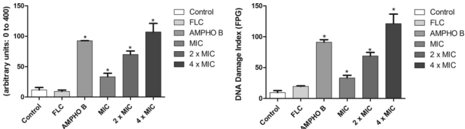
DNA damage.
Figure 5
shows the amount of damage to the
DNA of fluconazole-resistant
C. albicans
strains induced by
ber-berine. Analysis of single cells for the levels of DNA damage (
Fig.
5
) showed that fluconazole induced low levels of DNA damage. In
contrast, after 24 h of incubation, exposure of
C. albicans
to
ber-berine resulted in a significant increase (
P
⬍
0.05) in DNA strand
break levels (
Fig. 5
). Cells treated with berberine at the MIC, 2
⫻
MIC, and 4
⫻
MIC for 24 h exhibited damage index values
(arbi-trary units) of 32
⫾
5.68, 71
⫾
5.8, and 104
⫾
14.11, respectively.
Amphotericin B, used as a positive control, induced high DNA
strand break levels. Furthermore, the compounds also promoted
significant (
P
⬍
0.05) increases in the amounts of oxidized purine
and pyrimidine (
Fig. 6
).
Effect of berberine on the formed biofilm.
According to the
results presented in
Fig. 7
, the berberine MIC for the
C. tropicalis
2
isolate was less than 37.5
g/ml. The antifungal effects of
ber-berine caused a statistically significant reduction in the cellular
activity of biofilm cells (
P
⬍
0.05), with the MIC being
approxi-mately four times higher than the MIC obtained with the same
cells in the planktonic growth mode.
Cytotoxic activity of berberine against mammalian L929
cells.
The cytotoxic activity of berberine against mammalian L929
FIG 1Effect of berberine on the number of viable cells of aC. albicansFLC-resistant strain. *,P⬍0.05 compared to the results for the control, determined by ANOVA followed by the Newman-Keuls test.
FIG 2Effect of berberine at the MIC, 2⫻MIC, and 4⫻MIC on the stability of the cell membranes of theC. albicansFLC-resistant strains tested. *,P⬍0.05 compared to the results for the control, determined by ANOVA followed by the Newman-Keuls test.
FIG 3Assessment of the⌬m of fluconazole-resistantC. albicansstrains. Cells were labeled with rhodamine 123 (50 nM). The graph shows the results for strains incubated for 24 h with RPMI 1640 (control), FLC (64g/ml), amphotericin B (4 g/ml), and berberine at the MIC, 2⫻MIC, and 4⫻MIC. The percentage of cells of representative FLC-resistantCandidastrains with mitochondrial dysfunction was evaluated for 24 h. *,P⬍0.05 compared to the results for the control, deter-mined by ANOVA followed by the Newman-Keuls test.
FIG 4Phosphatidylserine externalization, which is observed at an early stage of apoptosis, shown by annexin V staining. The probe enabled the detection of alter-ation of phosphatidylserine localizalter-ation from the inner membrane to the outer membrane. The fluorescence intensity indicates the amount of exposed phospha-tidylserine in cells treated with berberine at the MIC, 2⫻MIC, and 4⫻MIC. The percentage of annexin V-positive cells of representative FLC-resistantC. albicans
strains was evaluated for 24 h. *,P⬍0.05 compared to the results for the control, determined by ANOVA followed by the Newman-Keuls test.
on June 14, 2016 by UNIVERSIDADE FEDERAL DO CEARÕ
http://aac.asm.org/
cells was assessed. It is interesting to highlight that berberine
showed low cytotoxicity, with the 95% confidence interval of the
MIC being above 100
g/ml in comparison to that for untreated
cells (
P
⬍
0.05), as analyzed by the MTT assay.
DISCUSSION
The results of this study show that berberine has antifungal
activ-ity against fluconazole-resistant
Candida
and
Cryptococcus
neofor-mans
strains. According to some studies, berberine showed
signif-icant antifungal activity against
Candida
strains, which was
consistent with the findings of this study (
15
,
38
). An MIC of 16
g/ml was obtained for the fluconazole-resistant
Cryptococcus
neoformans
strains. Some studies that used berberine derivatives,
such as
a
9-
O
-butyl-13-(4-isopropylbenzyl)berberine, have
re-ported that berberine has antifungal activity against
C. neoformans
strains and obtained significant MICs (
39
,
40
).
When strains were exposed to berberine in the presence of the
cytofluorometric marker propidium iodide (PI), a decrease in the
number of viable cells compared to that for the control was observed
at the concentrations used for treatment (the MIC, 2
⫻
MIC, and 4
⫻
MIC). Thus, this finding indicates that berberine causes cell
mem-brane damage and the possible impairment of cell function. The data
corroborate those obtained by Dhamgaye et al. (
15
), who showed that
berberine could cause a loss of membrane integrity, resulting in
in-creased cell membrane permeability. Inin-creased PI absorption in
flu-conazole-resistant
Candida
cells is an indication that berberine
com-pounds promote cell death, considering that this marker can bind
only to the nuclear DNA of dead cells (
41
).
The results of this study also indicate that cells treated with
berberine promote
⌬
m changes, suggesting that berberine may
affect the mitochondrial respiratory function, causing
⌬
m
breakdown and the lack of accumulation of rhodamine 123 in the
mitochondria (
42
). The collapse of
⌬
m may lead to transient
pore openings in the membranes and the release of mitochondrial
proapoptotic factors into the cytosol (
43
). Xu et al. (
44
) showed
that treatment with fluconazole and berberine resulted in an
in-crease in
⌬
m in fluconazole-resistant
Candida albicans
,
suggest-ing that this increase is coupled with a low ATP level when ATP
synthase activity is inhibited.
In the late stage of apoptosis, morphological changes, such as
DNA fragmentation, are considered a late marker of this type of
cell death (
45
). Thus, whether berberine triggers the apoptotic
mechanism in fluconazole-resistant
Candida albicans
cells was
in-vestigated.
The alkaline version of the comet assay (standard protocol) is a
sensitive procedure used to quantify different types of DNA
dam-age to the cell, including single- and double-strand breaks (
46
).
The results of this study showed that the DNA strands of cells
treated with berberine at the MIC, 2
⫻
MIC, and 4
⫻
MIC showed
total breaks, and it was found that the DNA strand breaks were
more significant with increasing concentrations of berberine. In
order to further assess the oxidative damage to DNA, the alkaline
version of the comet assay was conducted in the presence of FPG
(
27
,
47
). The results of this study confirm that significant DNA
damage, which is expressed as an index of DNA damage after
treatment with the FPG enzyme compared to that for the negative
control (untreated cells), is caused by treatment with berberine.
The increase in DNA damage was probably caused by the
ca-pacity of the FPG enzyme to recognize purine bases (adenine and
guanine) within the DNA. Li et al. (
14
) demonstrated that
ber-berine has a strong antifungal effect on
C. albicans
, causing cell
cycle arrest and DNA damage. Other studies have also suggested
that the berberine can bind to DNA, affecting DNA replication
and transcription and the cell cycle (
48
,
49
).
DNA condensation and fragmentation represent irreversible
steps in cell death (
43
). The detection of apoptosis at an early stage
can be determined using annexin V. In the presence of Ca
2⫹, this
marker binds with a high affinity to phosphatidylserine in
apop-totic cell membranes (
43
). However, double staining with
FITC-conjugated annexin V and PI allows discrimination between early
apoptosis and necrosis (
50
). According to the data obtained in this
study, berberine causes the death of fluconazole-resistant
C.
albi-cans
cells by apoptosis.
Biofilm formation represents a major problem in hospitals,
because, besides being able to form biomass in host tissue,
Can-dida
species can also grow on implanted medical devices in
hos-pitalized patients (
51
). An important characteristic of
Candida
biofilms is resistance to antifungal agents, which can be intrinsic
or acquired by the transfer of genetic material among biofilm cells
(
52
), making new therapies necessary.
The results of this study showed that the berberine
concentra-tion necessary to inhibit both planktonic cells and preformed
bio-FIG 5Effect of 24 h of incubation with RPMI 1640 (control), FLC (64g/ml),amphotericin B (4g/ml), and berberine at the MIC, 2⫻MIC, and 4⫻MIC on the DNA damage index. *,P⬍0.05 compared to the results for the control, determined by ANOVA followed by the Newman-Keuls test.
FIG 6Effects of different treatments on the distribution of oxidative DNA damage in FLC-resistantC. albicansstrains determined using the modified alkaline version (with FPG) of the comet assay. Yeasts were exposed to RPMI 1640 (control), FLC (64g/ml), amphotericin B (4g/ml), and berberine at the MIC, 2⫻MIC, and 4⫻MIC. *,P⬍0.05 compared to the results for the control, determined by ANOVA followed by the Newman-Keuls test.
on June 14, 2016 by UNIVERSIDADE FEDERAL DO CEARÕ
http://aac.asm.org/
film cells is similar. This indicates that berberine may reduce the
growth of planktonic cells and inhibit the viability of cells in
pre-formed biofilms at concentrations of 8
g/ml and 37.5
g/ml,
respectively. This finding is relevant because biofilms are
fre-quently associated with reduced sensitivity to conventional
anti-fungal agents.
In tests of the cytotoxicity of berberine for mammalian L929 cells
performed using the MTT assay, berberine showed low levels of
cy-totoxicity. The results of this study are in agreement with the findings
of Gu et al. (
53
): berberine further altered the morphology of L929
cells only at concentrations greater than 100
g/ml. Within this
con-text, the antimicrobial activity and the low cytotoxic potential
dem-onstrated by this compound reveal that it is a promising chemical
compound for development as a new antimicrobial.
Conclusion.
Studies related to the development of
phytoprod-ucts have been lacking, but this study has shown that treatment of
fluconazole-resistant strains with one such phytoproduct,
ber-berine, promoted alterations to the integrity of the plasma and
mitochondrial membranes, possibly acting at specific sites near
cell DNA, leading to death by apoptosis. The study also showed
that berberine may reduce the viability of biofilms formed by
flu-conazole-resistant
Candida tropicalis
cells grown
in vitro
.
There-fore, because of its antimicrobial activity, berberine is a promising
source of molecules with antifungal properties.
FUNDING INFORMATION
This work was supported by grants and fellowships from the National Council of Technological and Scientific Development (CNPq), Coordi-nation for the Improvement of Higher Level or Education Personnel (CAPES/Brazil), and the Foundation of Ceara Support for Scientific and Technological Development (FUNCAP/Ceara).
REFERENCES
1.Storti LR, Pasquale G, Scomparim R, Galastri AL, Alterthum F, Gam-bale W, Rodrigues Paula C.2012.Candidaspp. isolated from inpatients, the environment, and health practitioners in the Pediatric Unit at the Universitary Hospital of the Jundiaí Medical College, state of São Paulo, Brazil. Rev Soc Bras Med Trop45:225–231.http://dx.doi.org/10.1590 /S0037-86822012000200017.
2.Sardi JCO, Scorzoni L, Bernardi T, Fusco-Almeida AM, Mendes MJSG.
2013.Candidaspecies: current epidemiology, pathogenicity, biofilm for-mation, natural antifungal products and new therapeutic options. J Med Microbiol62:10 –24.http://dx.doi.org/10.1099/jmm.0.045054-0. 3.Arnold HM, Micek ST, Shorr AF, Zilberberg MD, Labelle AJ, Kothari S.2010.
Hospital resource utilization and costs of inappropriate treatment of candidemia. Pharmacotherapy30:361–368.http://dx.doi.org/10.1592/phco.30.4.361.
4.Nucci M, Queiroz-Telles F, Alvarado-Matute T, Tiraboschi IN, Cortes J, Zurita J.2013. Epidemiology of candidemia in Latin Amer-ica: a laboratory-based survey. PLoS One8:e59373.http://dx.doi.org /10.1371/journal.pone.0059373.
5.World Health Organization.2011. Rapid advice: diagnosis, prevention and management of cryptococcal disease in HIV-infected adults, adoles-cents and children. World Health Organization, Geneva, Switzerland. 6.Park BJ, Wannemuehler KA, Marston BJ, Govender N, Pappas PG,
Chiller TM.2009. Estimation of the current global burden of cryptococcal meningitis among persons living with HIV/AIDS. AIDS23:525–530.http: //dx.doi.org/10.1097/QAD.0b013e328322ffac.
7.Pfaller MA.2012. Antifungal drug resistance: mechanisms, epidemiology and consequences for treatment. Am J Med125:S3–S13.http://dx.doi.org /10.1016/j.amjmed.2011.11.001.
8.Zhang H, Gao A, Li F, Zhang G, Ho HI, Liao W.2009. Mechanism of action of tetrandrine, a natural inhibitor of Candida albicans drug efflux pumps. Yakugaku Zasshi129:623– 630.http://dx.doi.org/10.1248/yakushi .129.623.
9.Misik V, Bezakova L, Malekova L, Kostalova D. 1995. Lipoxygenase inhibition and antiantitoxina properties of protoberberine and aporphine alkaloids isolated from Mahonia aquifolium. Planta Med61:372–373. 10. Bezakova L, Misik V, Malekova L, Svajdlenka E, Kostalova D.1996.
Lipoxygenase inhibition and antioxidant properties of bisbenzylisoquno-line alkaloids isolated from Mahonia aquifolium. Pharmazie51:758 –761. 11. Chen J, Zhao H, Wang X, Lee FS, Yang H, Zheng L.2008. Analysis of major alkaloids in Rhizoma coptidis by capillary electrophoresis-electrospray-time of flight mass spectrometry with different background electrolytes. Electrophoresis 29:2135–2147. http://dx.doi.org/10.1002 /elps.200700797.
12. Tan W, Li Y, Chen M, Wang Y.2011. Berberine hydrochloride: antican-cer activity and nanoparticulate delivery system. Int J Nanomedicine
6:1773–1777.http://dx.doi.org/10.2147/IJN.S22683.
13. Iwazaki RS, Endo EH, Ueda-Nakamura T, Nakamura CV, Garcia LB, Filho BP.2010.In vitroantifungal activity of the berberine and its syner-gism with fluconazole. Antonie Van Leeuwenhoek97:201–205.http://dx .doi.org/10.1007/s10482-009-9394-8.
14. Li DD, Xu Y, Zhang DZ, Quan H, Mylonakis E, Hu DD, Li MB, Zhao LX, Zhu LH, Wang Y, Jiang YY.2013. Fluconazole assists berberine to kill fluconazole-resistantCandida albicans. Antimicrob Agents Che-mother57:6016 – 6027.http://dx.doi.org/10.1128/AAC.00499-13. 15. Dhamgaye S, Devaux F, Vandeputte P, Khandelwal NK, Sanglard D,
Mukhopadhyay G, Prasad R.2014. Molecular mechanisms of action of herbal antifungal alkaloid berberine, inCandida albicans. PLoS One
9:e104554.http://dx.doi.org/10.1371/journal.pone.0104554.
16. Warner SAJ.1996. Genomic DNA isolation and lambda library construc-tion, p 51–73.InFoster GD, Twell D (ed), Plant gene isolation: principles and practice. John Wiley & Sons, West Sussex, United Kingdom. 17. White T, Bruns T, Lee S, Taylor J. 1990. Amplification and direct
sequencing of fungal ribosomal RNA genes for phylogenetics, p 315–322.
InInnis MA, Gefland DH, Sninsky JJ, White TJ (ed), PCR protocols: a guide to methods and applications. Academic Press, San Diego, CA. 18. Sambrook J, Fritsch EF, Maniatis T.1989. Molecular cloning: a
labora-FIG 7Effect of different concentrations of berberine (A) and fluconazole (B) on the metabolic activity of growing and mature biofilms ofC. tropicalis, analyzed by the MTT reduction assay. *,P⬍0.05 compared to the results for the control, determined by ANOVA followed by the Newman-Keuls test.
on June 14, 2016 by UNIVERSIDADE FEDERAL DO CEARÕ
http://aac.asm.org/
tory manual, 2nd ed. Cold Spring Harbor Laboratory Press, Cold Spring Harbor, NY.
19. Ewing B, Green P.1998. Base-calling of automated sequencer traces using Phred. II. Error probabilities. Genome Res8:186 –194.
20. Ewing B, Hillier L, Wendl MC, Green P.1998. Base-calling of auto-mated sequencer traces using Phred. I. Accuracy assessment. Genome Res8:175–185.
21. Gordon D, Abajian C, Green P. 1998. Consed: a graphical tool for sequence finishing. Genome Res8:195–202.http://dx.doi.org/10.1101/gr .8.3.195.
22. Santamaria M, Fosso B, Consiglio A, De Caro G, Grillo G, Licciulli F, Liuni S, Marzano M, Alonso-Alemany D, Valiente G, Pesole G.2012. Reference databases for taxonomic assignment in metagenomics. Brief Bioinform13:682– 695.http://dx.doi.org/10.1093/bib/bbs036.
23. Keller A, Schleicher T, Schultz J, Müller T, Dandekar T, Wolf M.2009. 5.8S-28S rRNA interaction and HMM-based ITS2 annotation. Gene430:
50 –57.http://dx.doi.org/10.1016/j.gene.2008.10.012.
24. Altschul SF, Gish W, Miller W, Myers EW, Lipman DJ.1990. Basic local alignment search tool. J Mol Biol215:403– 410.http://dx.doi.org/10.1016 /S0022-2836(05)80360-2.
25. Clinical and Laboratory Standards Institute.2008. Reference method for broth dilution antifungal susceptibility testing of yeasts. Approved stan-dard M27-A3. Clinical and Laboratory Stanstan-dards Institute, Wayne, PA. 26. Clinical and Laboratory Standards Institute.2012. Reference method for
broth dilution antifungal susceptibility testing of yeasts; 4th informational supplement. CLSI document M27-S4. Clinical and Laboratory Standards Institute, Wayne, PA.
27. Da Silva CR, De Andrade Neto JB, Sidrim JJC, Ângelo MRF, Magalhães HIF, Cavalcanti BC, Brilhante RSN, Macedo DS, De Moraes MO, Pinto Lobo MD, Grangeiro TB, Nobre Junior HV.2013. Synergistic effects of amiodarone and fluconazole on Candida tropicalis resistant to fluconazole. Antimicrob Agents Chemother57:1691–1700.http://dx.doi.org/10.1128/AAC.00966-12. 28. Da Silva CR, de Andrade Neto JB, de Sousa Campos R, Figueiredo NS,
Sampaio LS, Magalhães HI, Cavalcanti BC, Gaspar DM, de Andrade GM, Lima IS, de Barros Viana GS, de Moraes MO, Lobo MD, Grangeiro TB, Nobre Júnior HV.2014. Synergistic effect of the flavonoid catechin, querce-tin, or epigallocatechin gallate with fluconazole induces apoptosis inCandida tropicalisresistant to fluconazole. Antimicrob Agents Chemother58:
1468 –1478.http://dx.doi.org/10.1128/AAC.00651-13.
29. Andrade Neto JB, da Silva CR, Neta MAS, Campos RS, Siebra JT, Silva RA, Gaspar DM, Magalhães HI, de Moraes MO, Lobo MD, Grangeiro TB, Carvalho TS, Diogo EB, da Silva Júnior EN, Rodrigues FA, Cavalcanti BC, Júnior HV.2014. Antifungal activity of naphthoquinoidal compoundsin vitroagainst fluconazole-resistant strains of differentCandidaspecies: a spe-cial emphasis on mechanisms of action onCandida tropicalis. PLoS One
9:e93698.http://dx.doi.org/10.1371/journal.pone.0093698.
30. Andrade Neto JB, Silva CR, Campos RS, Nascimento FBSA, Freitas DD, Josino MAA, Andrade LND, Gonçalves TB, Mesquita JRL, Magalhães HIF, Rodrigues FAR, Gaspar DM, Moraes MO, Lobo MDP, Moreno FBMB, Grangeiro TB, Gomes AOCV, Nascente LC, Romeiro LAS, Cavalcanti BC, Nobre Júnior HV.2015. Effects of piperonal nitro deriv-atives onCandidaspecies: antifungal activity against fluconazole-resistant strains is associated with oxidative DNA damage. Int J Curr Microbiol Appl Sci4:777–792.
31. Miloshev G, Mihaylov I, Anachkova B.2002. Application of the single cell electrophoresis on yeast cells. Mutat Res513:69 –74.http://dx.doi.org /10.1016/S1383-5718(01)00286-8.
32. Pinkerton DM, Banwell MG, Garson MJ, Kumar N, De Moraes MO, Cavalcanti BC, Barros FWA, Pessoa C.2010. Antimicrobial and cytotoxic activities of synthetically derived tambjamines C and E-J, BE-18591, and a related alkaloid from the marine bacterium Pseudoalteromonas tunicate. Chem Biodivers7:1311–1324.http://dx.doi.org/10.1002/cbdv.201000030. 33. Collins AR.2004. The comet assay for DNA damage and repair:
princi-ples, applications, and limitations. Mol Biotechnol26:249 –261.http://dx .doi.org/10.1385/MB:26:3:249.
34. Pierce CG, Uppuluri P, Tristan AR, Wormley FL, Jr, Mowat E, Ramage G, Lopez-Ribot JL.2008. A simple and reproducible 96-well plate-based method for the formation of fungal biofilms and its application to anti-fungal susceptibility testing. Nat Protoc3:1494 –1500.http://dx.doi.org /10.1038/nprot.2008.141.
35. Hawser SP, Douglas LJ.1994. Biofilm formation byCandidaspecies on the surface of catheter materials in vitro. Infect Immun62:915–921.
36. Rajput SB, Karuppayil SM.2013.-Asarone, an active principle ofAcorus calamusrhizome, inhibits morphogenesis, biofilm formation and ergos-terol biosynthesis inCandida albicans. Phytomedicine20:139 –142.http: //dx.doi.org/10.1016/j.phymed.2012.09.029.
37. Cavalcanti BC, Bezerra DP, Magalhães HI, Moraes MO, Lima MA, Silveira ER, Câmara CA, Rao VS, Pessoa C, Costa-Lotufo LV.2009. Kauren-19-oic acid induces DNA damage followed by apoptosis in human leukemia cells. J Appl Toxicol29:560 –568.http://dx.doi.org/10.1002/jat.1439.
38. Zhao M, Zhou ZT, Zhang WD.2006. Antifungal susceptibility testing and antifungal traditional Chinese medicines screening of oral Candida isolated from head and neck cancer patients with radiotherapy or chemo-therapy. Hua Xi Kou Qiang Yi Xue Za Zhi24:131–134.
39. Park KD, Cho SJ, Moon JS, Kim SU.2010. Synthesis and antifungal activity of a novel series of 13-(4-isopropylbenzyl)berberine derivatives. Bioorg Med Chem Lett20:6551– 6554.http://dx.doi.org/10.1016/j.bmcl .2010.09.045.
40. Bang S, Kwon H, Hwang HS, Park KD, Kim SU, Bahn YS. 2014. 9-O-Butyl-13-(4-isopropylbenzyl)berberine, KR-72, is a potent antifun-gal agent that inhibits the growth of Cryptococcus neoformans by regu-lating gene expression. PLoS One9:e109863.http://dx.doi.org/10.1371 /journal.pone.0109863.
41. Xu C, Wang J, Gao Y, Lin H, Du L, Yang S, Long S, She Z, Cai X, Zhou S, Lu Y.2010. The anthracenedione compound bostrycin induces mitochon-dria mediated apoptosis in the yeastSaccharomyces cerevisiae. FEMS Yeast Res10:297–308.http://dx.doi.org/10.1111/j.1567-1364.2010.00615.x. 42. Ludovico P, Sansonetty F, Côrte-Real M.2001. Assessment of
mitochon-drial membrane potential in yeast cell populations by flow cytometry. Micro-biology147:3335–3343.http://dx.doi.org/10.1099/00221287-147-12-3335. 43. Hwang I, Lee J, Jin HG, Woo ER, Lee DG. 2012. Amentoflavone
stimulates mitochondrial dysfunction and induces apoptotic cell death in
Candida albicans. Mycopathologia 173:207–218. http://dx.doi.org/10 .1007/s11046-011-9503-x.
44. Xu Y, Wang Y, Yan L, Liang RM, Dai DD, Tang RJ, Gao PH, Jiang YY.
2009. Proteomic analysis reveals a synergistic mechanism of fluconazole and berberine against fluconazole-resistant Candida albicans: endoge-nous ROS augmentation. J Proteome Res8:5296 –5304.http://dx.doi.org /10.1021/pr9005074.
45. Schiller M, Blank N, Heyder P, Herrmann M, Gaipl US, Kalden JR, Lorenz HM.2005. Induction of apoptosis by spermine-metabolites in primary human blood cells and various tumor cell lines. Apoptosis10:
1151–1162.http://dx.doi.org/10.1007/s10495-005-1188-5.
46. Burlinson B, Tice RR, Speit G, Agurell E, Brendler-Schwaab SY, Collins AR, Escobar P, Honma M, Kumaravel TS, Nakajima M, Sasaki YF, Thybaud V, Uno Y, Vasquez M, Hartmann A.2007. Fourth Interna-tional Workgroup on Genotoxicity Testing: results of thein vivocomet assay workgroup. Mutat Res627:31–35.http://dx.doi.org/10.1016/j .mrgentox.2006.08.011.
47. Cavalcanti BC, Barros FW, Cabral IO, Ferreira JR, Magalhaes HI, Junior HV, Da Silva Junior EN, De Abreu FC, Costa CO, Goulart MO, Moraes MO, Pessoa C. 2011. Preclinical genotoxicology of nor-betalapachonein human cultured lymphocytes and Chinese hamster lung fibroblasts. Chem Res Toxicol24:1560 –1574.http://dx.doi.org/10.1021 /tx200180y.
48. Bhadra K, Kumar GS.2011. Therapeutic potential of nucleic acid binding isoquinoline alkaloids: binding aspects and implications for drug design. Med Res Rev31:821– 862.http://dx.doi.org/10.1002/med.20202. 49. Yadav RC, Kumar GS, Bhadra K, Giri P, Sinha R, Pal S, Maiti M.2005.
Berberine, a strong polyriboadenylic acid binding plant alkaloid: spectro-scopic, viscometric, and thermodynamic study. Bioorg Med Chem13:
165–174.http://dx.doi.org/10.1016/j.bmc.2004.09.045.
50. Kojic EM, Darouiche RO.2004.Candidainfections of medical devices. Clin Microbiol Rev17:255–267.http://dx.doi.org/10.1128/CMR.17.2.255 -267.2004.
51. Eisenberg T, Carmona-Gutierrez D, Buttner S, Tavernarakis N, Madeo F.2010. Necrosis in yeast. Apoptosis15:257–268.http://dx.doi.org/10 .1007/s10495-009-0453-4.
52. Fanning S, Mitchell AP.2012. Fungal biofilms. PLoS Pathog8:e1002585.
http://dx.doi.org/10.1371/journal.ppat.1002585.
53. Gu M, Xu J, Han C, Kang Y, Liu T, He Y, Huang Y, Liu C.2015. Effects of berberine on cell cycle, DNA, reactive oxygen species, and apoptosis in L929 murine fibroblast cells. Evid Based Complement Alternat Med2015:
796306.http://dx.doi.org/10.1155/2015/796306.