The Clinical Significance of Antibody Determination
to Cyclic Citrullinated Peptides in Systemic Sclerosis
Bojana Stamenković1, Aleksandra Stanković1, Aleksandar Dimić1, Nemanja Damjanov2,
Jovan Nedović1, Sonja Stojanović1, Vojin Savić3, Dragan Djordjević1 1Institute for Treatment and Rehabilitation ”Niška Banja”, Niška Banja, Serbia; 2Institute for Rheumatology, Belgrade, Serbia;
3Institute for Biomedical Research, School of Medicine, University of Niš, Niš, Serbia
INTRODUCTION
Systemic sclerosis (SSc) is a chronic autoim-mune disease of unknown origin, character-ized by increased production of matrix proteins by fibroblasts, their precipitation in the walls of blood vessels, skin and internal organs leading to fibrosis of the skin and internal organs, con-currently with the activation of the immune sys-tem and significant vascular damage.
The basic classification was defined accord-ing to the degree and location of skin thicken-ing: limited disease (lSSc) and diffuse systemic sclerosis (dSSc) with different severity and prognosis [1].
Immunopathogenesis of systemic sclerosis is complex. Cellular immunity has the main role, while the activity of humoral immunity has been observed, which is characterized by the formation of disease-characteristic antibodies. Quantification of total antinuclear antibodies (ANA) and especially specific, anticentromere (ACA) and antibodies to topoisomerase 1 (ATA) is useful in the diagnosis, classification and prognosis of systemic sclerosis [1, 2].
During the evolution of the disease, a large number of patients have arthralgia, myalgia, muscle atrophy, arthritis with or without flex-ion contracture, which can lead to perma-nent damage of joint function and disability
[3]. The literature identifies the different fre-quency of articular manifestations in 24-97% patients [4, 5].
The most serious disorders in the joint damage are flexion contractures of joints that usually occur in later phases of the disease, usu-ally affecting wrist, fingers and elbow joints, followed by the restriction of movements, functional incapacity and limitation of daily activities. There are no clearly defined con-clusions on whether the contractures occur due to periarticular fibrosis, synovitis, or as a solid connection between thickened and tough fibrous skin with subcutaneous tissue [6-9].
Articular damage on the hands is usually diagnosed by physical examination and radi-ography, which can help in the visualization of different bone abnormalities, divided into two groups: extraarticular (subcutaneous calcino-sis, digital tuft resorption) and articular (joint space narrowing, juxtaarticular osteoporosis, marginal bone erosions) [6].
The analysis of acute phase reactants of inflammation (C reactive protein, rheumatoid factor, sedimentation rate, antinuclear antibod-ies) in joint damage assessment is useful but insufficient. During the last ten years an impor-tant role of antibodies to cyclic citrullinated peptide was proven. It is an important diag-nostic marker for rheumatoid arthritis (RA)
SUMMARy
Introduction Anticitrullinated peptides antibodies (ACPA) are present in 80% of sera of rheumatoid
arthritis (RA) patients with high specificity for diagnosis and prediction for the development of early erosive arthritis. A few studies have reported a low frequency ACPA in systemic sclerosis (SSc) patients with the presence of arthritis.
Objective The aim of our study was to determine the frequency of ACPA in systemic sclerosis (SSc)
patients, their correlation with clinical manifestations and radiographic features.
Methods The study included 82 patients with SSc, mean age 54.4 years, 59 with the limited (lSSc) and 23
with the diffuse (dSSc) form of the disease. The control group included 28 healthy age and sex matched subjects. ACPA and rheumatoid factor (RF) were determined in all SSc patients and healthy subjects in whom standard radiography of hands and wrists was also done.
Results The presence of ACPA was detected in 11 (13.4%) of SSc patients. Their level was not increased
in any of the controls. Positive RF was found in 15.9% of SSc patients. Arthritis was present in 17.1%, as well as marginal bone erosions. There was a statistically significant association between positive ACPA and arthritis (p<0.0001) and positive ACPA and marginal bone erosions (p=0.0002).
Conclusion The research confirmed the correlation between ACPA with clinical signs of arthritis and
radiographic damage of hand joints. ACPA is a useful diagnostic marker in the identification of SSc patients with arthritis and anatomic bone damage enabling the use of adequate therapy in order to prevent joint damage and poor quality of life.
keywords: anticitrullinated peptide antibodies; systemic sclerosis; arthritis; erosions
Correspondence to:
Bojana STAMENKOVIĆ Jug Bogdanova 26a, 18000 Niš Serbia
with the frequency of about 80% of patients with extreme specificity. It is now believed that ACPA could be a possi-ble predictor for the occurrence of severe, destructive and erosive form of the disease [10, 11].
Recently, the presence of ACPA has been proven in a small number of SSc patients with limited and diffuse forms of systemic sclerosis with some joint manifestations, which leads to a conclusion that a high titer of these anti-bodies can predict patients with overlap syndrome – SSc and RA. However, we still do not have data about the sig-nificance of ACPA in predicting joint damage in SSc [12, 13, 14].
OBjECTIvE
The aim of this study was to determine the frequency of ACPA in systemic sclerosis and its relationship with clini-cal manifestations and radiographic features of the disease.
METHODS
The study included 82 patients that fulfilled the ACR (American College of Rheumatology) criteria for the diag-nosis of systemic sclerosis [15]. Fifty-nine patients had lSSc and 23 dSSc, according to the classification of Le Roy and associates [16]. The control group included 28 healthy age and sex matched controls.
All the patients underwent a physical examination, laboratory testing, standard radiography of hands and were evaluated by clinical manifestations of the disease. Damage to internal organs was defined by the above crite-ria [17]. Pulmonary involvement was defined by the pres-ence of bibasilar fibrosis on standard radiography of the heart and lungs and/or high-resolution computed tomog-raphy (HRCT), pulmonary function tests (FVC, DLCO/ VA) and/or the presence of pulmonary arterial hyperten-sion detected by colour Doppler echocardiography.
Esophagus involvement was defined in the case of hypomotility shown by barium radiography. Cardiac involvement was defined by the presence of pericarditis, complex arrhythmias, conduction disturbances, diastolic dysfunction and the presence of reduced left ventricular ejection fraction. Renal involvement was defined by the occurrence of renal hypertension, renal crisis in the his-tory of the disease, proteinuria and/or a rapid decline of renal function.
Joint involvement was defined by the detection of arthralgias, arthritis and/or flexion contracture, and skin involvement was assessed by the modified Rodnan skin thickness score [18].
The presence of ACPA was determined by enzyme-linked immunosorbent assay (ELISA) method using the first gen-eration anti-CCP ELISA kit, Imtec, Immunodiagnostics, Berlin, Germany. The samples were classified as positive if the value was >25 U/ml.
Antinuclear (ANA) and anticentromere antibodies (ACA) were determined by indirect immunofluorescence
on HEP-2 cells (Immunoconcepts, Sacramento, California, USA). As a borderline, ANA titer (cut off titer, i.e., the titer of which the lowest value is accepted as a positive test result) was taken, i.e., a titer of 1:40, the titer which in prac-tice is recommended for screening. Antitopoisomerase I antibodies (ATA) were determined by CIE (counter immu-noelectrophoresis), Imtec, Immunodiagnostics, Berlin, Germany.
Standard radiography of the hands and wrists was per-formed in all patients. The changes were evaluated by radi-ologist without access to clinical and serological data of patients. The presence of extraarticular changes (digital tuft resorption, subcutaneous calcinosis) or articular man-ifestations (juxta-articular osteoporosis and marginal bone erosions) was recorded.
The data were statistically analyzed using SPSS 10.0 for Windows. The frequency of categories of described char-acteristics was tested by Pearson χ2-test or Fisher exact
probability test when some of the expected frequencies were less than 5.
RESULTS
The study included 77 women and 5 men with SSc, mean age 54.4±12.6 years (range from 26-75 years). Fifty-nine patients had lSSc and 23 dSSc (Table 1). The control group included 28 healthy age and sex matched controls.
Pulmonary arterial hypertension was found in 17 (20.7%) and pulmonary fibrosis in 42 (51.2%) SSc patients. Restrictive disorders of pulmonary ventilation (FVC<75%) was demonstrated in 15 (20.5%) and reduced transfer fac-tor for carbon monoxide (DLco/VA <75%) in 27 (37%) SSc patients.
Oesophageal involvement was demonstrated in 28 (54%) of 51 SSc patients, who had undergone barium radi-ography. Cardiac involvement was found in 38 (46.3%) and renal failure in 3 (3.7%) SSc patients.
Clinical findings of changes in the joints showed the presence of arthralgia in 58 (70.7%), arthritis was detected in 14 (17.1%), while 28 (34.1%) SSc patients had flexion contractures of joints. The presence of ACPA was registered in 11 (13.4%), while positive RF was found in 13 (15.9%) SSc patients. The mean value of the modified score-Rodnan skin thickness was 10.91. Table 1 shows the characteristics of the whole SSc group and in different SSc forms.
Radiological changes of hands and wrists are given in Table 2. We found marginal bone erosion in 14 (17.1%) SSc patients, mostly of capitate and lunate carpal bones or MCP 2, 3 joints; digital tuft resorption in 22 (26.8%), radiological demineralization in 24 (29.3%) and calcinosis in 31 (37.8%) SSc patients.
ACPA were present in 11/82 (13.4%) SSc patients. In the control group of healthy subjects, there were no patients with positive findings for anti-CCP. Positive RF was found in 13 (15.9%) SSc patients.
Table 1. Characteristics of the patients with systemic sclerosis (SSc), limited form (lSSc) and diffuse form (dSSc)
Parameter (n=82)SSc (n=59)lSSc (n=23)dSSc
Sex (F/M) 77/5 58/1 19/4
Age, mean±SD (years) 54.4±12.6 55.5±11.2 51.7±15.6
Pulmonary arterial hypertension 17 (20.7%) 11 (18.6%) 6 (26.1%)
Bibasilar fibrosis 42 (51.2%) 27 (45.8%) 15 (65.2%)
Oesophagus involvement 28/51 (54.9%) 17/33 (51.5%) 11/18 (61.1%)
Cardiac involvement 38 (46.3%) 24 (40.7%) 14 (60.9%)
Renal involvement 3 (3.7%) 1 (1.7%) 2 (8.7%)
Positive antinuclear antibodies 63 (76.9%) 43 (72.9%) 20 (87%)
AntiScl70 antibodies 15 (18.3%) 3 (5.1%) 12 (52.2%)
Anticentromere antibodies 23 (28.1%) 20 (33.9%) 3 (13%)
Rheumatoid factor 13 (15.9%) 9 (15.2%) 4 (17.4%)
AntiCCP antibodies 11 (13.4%) 4 (6.8%) 7 (30.4%)
Arthralgias 58 (70.7%) 43 (72.9%) 15 (65.2%)
Arthritis 14 (17.1%) 5 (8.5%) 9 (39.1%)
Flexion contractures 28 (34.1%) 16 (27.1%) 12 (59.2%)
Modified Rodnan (skin thickness score) 10.91 8.64 16.74
Decreased FVC* 15/73 (20.5%) 6/51 (11.8%) 9/22 (40.9%)
Decreased DLCO/VA** 27/73 (37%) 15/51 (29.4%) 12/22 (54.5%)
* <75% normal value; ** <75% normal value
Table 3. Relationship between serum anticitrullinated peptides anti bodies (ACPA), arthritis and marginal bone erosions in 82 patients with systemic sclerosis
Parameter
ACPA positive
(n=11)
ACPA negative
(n=71) p
With arthritis 8 3 <0.0001
Without arthritis 3 68
With marginal bone erosions 7 7 0.0002 Without marginal bone
erosions 4 64
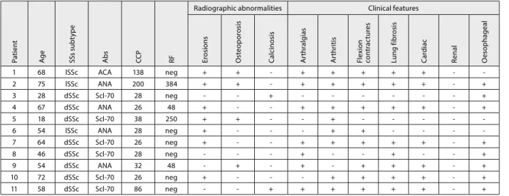
Table 4. Clinical features of 11 female patients with systemic sclerosis and positive ACPA
P
a
tien
t
Ag
e
SSs s
u
b
ty
p
e
A
bs
CC
P
RF
Radiographic abnormalities Clinical features
Er
o
si
o
n
s
O
st
e
o
p
o
ro
sis
C
a
lci
n
o
sis
Ar
th
ral
gi
a
s
A
rth
ri
ti
s
F
le
x
ion
co
n
tr
a
c
tur
e
s
L
u
n
g
f
ib
ro
sis
C
ardi
a
c
R
e
nal
O
e
so
p
ha
g
e
al
1 68 lSSc ACA 138 neg + + + + + + +
2 75 lSSc ANA 200 384 + + + + + + + +
3 28 dSSc Scl70 28 neg + +
4 67 dSSc ANA 26 48 + + + + + + +
5 18 dSSc Scl70 38 250 + + +
6 54 lSSc ANA 28 neg + + +
7 64 dSSc Scl70 26 neg + + + + + + +
8 46 dSSc Scl70 28 neg + + +
9 54 dSSc ANA 32 48 + + + + + +
10 72 dSSc Scl70 26 neg + + + + + +
11 58 dSSc Scl70 86 neg + + + + + + +
ACA – anticentromere antibodies; ANA – antinuclear antibodies; Scl70 – anti topoisomerase I antibodies; CCP – cyclic citrullinated peptides; RF – rheumatoid factor
Table 2. Radiographic abnormalities of the hands in systemic sclerosis
Radiological abnormalities
SSc (n=82)
lSSc (n=59)
dSSc (n=23) Marginal bone erosions 14 (17.1%) 5 (8.5%) 9 (39.1%) Digital tuft resorption 22 (26.8%) 13 (22.0%) 9 (39.1%) Radiological
demineralisation 24 (29.3%) 16 (27.1%) 8 (34.8%) Calcinosis 31 (37.8%) 23 (39.0%) 8 (34.8%)
between ACPA positivity and the presence of marginal bone erosions (p=0.0002). We showed the connection between ACPA positivity with arthritis and marginal bone erosions in SSc patients in Table 3.
No statistically significant association was found between positive ACPA and internal organ involvement: with pulmonary fibrosis (p=0.99); pulmonary hyperten-sion (p=0.08); cardiac involvement (p=0.42; oesophageal involvement (p=0.99).
Clinical, laboratory and radiological characteristics of patients with SSc and positive ACPA are shown in Table 4.
DISCUSSION
Joint damage in SSc leads to functional disability, which is caused by arthritis, thickening and hardening of the skin, flexion contractures, which is a challenge to the clinician to precisely define it. In addition to the involvement of syno-vial tissue, which is clinically manifested by arthritis, SSc patients are quite often discussed in regard to generalized arthralgia and stiffness [7, 9, 19]. Joint pain, researched by numerous authors, was found in 24-97% SSc patients [7, 8, 20, 21]. It could be due to synovial fibrosis, without prior synovitis in the later stages of the disease.
High incidence of joint involvement was found in our research of SSc, which is in agreement with other authors [4, 5, 8, 21]. 70,7% of our SSc patients suffered joint pain and joint stiffness, arthritis was found in 17.1% and 34.1% SSc patients had flexion contracture.
Recently published results of ultrasound and MRI exam-ination of the hands and wrists showed the importance of these tests in the precise diagnosis of hand and wrist synovitis in patients with SSc [22, 23, 24]. The presence of arthritis may be associated with overlapping SSc and RA, or, more likely, in the primary erosive arthropathy specific for SSc [25]. Results of the study of 120 patients with SSc showed radiographic presence of erosive arthri-tis in 18% of patients [8]. Only 2 patients in this study met the ACR criteria for classical RA. These results are sup-ported by findings of antibodies to cyclic citrullinated in 1.5-10.6% scleroderma patients, which are highly specific for RA [12, 13, 14].
Our research showed that ACPA may be present in SSc, although their frequency is much lower compared to RA, which can be found in 76% of patients [11]. In our study, these antibodies were detected in 13.4% of SSc patients, and with further analysis of clinical and radiological fea-tures we found a significant correlation between ACPA with the presence of arthritis and marginal bone erosions. Data from the literature [11, 12, 13, 25] indicate that the
clinical findings of similar changes in RA on radiography of the hand and wrist (juxtaarticular osteoporosis, ero-sions) in patients with SSc and high titers of ACPA could be defined as an overlap syndrome of scleroderma and RA.
The prevalence of marginal bone erosions in SSc was estimated to 5-40% in previously published studies [4, 6, 7, 8, 12, 21, 22, 24, 26]. Our results, based on the presence of erosions of the wrists found in 17.1% of SSc patients, are consistent with the above.
By previous research, determined prevalence of over-lap syndrome SSc-RA was 4.3% to 5.2% [25, 27]. The pub-lished data indicate a higher incidence of RA in SSc than in the general population [27].
Early diagnosis of RA in patients with SSc is diffi-cult, because arthritis can be present in both diseases. Symmetric polyarthritis and joint contractures are often present in both diseases; however, some of the clinical and pathological findings of joint damage are different in these diseases. Radiological findings of joint destruction are usu-ally more difficult in RA than in SSc.
CONCLUSION
Many patients with SSc have musculoskeletal signs and symptoms, such as joint pain, swelling and/or restriction of movement, but according to the skin involvement it is difficult to localize the origin of joint symptoms and to evaluate the role of joint inflammation. This inflamma-tion may be caused by SSc itself or could be a sign of RA associated with scleroderma (overlap syndrome SSc-RA), especially in patients with a high level of ACPA and ero-sive arthritis. These findings are crucial in the therapy of SSc, because they can provide adequate treatment to pre-vent further joint damage in patients who already have poor quality of life.
REFERENCES
1. Allanore Y, Avouac J, Wipff J, Kahan A. New therapeutic strategies in the management of systemic sclerosis. Expert Opin Pharmacother. 2007; 8:60715.
2. Damjanov N. Systemic sclerosis: clinical features and early diagnosis. Acta Rheum Belgrad. 2005; 35(Supll 2):1315.
3. Sandqvist G, Eklund M, Akesson A, Nordenskiold U. Daily activities and hand function in women with scleroderma. Scand J Rheumatol. 2004; 33:1027.
4. Blocka KLN, Basset LW, Furst DE, Clements PJ, Paulus HE. The arthropathy of advanced progressive systemic sclerosis. A radiographic survey. Arthritis Rheum. 1981; 24(7):874884. 5. Misra R, Darton K, Jewkes RF, Black CM, Maini RN. Arthritis in
scleroderma. Br J Rheumatol. 1995; 34:8317.
6. La Montagna G, Sodano A, Capurro V, Malesci D, Valentini G. The arthropathy of systemic sclerosis: a 12 month prospective clinical and imaging study. Skeletal Radiol. 2005; 34(1):3541.
7. Baron M, Lee P, Keystone EC. The articular manifestations of progressive systemic sclerosis (scleroderma). Ann Rheum Dis. 1982; 41:14752.
8. Avouac J, Guerini H, Wipff J, Assous N, Chevrot A, Kahan A, et al. Radiological hand involvement in systemic sclerosis. Ann Rheum Dis. 2006; 65:108892.
9. Lovell CR, Jayson MI. Joint involvement in systemic sclerosis. Scand
J Rheumatol. 1979; 8:15460.
10. Schellekens GA, Visser H, De Jong BAW, van de Hoogen FHJ, Hazea JMW, Breedveld FC, et al. The diagnostic properties of rheumatoid arthritis antibodies recognizing a cyclic citrullinated peptide. Arthritis Rheum. 2000; 43(1):15563.
11. Avouac J, Gossec L, Dougados M. Diagnostic and predictive value of anticyclic citrullinated protein antibodies in rheumatoid arthritis: a systematic literature review. Ann Rheum Dis. 2006; 65:84551.
12. Ingegnoli F, Galbiati V, Zeni S, Meani L, Zahalkova L, Lubatti C, et al. Use of antibodies recognizing cyclic citrullinated peptide in the differential diagnosis of joint involvement in systemic sclerosis. Clin Rheumatol. 2007; 26:5104.
13. Morita Y, Muro Y, Sugiura K, Tomita Y. Anticyclic citrullinated peptide antibody in systemic sclerosis. Clin Exp Rheumatol. 2008; 26:5427.
14. Marrone M, Chiala A, Tampoia M, Iannone F, Raho L, Covelli M, et al. Prevalence of antiCCP antibodies in systemic sclerosis.
Reumatismo. 2007; 59:204.
16. LeRoy EC, Black CM, Fleischmajer R, Jablonska S, Krieg T, Medsger TA Jr, et al. Scleroderma (systemic sclerosis): classification, subsets and pathogenesis. J Rheumatol. 1988; 15:2025.
17. Ferri C, Valentini G, Cozzi F, Sebastiani M, Michelassi C, La Montagna G, et al. Systemic sclerosis: demographic, clinical, and serologic features and survival in 1,012 Italian patients. Medicine (Baltimore). 2002; 81:13953.
18. Clements P, Lachenbruch P, Siebold J, White B, Weiner S, Martin R, et al. Intra and interobserver variability of total skin thickness score (modified Rodnan) in systemic sclerosis (SSc). J Rheumatol. 1955; 22:12815.
19. Resnick D. Scleroderma (progressive systemic sclerosis). In: Diagnosis of Bone and Joint Disorders. 2nd ed. Philadelphia: WB Saunders Company; 1988. p.1191216.
20. Schumacher HR Jr. Joint involvement in progressive systemic sclerosis (scleroderma): a light and electron microscopic study of synovial membrane and fluid. Am J Clin Pathol. 1973; 60:593600. 21. Avouac J, Walker U, Tyndall A, Kahan A, MatucciCerinic M, and
EUSTAR. Characteristics of joint involvement and relationships with systemic inflammation in systemic sclerosis: results from the EULAR
Scleroderma Trial and Research Group (EUSTAR) Database. J Rheumatol. 2010; 37:7.
22. Low AH, Lax M, Johnson SR, Lee P. Magnetic resonance imaging of the hand in systemic sclerosis. J Rheumatol. 2009; 36:9614. 23. Allanore Y, Secor R, Chevrot A, Kahan A, Drape JL. Hand vascular
involvement assessed by magnetic resonance angiography in systemic sclerosis. Arthritis Rheum. 2007; 56:274754.
24. Cuomo G, Zappia M, Abignano G, Iudici M, Rotondo A, Valentini G. Ultrasonographic features of the hand and wrist in systemic sclerosis. Rheumatology. 2009; 48:14147.
25. Szucs G, Szekanecz Z, Zilahi E, Kapitany A, Barath S, Szamosi S, et al. Systemic sclerosisrheumatoid arthritis overlap syndrome: a unique combination of features suggests a distinct genetic, serological and clinical entity. Rheumatology. 2007; 46:98993.
26. Avouac J, Mogavero G, Guerini H, Drapé JL, Mathieu A, Kahan A, et al. Predictive factors of hand radiographic lesions in systemic sclerosis: a prospective study. Ann Rheum Dis. 2011; 70(4):6303. 27. Jinnin M, Ihn H, Yamane K, Asano Y, Yazawa N, Tamaki K. Clinical
features of patients with systemic sclerosis accompanied by rheumatoid arthritis. Clin Exp Rheumatol. 2003; 21:914.
КРАТАК САДРжАЈ
Увод Ан ти те ла на ци клич ни ци тру ли ни са ни пеп тид (an ti-CCP) ви со ко су спе ци фич на за ре у ма то ид ни ар три тис и ва жан прог но стич ки мар кер за раз вој еро зив ног ар три ти са. Не ке сту ди је су до ка за ле њи хо ву ма лу уче ста лост код осо ба обо ле лих од си стем ске скле ро зе (СС) с ар три ти сом.
Циљ ра да Циљ ра да био је да се утвр ди уче ста лост an ti-CCP
ан ти те ла код бо ле сни ка са СС и њи хо ва по ве за ност с кли нич ким ма ни фе ста ци ја ма и ра ди о граф ским зна ци ма, по себ но на згло бо ви ма ша ка.
Ме то де ра да Ис пи ти ва ње је об у хва ти ло 82 бо ле сни ка са
СС (59 са ли ми ти ра ном СС и 23 са ди фу зним об ли ком СС), про сеч не ста ро сти од 54,4 го ди не, и 28 здра вих ис пи та ни ка исте ста ро сти, ко ји су чи ни ли кон трол ну гру пу. Свим ис пи та ни ци ма од ре ђе ни су an ti-CCP ан ти те ла и ре у ма то ид ни фак тор (РФ) и на чи њен ра ди о граф ски сни мак ша ка с руч ним згло бо ви ма.
Ре зул та тиAn ti-CCP ан ти те ла су утвр ђе на код 11 бо ле сни ка
(13,4%), док код ис пи та ни ка кон трол не гру пе ни су на ђе на. По зи ти ван РФ утвр ђен је код 15,9% бо ле сни ка. Ар три тис је ди јаг но сти ко ван код 17,1% бо ле сни ка, а мар ги нал не еро зи је (ра ди о граф ски) код истог бро ја бо ле сни ка. Утвр ђе на је ста ти стич ки зна чај на по ве за ност по зи тив них an ti-CCP ан ти те ла с ар три ти сом (p<0,0001) и мар ги нал ним еро зи ја ма (p=0,0002).
За кљу чакAn ti-CCP ан ти те ла су по ве за на с кли нич ким зна ци
ма ар три ти са и ра ди о граф ским оште ће њем згло бо ва ша ка. Ко ри стан су мар кер у пре по зна ва њу осо ба обо ле лих од СС с ар три ти сом и ана том ским оште ће њем ко сти, ра ди при ме не од го ва ра ју ћег ле че ња, чи ји је циљ пре вен ци ја оште ће ња згло бо ва и ло шег ква ли те та жи во та бо ле сни ка.
Кључ не ре чи: ан ти те ла на ци клич ни ци тру ли ни са ни пеп
тид; си стем ска скле ро за; ар три тис; еро зи је
Клинички значај одређивања антитела на цикличне цитрулинисане
пептиде у системској склерози
Бојана Стаменковић1, Александра Станковић1, Александар Димић1, Немања Дамјанов2, Јован Недовић1,
Соња Стојановић1, Војин Савић3, Драган Ђорђевић1
1Институт за лечење и рехабилитацију „Нишка Бања”, Нишка Бања, Србија; 2Институт за реуматологију, Београд, Србија;
3Институт за биомедицинска истраживања, Медицински факултет, Универзитет у Нишу, Ниш, Србија