Clinical and radiographic study of
orofacial alterations in patients with
systemic sclerosis
Abstract: Systemic sclerosis (SS) is an autoimmune disease with great repercussions on the hard and soft tissues of the orofacial region. The aim of this study was to investigate the relationship between mandibu-lar osteolysis and mouth opening measurements, duration of disease and presence/absence of teeth. Twenty-i ve subjects were selected: 15 diag-nosed with systemic sclerosis and 10 healthy controls. The SS patients were grouped according to the presence (group I) or absence (group II) of mandibular osteolysis. The healthy subjects served as the control group (III). All of them underwent panoramic radiography on Orto-phos® equipment (Siemens) and were clinically examined, with mouth opening measurement. We observed that group I had a longer duration of the disease than group II (p = 0.003). Groups I and II presented the same mean mouth opening. There was an increasing correlation between mouth opening and duration of the disease in group I (p = 0.095), but this was not observed in group II (p = 0.596). There was no correlation between presence/absence of teeth and osteolysis (p > 0.999), or between presence/absence of teeth and side of osteolysis (p = 0.143). We could conclude that osteolysis seemed to develop in patients with a longer dura-tion of the disease, but did not modify the degree of mouth opening in relation to patients without osteolysis, and the presence/absence of teeth was not signii cant. On the other hand, in the osteolysis cases, the longer the duration of the disease, the greater the opening of the mouth.
Descriptors: Scleroderma, systemic; Clinical trial; Panoramic radiography.
Marcelo Marcucci(a)
Nitamar Abdala(b)
(a) PhD, Department of Stomatology and
Oral & Maxillo Facial Surgery, Heliópolis Hospital, São Paulo, SP, Brazil.
(b) PhD, Professor of Radiology, Department of
Imaging Diagnostics, Federal University of São Paulo (UNIFESP), São Paulo, SP, Brazil.
Corresponding author:
Marcelo Marcucci Rua Pirapora, 248, Paraíso São Paulo - SP - Brazil CEP: 04008-060
E-mail: [email protected]
Introduction
Systemic sclerosis (SS) is a chronic inl ammatory disease of unknown origin and autoimmune nature characterized by excessive deposition of collagen and glycosaminoglycans in the conjunctive tissue of the dermis and internal organs.1,2 It is a disease of low incidence, with an average of 4 to 19 new cases per million inhabitants and preferentially affects fe-males (4:1).1 The age group most affected is between the third and i fth decades of life.3,4
Its etiopathogenesis has not been completely clarii ed. The conjunctive tissue undergoes a i bro-sis process that is probably mediated by cytokines produced by lymphocytes and inl ammatory cells, particularly the transforming growth factor beta (TGF-β). The microcirculation undergoes a process of primary vasculitis, which in conjunction with dif-fering vessel thicknesses may cause total oblitera-tion of the vessel due to collagen deposioblitera-tion, thereby leading to clinical manifestations of varying degrees of severity.1,2,3,5
This disease presents a variety of clinical fea-tures. Raynaud’s phenomenon is usually its i rst manifestation, and skin thickening, esophageal dys-motility, restrictive pulmonary disease, pulmonary hypertension, arthralgia, myopathy, myocardiopa-thy and progressive renal insufi ciency are also ob-served.1-3 The thickening of the skin (scleroderma) presents an initial phase of edema that makes the skin swollen and shiny, followed by a hardening phase in which the skin starts to have a dried-out, rough and inelastic appearance, and a third phase in which, in most patients, the skin gradually starts to take on an atrophied and thin appearance.1,3
The orofacial manifestations include stiffness and atrophy of the facial skin that gives the face a mask-like appearance; progressive limitation of mouth opening; skin and mucosa pigmentation (melanoleukoderma) and telangiectasia; hardening and loss of elasticity of the oral mucosa; harden-ing of the tongue and soft palate; varyharden-ing degrees of xerostomy; periodontitis; and difi culty in chewing, speaking and swallowing.6-10
Radiographically, some characteristic alterations
the periodontal ligament and areas of osteolysis in the mandible. These areas coincide with the inser-tion zones of the lateral pterygoid, temporal and, particularly, the masseter muscle.11
The aim of the present study was to correlate the clinical variables during the evolution of the disease, mouth opening measurements, presence/absence of teeth and presence/absence of mandibular osteolysis among patients with SS. By doing so, we might be able to i nd evidence enabling a better comprehen-sion of the alterations to the stomatognathic system and, in the future, institute preventive and/or cura-tive measures that could improve the quality of life of SS patients.
Material and Methods
For this study, which was developed in the De-partment of Imaging Diagnostics, Federal Univer-sity of São Paulo (UNIFESP), 25 patients of both sexes were selected. Fifteen of these individuals (mean age 43.72 ± 7.59 years) had diffuse SS and the other ten (mean age 31.82 ± 12.64 years) were normal. Patients who presented SS in a limited form or in association with other rheumatic diseases were excluded. This study obtained prior approval by the Research Ethics Committee, UNIFESP.
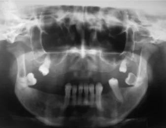
All of the 25 subjects underwent oral clinical evaluation and panoramic radiography. The latter was performed on an orthopantomograph (Orto-phos Plus PS®, Siemens – Sirona Dental Company, Bensheim, Germany), working at 14 mA and vari-able kVp of 60.16 for women and 64.14 for men, at a mean magnii cation of 30%. The images were interpreted by the examiner and the 15 patients were distributed into two groups according to the radiographic i ndings: with or without the presence of mandibular osteolysis (Figure 1).
had had the disease was obtained from their medi-cal records.
Following this, the patients underwent clinical examination to measure their mouth opening. In a comfortably seated position, with the head support-ed on a hard surface at 90° to the l oor, the patients were asked to open their mouths as wide as possible, three times. Each movement was measured, and only the largest measurement was used. The same procedure was used to obtain this measurement for the healthy individuals.
To make the measurements of maximum mouth opening, we used an adaptation of the linear
interin-cisal distance method.12 The points of a dry-point compass were positioned on the middle third of the incisal aspect of the upper and lower right central incisors at maximum mouth opening, and this mea-surement was transferred to a ruler graduated in millimeters (Figure 2).
The patient was then asked to perform the in-tercuspidation movement, from which the vertical trespass distance was obtained and subtracted from the compass measurement. If the right central inci-sors were absent, the left central inciinci-sors, right lat-eral incisors or left latlat-eral incisors would be used, in that order. For the inferential analysis of the clinical variables, Pearson’s linear correlation coefi cient, Student’s t test and Fisher’s exact test were used. The rejection level adopted for the nullity hypoth-esis was 0.05 (5%).
Results
The arithmetic mean and respective standard de-viation for the age of the 25 patients was 39.71 ± 7.91 years. The results from the clinical variables studied among the patients with systemic sclerosis can be seen in Tables 1 and 2.
Graph 1 shows the distribution of the groups in relation to the variable length of time with the dis-ease. With the aim of investigating whether group I had had the disease for the same length of time as group II, Student’s t test was applied. The result from this showed that group I had had the disease Figure 1- Panoramic radiograph of the mandible
present-ing bilateral osteolysis of the angle, ascendpresent-ing branch and condyles.
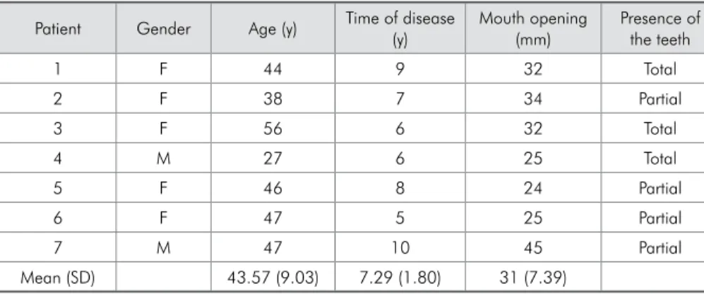
Patient Gender Age (y) Time of disease (y)
Mouth opening (mm)
Presence of the teeth
1 F 44 9 32 Total
2 F 38 7 34 Partial
3 F 56 6 32 Total
4 M 27 6 25 Total
5 F 46 8 24 Partial
6 F 47 5 25 Partial
7 M 47 10 45 Partial
Mean (SD) 43.57 (9.03) 7.29 (1.80) 31 (7.39)
SD = standard deviation; F = female; M = male; y= years; mm= millimeters.
Table 1 - Clinical values of group I: patients with osteolysis.
Patient Gender Age (y) Time of disease (y)
Mouth opening (mm)
Presence of the teeth
8 F 35 7 23 Partial
9 F 46 5 26 Total
10 F 35 4 41 Partial
11 F 47 7 44 Total
12 F 52 8 38 Partial
13 F 44 4 40 Partial
14 F 43 6 27 Total
15 F 49 5 49 Total
Mean (SD) 43.88 (6.15) 5.75 (1.49) 36 (9.47)
SD = standard deviation; F = female; M = male; y= years; mm= milimeters.
Table 2 - Clinical values of group II: patients without osteolysis.
T
im
e
of
the
di
seas
e
(y
ear
s)
N = 3 4 5 6 7
Group I 7 9
8
Group II 8 10
11
Graph 1 - Boxplot of time with the disease (years) accord-ing to the groups.
Mouth opening (mm)
N =
Group I 7
Group II 8 60
50
40
30
20
Graph 2 - Boxplot of mouth opening (mm) according to the groups.
for a longer time than group II (p = 0.003). The re-sults relating to the variable of mouth opening
opening and length of time with the disease accord-ing to the groups was investigated by estimataccord-ing Pearson’s linear correlation coefi cient, as observed in dispersion plots. Pearson’s linear correlation co-efi cient was found to be 0.676 (p = 0.095), and therefore it was considered that for the individuals in group I there was a moderate increasing linear correlation between mouth opening and length of time with the disease (Graph 3). For group II, the distribution of information on mouth opening and length of time with the disease did not show any vi-sual relationship (Graph 4). This was also coni rmed by the i nding that Pearson’s linear correlation coef-i ccoef-ient was 0.223 (p = 0.596).
With regard to the presence of teeth, the patients were classii ed as possessing a full or a partial set of teeth. Patients were taken to have a partial set of teeth if they did not present a row of back teeth, which could be on one or both sides, in the upper and/or low jaw. Fisher’s exact test for this associa-tion showed that there was no relaassocia-tionship between presence/absence of teeth and group (p > 0.999).
Discussion
Limitation of mouth opening is a common i nding in SS cases and is possibly related to the skin thickening that is characteristic of the dis-ease.6,7,10,13,14 The progressive loss of elasticity of the cheeks and lips makes these regions hardened and causes a gradual decrease in the lip perimeter and
maximum mouth opening.7 In our study, this limi-tation was the patients’ main complaint in relation to the stomatognathic system, because of their dif-i cultdif-ies dif-in chewdif-ing or performdif-ing oral hygdif-iene ma-neuvers,10 or even in relation to undergoing dental treatment. Some techniques involving active exercis-es have been indicated with some succexercis-ess in achiev-ing expansion of the mandibular movements.7,13,14
Only a few studies have sought to objectively quantify this reduction in mouth opening in sys-temic sclerosis cases.6,10,15 In our study, a mean measurement of 47 ± 5.1 mm was obtained among the healthy controls. Among the patients with oste-olysis, the mean opening was 31 ± 7.39 mm, while among the patients without osteolysis, the mean was 36 ± 9.47 mm. In both the latter groups, the mean mouth opening was smaller than in the con-trol group (p = 0.003 and p = 0.036 respectively). Comparing only the two groups with SS, the group with osteolysis presented the same mean opening as did the group without osteolysis (p = 0.280). These i ndings do not agree with those found in the litera-ture, in which the patients with osteolysis present-ed a slight decrease in opening in relation to those without osteolysis.10,11
On the other hand, when the variable of length of time with the disease was analyzed, it was seen that the patients with osteolysis had a moderate ten-dency towards increased mouth opening over the course of the years (p = 0.095), while among the
pa-Mouth opening (mm)
Time of the disease (years) 50
40
30
20
4 5 6 7 8 9 10 11
Graph 3 - Distribution of mouth opening and time of the disease: Group I.
Mouth opening (mm)
Time of the disease (years) 50
40
30
20
3 4 5 6 7 8 9
tients without osteolysis this trend was not observed (p = 0.596). One possible explanation for this i nd-ing could be the fact that the most intense skin thickening occurs within the i rst 18 to 36 months of the disease, which is then followed by a variable period of stability, after which there is a slow and continuous decline,3 such that the skin takes on an atrophied and thin appearance.1,16 Other factors, such as the number of patients studied, length of time with the disease and method used for measur-ing the mouth openmeasur-ing may cause variations in these results.6,17 In our sample, the patients reported tran-sitory improvement following the use of an endove-nous infusion of 2% lidocaine hydrochloride, which was used to attenuate the skin thickening.1
Previous studies did not i nd any direct rela-tionship between the presence of osteolysis and the length of the course of the disease.6,11,18 However, osteolysis is detected between the i fth and seventh year after diagnosing SS.19 In our study, we ob-served that the patients with osteolysis presented a longer mean time with the disease (7.29 ± 1.8 years) than did the patients without osteolysis (5.75 ± 1.49 years) (p = 0.003). Considering that the systemic im-pairment caused by the disease is greater over the i rst i ve years of the disease1 and the skin involve-ment is most intense over the i rst three years,3 it can be inferred that the emergence of mandibular osteolysis may be related both to the peak of skin involvement and to the visceral involvement, since osteolysis tends to occur in patients with greater vis-ceral involvement.19
The extent of the osteolysis was not quantii ed in our study but, rather, only its presence or absence. The rarity of the disease and the fact that mandib-ular osteolysis is present in a mean of 25% of the patients10,11,17,18 makes it difi cult to standardize the
grades of osteolysis. For this reason, the samples in the literature have included osteolysis to varying ex-tents.6,17,18 Moreover, some patients with the disease for a long time may present only slight degrees of osteolysis, while others who have just had the dis-ease for a short time may present great destruction.8 Concerning the presence/absence of teeth, the re-sults showed that this was unrelated to the presence of osteolysis. That is, osteolysis may develop both in patients with a full set of teeth and in those with only a partial set (p > 0.999). This i nding coni rms the results from previous studies.17,18 In addition, we investigated the relationship between the side of the osteolysis and the presence/absence of teeth, and found that there was no association (p = 0.143). We did not i nd any similar study with which we could compare or contrast this information.
We believe that new studies with different sam-ples and methods are needed in order to bring out more evidence regarding the involvement of the sto-matognathic system in patients with SS. Thus, longi-tudinal studies may provide predictive information about orofacial involvement in relation to the course of the disease.
Conclusions
According to our results, the patients with osteol-ysis presented longer times with the disease than did the group without osteolysis, but the presence of oste-olysis did not inl uence the mouth opening measure-ment. We also observed that there was an increasing correlation between mouth opening and length of time with the disease among the patients with osteol-ysis, whereas this did not occur in the group without osteolysis. There was no relationship between ence/absence of teeth and osteolysis, or between pres-ence/absence of teeth and side of osteolysis.
References
1. Kayser C, Andrade LEC. Esclerose Sistêmica. In: Sato E. Guias de Medicina Ambulatorial e Hospitalar – Reumatologia. São Paulo: Manole; 2004. p. 111-20.
2. Mayes MD, Lacey Jr JV, Beebe-Dimmer J, Gillespie B, Cooper B, Timothy JL et al. Prevalence, incidence, survival and disease
3. Marques Neto JF, Sampaio-Barros PD. Reumatologia: diagnós-tico e tratamento. Rio de Janeiro: Medsi; 2000. p. 465-80. 4. Spackman GK. Scleroderma: what the general dentist should
know. Gen Dent. 1999;47(6):576-9.
6. Marmary Y, Glaiss R, Pisanty S. Scleroderma: oral manifesta-tions. Oral Surg Oral Med Oral Pathol. 1981;52(1):32-7. 7. Naylor PW. Oral management of the scleroderma patient.
JADA. 1982;105(5):814-7.
8. Ramón Y, Samra H, Oberman M. Mandibular condylosis and apertognathia as presenting symptoms in progressive systemic sclerosis. Oral Surg. 1987;63(3):269-74.
9. Weisman RA, Calcaterra TC. Head and neck manifestations of scleroderma. Ann Otol. 1978;87(3):332-9.
10. Wood RE, Lee P. Analysis of the oral manifestations of sys-temic sclerosis (scleroderma). Oral Surg Oral Med Oral Pathol. 1988;65(2):172-8.
11. Seifert MH, Steigerwald JC, Cliff MY. Bone resorption of the mandible in progressive systemic sclerosis. Arthritis and Rheum. 1975;18(5):507-12.
12. Wood GD, Branco JA. A comparison of three methods of measuring maximal opening of the mouth. J Oral Surg. 1979;37(3):175-7.
13. Defabianis P. Scleroderma: a case report of possible cause of restricted movement of the temporomandibular joint with effects on facial development. J Clin Pediatr Dent. 2003;28(1):33-8.
14. Pizzo G, Scardina GA, Messina P. Effects of a nonsurgical exercise program on the decreased mouth opening in patients with systemic scleroderma. Clin Oral Invest. 2003;7(3):175-8.
15. Nagy G, Kovacs J, Zeher M, Czirjak L. Analysis of the oral manifestations of systemic sclerosis. Oral Surg Oral Med Oral Pathol. 1994;77(2):141-6.
16. Akesson A, Fiori G, Krieg T, van den Hoogen FHJ, Seibold JR. Assessment of skin, joint, tendon and muscle involvement. Clin Exp Rheumatol. 2003;21(suppl. 29), S5-S8.
17. White SC, Frey NW, Blaschke DD, Ross MD, Clements PJ, Furst DE et al. Oral radiographic changes in patients with progressive systemic sclerosis. JADA. 1977;94(6):1178-82. 18. Ramirez JC, Cobos LS, Jelic PM. Reabsorción Osea Patológica
de la Mandíbula y Ensanche Del Espacio Peridontal Dentá-rio en Pacientes con Esclerosis Sistêmica. Rev Méd Chile. 1984;112(1):13-9.