369
Revista da Sociedade Brasileira de Medicina Tropical 34(4): 369-372, jul-ago, 2001.
ARTIGO
Radiographic features of pulmonary tuberculosis in patients infected
by HIV: is there an objective indicator of co-infection?
Características radiológicas da tuberculose pulmonar em doentes infectados
pelo HIV: existe um indicador objetivo da co-infecção?
Maria de Fátima Militão de Albuquerque1, Silvio Cavalcanti de Albuquerque1,
Antonio Roberto Leite Campelo1, Marta Cruz1, Wayner V. de Souza2,
Ricardo A.A. Ximenes1 and Roberta A.S. de Souza1
Abstract This study aimed to compare the radiographic characteristics of patients with pulmonary tuberculosis (TB) and human immunodeficiency virus (HIV) infection with those of HIV-negative patients. In all, 275 TB patients attending the outpatients clinics at the University Hospital/UFPE, were studied from January 1997 to March 1999. Thirty nine (14.2%) of them were HIV+, with a higher frequency of males in this group (p=0.044).
Seventy-five percent of the HIV+ patients and 19% of the HIV- had a negative tuberculin test (PPD) (p < 0.001).
The proportion of positive sputum smears in the two groups was similar. The radiological finding most strongly associated with co-infection was absence of cavitation (p < 0.001). It may therefore be concluded that the lack of cavitation in patients with pulmonary TB may be considered a useful indicator of the need to investigate HIV infection. This approach could contribute to increasing the effectiveness of local health services, by offering appropriate treatment to co-infected patients.
Key-words: Tuberculosis. HIV/AIDS. Radiographic features.
Resumo Este trabalho objetivou comparar características radiológicas de doentes com tuberculose (TB) pulmonar, soropositivos para o vírus da imunodeficiência humana (HIV), com aquelas de doentes HIV-.
Estudou-se, prospectivamente, 275 doentes com TB pulmonar, atendidos no Hospital das Clínicas da UFPE, entre janeiro de 1997 e março de 1999. Destes, 39 (14,2%) eram HIV+, com um predomínio maior de homens neste
grupo (p=0,044). Setenta e cinco por cento dos doentes HIV+ e 19% dos HIV- apresentavam reação negativa
ao teste tuberculínico (PPD) (p<0,001). A proporção de baciloscopias positivas nos dois grupos foi semelhante. A característica radiológica mais fortemente associada à co-infecção tuberculose pulmonar/HIV+ foi a ausência
de cavidades (p<0,001). Conclui-se que, diante de doentes com TB pulmonar, a ausência de cavidades na telerradiografia de tórax constitui-se num indicador útil da necessidade de se investigar a infecção pelo HIV. Esta conduta pode contribuir para aumentar a efetividade dos serviços locais de saúde, permitindo oferecer tratamento adequado a estes doentes.
Palavras-chaves: Tuberculose. HIV/AIDS. Achados radiológicos.
1. Departamento de Clínica Médica da Universidade Federal de Pernambuco, Recife, PE. 2. Centro de Pesquisas Aggeu Magalhães da Fundação Oswaldo Cruz. Recife, PE. Suporte Financeiro: FACEPE.
Address to: Dra. Maria de Fátima Militão Albuquerque. Deptº de Clínica Médica/Hospital das Clínicas/UFPE. Av. Prof. Moraes Rêgo s/n, Bloco A, Cidade Universitária,50670-420 Recife, PE.
Recebido para publicação em 21/9/2000.
The pandemic of the Human Immunodeficiency Virus (HIV) has been pointed out as one of the major causes of the worldwide increase in tuberculosis cases. This is due to the fact that tuberculosis is one of the most common infections to occur in the course of HIV infection, either because of the reactivation of latent TB or as a result of new infection by the tuberculosis mycobacterium, with a faster progression towards the active disease. When tuberculosis is associated with HIV infection, the difficulty in establishing a diagnosis using traditional parameters contributes to the resurgence of this disease12.
In Brazil, as in most developing countries, the diagnosis of pulmonary tuberculosis depends on the clinical symptoms, chest radiography, tuberculin test and sputum smear examination for acid-fast bacilli (AFB). The culture for M. tuberculosis is not routinely performed due to the cost and delay in obtaining results11. In cases of pulmonar y tuberculosis
associated with HIV infection, the results of these examinations are often uncharacteristic, which delays the diagnosis15. At present, HIV testing is not routinely
370
Albuquerque MFM et al
However, the relatively high frequency of HIV infection among new cases of tuberculosis in some Brazilian cities (9.6%) indicates the need to investigate the presence of this association, in order to offer these patients appropriate treatment for both infections10. In 1996, it was
estimated that in Brazil the greater number of individuals infected by HIV would result in a 20% increase in the incidence of tuberculosis by the end of the decade.
Among the diagnostic tools traditionally used for the diagnosis of pulmonary tuberculosis, the chest x-ray plays a fundamental role, mainly because it is widely available within the public health system, even in poor areas. The study of the radiographic features of pulmonary tuberculosis associated with HIV infection is therefore justified in seeking objective indicators that may suggest a higher probability of the presence of co-infection.
MATERIAL AND METHODS All patients diagnosed as having pulmonar y
tuberculosis who attended the tuberculosis out-patients clinic at the University Hospital of the Federal University of Pernambuco (HC/UFPE) from January 1997 to May 1999, who had a chest x-ray taken and anti-HIV test performed were studied prospectively. All the patients had three sputum smear exams for acid-fast bacilli carried out using the Ziehl-Neelsen technique. The tuberculin test was performed with 0.1ml of PPD RT23, according to a standardized technique.
The chest radiographs were analyzed by a single radiologist of the HC/UFPE, a member of the research team, who filled out a standardized for m. The radiographic features were summarized in categories previously discussed (as described in Table 2). The
radiologist was aware neither of the HIV status of the patient nor the previous radiographic reading, carried out by another radiologist from the hospital staff.
ELISA 1 and 2, immunofluorescence and Western-blot techniques were used for the diagnosis of HIV. When ELISA 1 and 2 tests were negative the result was considered as non-reagent. Those that were positive for ELISA 1 and 2 underwent an immunofluorescence test and when positive were considered as reagent.
The association between the clinical and radiographic characteristics and the result of the anti-HIV test was studied and the significance of this association was tested by the chi-square or Fisher test, when indicated, and the p-value.
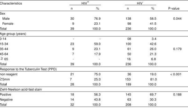
RESULTS Two hundred and seventy-five (275) patients with a diagnosis of pulmonary tuberculosis met the inclusion criteria. Thirty-nine (14.2%) of these were HIV+, with a
significantly larger proportion of men among HIV+
patients than among HIV- (p=0.044), whilst the age distribution of the two groups was similar (Table 1).
Seventy-five percent (75%) of the HIV+ patients had a negative tuberculin test (PPD=0mm), compared with 19% (36 out of 189) of the HIV- patients (p < 0.001). There was no statistically significant association between the result of the sputum smear examination and the result of the HIV test (p=0.188).
Table 1- Demographic and clinical characteristics of 275 pulmonary tuberculosis patients HIV+ and HIV-, Hospital das Clínicas - UFPE, January 1997 to March 1999.
Characteristics HIV+ HIV
n % n % P-value Sex
Male 30 76.9 138 58.5 0.044
Female 9 23.1 98 41.5
Total 39 100.0 236 100.0
Age group (years)
0-14 - - 08 3.4
15-34 23 59.0 100 42.6
35-44 9 23.1 61 26.0 0.179
45-64 7 17.9 50 21.3
65 - - 16 6.8
Total 39 100.0 236 100.0
Response to the Tuberculin Test (PPD)
non reagent 21 75.0 36 19.0 < 0.001
5mm 7 25.0 153 81.0
Total 28 100.0 189 100.0
Ziehl-Neelson acid-fast stain
Positive 18 56.3 145 69.7 0.188
Negative 14 43.8 63 30.3
Total 32 100.0 208 100.0
≥
371
Revista da Sociedade Brasileira de Medicina Tropical 34: 369-372, jul-ago, 2001
Table 2 shows the radiological findings of the 275 pulmonary tuberculosis patients. Focal infiltrate was the most frequent radiographic pattern observed.
The chest radiographic findings were grouped into eight categories: normal, focal infiltrate, diffuse infiltrate,
pulmonary nodules, miliary disease, lymphadenopathy, pleural effusion and absence of cavities and the results presented according to the HIV status. It was found that absence of cavities was significantly associated with HIV infection (p < 0.001) (Table 3).
Table 3 - Radiological patterns of pulmonary tuberculosis patients HIV+ and HIV-, Hospital das Clínicas - UFPE, January 1997 to March 1999.
HIV+ HIV
-Radiological pattern n % n % P-value
Normal 3 7.7 14 5.9 p=0.717*
Focal infiltrate 24 61.5 165 69.9 p=0.390**
Diffuse Infiltrate 2 5.1 24 10.2 p=0.552*
Nodules - - 7 2.9 p=0.598*
Miliary 3 7.7 4 1.7 p=0.061*
Lymphadenopathy 4 10.3 8 3.4 p=0.073*
Pleural effusion 6 15.4 19 8.1 p=0.141*
Absence of cavities 32 82.1 119 50.4 p=0.0005** *Fisher exact test
**cc2 Yates
DISCUSSION
There seems to be a consensus regarding the modifications of the clinical-epidemiological pattern of pulmonary tuberculosis when associated with HIV infection. Traditional diagnostic methods to a certain extent lose their specificity15, thus necessitating the
redefinition of their characteristics.
Regarding the age group, although a larger concentration of cases of co-infection among the individuals aged 20 to 39 years (74.4% of the cases) was observed, there was no statistically significant difference in the age distribution between the two groups HIV+ and HIV-.
The difference in the frequency of patients with pulmonary tuberculosis associated with HIV+ between
sexes and age groups depends on the specific prevalence of both infections by sex and age in the population. Pozniak et al15 found in Zimbabwe, Africa,
that the distribution of co-infection by sex was similar, whilst Awil et al1, in another area of Africa (Gulu, Uganda)
described a higher frequency of co-infection among women. In the present study there was a larger proportion of males among those infected by HIV (p=0.044).
With respect to age distribution, there is an overlap of the population groups affected by both infections. The prevalence of tuberculosis is greater among young adults, and the individuals most frequently affected by HIV infection are aged 15 to 49 years2. A survey carried
out in Rio de Janeiro showed that the highest frequency of HIV infection among tuberculosis patients, 7.4%, was found in the 15 to 39 age group7. In this study a larger
number of individuals in the 15 to 34 age group was found among co-infected patients (59%), but a similar finding was observed in those with only pulmonary tuberculosis (Table 1).
The association found between a negative reaction to the tuberculin test (PPD=0mm) and a positive anti-HIV test (p< 0.001) is in agreement with the literature15.
Table 2 - Radiological patterns of 275 pulmonar y tuberculosis patients HIV+ and HIV-, Hospital das Clínicas - UFPE, January 1997 to March 1999.
Radiological pattern N %
Normal 17 3.6
Focal infiltrate 189 40.4
Diffuse infiltrate 26 5.6
Single cavity 57 12.2
Multiple cavities 67 14.3
Miliary 7 1.5
Nodules 6 1.3
Lymphadenopathy 12 2.6
Pleural effusion 25 5.3
Others 62 13.2
372
In HIV+ patients, due to the absence of delayed-type
hypersensitivity response, lung cavitation is not usual, so the dissemination of bacilli may occur and result in atypical findings. Absence of cavitation has been related to a higher frequency of negative acid-fast bacilli sputum smears in co-infected patients6 15. There was a similar proportion of
positive sputum smears in the two groups in this study, which agrees with other reports in the literature4.
Some authors, analyzing the radiological pattern most frequently found among patients with pulmonar y tuberculosis associated with HIV, describe the following features: hilar adenopathy, no cavitary infiltrates and miliary disease13 14 15 16. In Brazil, few studies have been
carried out with the objective of studying the radiographic pattern of pulmonary tuberculosis associated with HIV infection3 5 9 17. Camera et al3 found, in a retrospective study
of 104 individuals with pulmonary tuberculosis /HIV+, that
the most frequent findings were interstitial infiltrate (71.2%)
and alveolar lesion (63.5%). However, there was no comparison group i.e. HIV-individuals.
In the present study, the absence of cavities was the radiographic characteristic most strongly associated with co-infection. Miliary disease and lymphadenopathy were more frequent in the HIV+ group, but the difference was
not statistically significant, possibly due to the sample size. The small percentage of diffuse infiltrates among the HIV+ patients may be due to the low sensibility of conventional radiology in detecting early interstitial disease.
It may therefore be concluded that absence of cavities may be considered a useful indicator for the need to investigate HIV infection in patients with a clinical or bacteriological diagnosis of pulmonary tuberculosis. This approach could contribute to increasing the effectiveness of local health services, which diagnose and treat tuberculosis cases, by providing appropriate treatment for co-infected patients, in addition to orientating them to prevent HIV transmission.
REFERENCES
1. Awil PO, Bowlin SJ, Daniel TM. Radiology of pulmonary
tuberculosis and human immunodeficiency virus infection in Gulu, Uganda. European Respiratory Journal 10: 615-618, 1997.
2. Barnes PF, Bloch AB, Davidson PT, Snider Jr DE. Tuberculosis
in patients with human immunodeficiency virus infection. New England Journal of Medicine 324:1644-1650, 1991.
3. Camera CS, Santos AAS, Moraes APP, Silva WAE, Camisão CC,
Oliveira CAB, Kischinhevski W. Radiologia do tórax em pacientes HIV-positivos com tuberculose; análise of 104 casos. Radiologia Brasileira 23: 241-247, 1990.
4. Finch D, Beaty CD. The utility of single sputum specimen in the
diagnosis of tuberculosis. Comparison between HIV-infected and non-infected patients. Chest 111:1174-1179, 1999.
5. Henn L, Nagel F, Dal-Pizzol F. Comparison between human
immunodeficiency virus positive and negative patients with tuberculosis in Southern Brazil. Memórias do Instituto Oswaldo Cruz 94: 377-381, 1999.
6. Jones BE, Ryu R, Yang Z, Dig MD, Pogoda JM, Otaya M, Barnes
PF. Chest radiographic findings in patients with tuberculosis with recent or remote infection. American Jounal Respiratory and Critical Care Medicine 155: 1270-1273, 1997.
7. Kritski AL, Werneck-Barroso E, Vieira MA, Car valho ACC,
Carvalho CE, Bravo-de-Souza R, Andrade GD, I Galvão-Castro B, Castilho EA, Hearst N. HIV infection in 567 active pulmonary tuberculosis patients in Brazil. Journal of Acquired Immune Deficiency Syndrome 6:1008-1012, 1993.
8. Kumar D, Watson JM, Charlett A, Nicholas S, Darbyshire JH.
Tuberculosis in England and Wales in 1993: results of a national survey. Thorax 52:1060-1067, 1997.
9. Kusano MS. Comparative study of patients with tuberculosis with
and without HIV infection in the Federal District. Revista Brasileira de Enfermagem 49:41-54, 1996.
10. Lima MM, Belluomini M, Almeida MMB, Arantes GR. Co-infecção HIV/Tuberculose: necessidade de uma vigilância mais efetiva. Revista de Saúde Pública 31: 217-220, 1997.
11. Ministério da Saúde. Manual de Normas de Tuberculose para o Controle da Tuberculose. Centro Nacional de Epidemiologia. 5 ª e d i ç ã o, C o o r d e n a ç ã o N a c i o n a l d e P n e u m o l o g i a Sanitária.Brasília, DF.
12. Macadams HP, Erasmus J, Winter JA. Radiologic manifestation of pulmonary tuberculosis. Radiologic Clinics of North America, 33: 655-678, 1995
13. Perlman AD, El-Sadr WM, Nelson ET, Matts JP, Telzak EE, Salomon N, ChirgwiN K, Hafner R. Variation of chest radiographic patterns in pulmonary tuberculosis by degree of human immunodeficiency virus-related immunosuppression. Clinical Infection Diseases 25: 242-246, 1997.
14. Pitchenik AE, Rubinson HA. The radiographic appearance of tuberculosis in patients with the acquired Immune Deficiency Syndrome (AIDS) and Pre-AIDS. Journal Medicine 307: 162-165, 1982.
15. Pozniak AL, MacLeod GA, Ndlovu D, Ross E, Mahari M, Weinberg J. Clinical and Chest Radiographic Features of Tuberculosis associated with Human Immunodeficiency Virus in Zimbabwe. American Jounal Respiratory and Critical Care Medicine 152 :1558-1561, 1995.
16. Shah RM, Kaji AV, Ostrum BJ, Friedman BC. interpretation of chest radiographs in AIDS patients. Scientific Exhibit 17: 47-57, 1997.
17. Trajman A, Neto EB, Belo MT, Teixeira EG, Selig L, Ferrari G, Branco MM. Pleural tuberculosis and human immunodeficiency virus co-infection. International Journal of Tuberculosis and Lung Diseases 1: 487, 1997.