____________________________
Corresponding author: Sandra Petrovic,Vinca Institute of Nuclear Sciences, University of Belgrade,M. Petrovica Alasa 12-14, 11001 Belgrade, Serbia,Phone: +381 11 340856,Email1:
UDC 575 DOI: 10.2298/GENSR1502695P
Original scientific paper
ASSESSMENT OF SINGLE NUCLEOTIDE POLYMORPHISMS IN SCREENING 52 DNA REPAIR AND CELL CYCLE CONTROL GENES IN FANCONI ANEMIA
PATIENTS
Sandra PETROVIC1, Andreja LESKOVAC1, Ivana JOKSIC1,2, Dragana VUJIC3, 4,
Ana Valenta SOBOT1, Jelena FILIPOVIC1 and Gordana JOKSIC1
1 Vinca Institute of Nuclear Sciences, University of Belgrade, Belgrade, Serbia 2Hospital of Gynecology and Obstetrics “Narodni Front”, Belgrade, Serbia
3Medical School, University of Belgrade, Belgrade, Serbia
4Mother and Child Health Care Institute of Serbia “Dr. Vukan Cupic”, Belgrade, Serbia
Petrovic S., A. Leskovac, I. Joksic2, D. Vujic, A. Valenta Sobot, J. Filipovic
and G. Joksic (2015): Assessment of single nucleotide polymorphisms in screening 52 DNA repair and cell cycle control genes in Fanconi anemia patients .- Genetika, Vol 47, No. 2, 695-710.
which may be of great importance for better clinical description of FA. In addition, the present report recommends the use of SNPs as predictive and prognostic genetic markers to individualize therapy of FA patients.
Key words: DNA repair, Fanconi anemia, single nucleotide polymorphisms
List of abbreviations
APEX APE1- APEX nuclease (multifunctional DNA repair enzyme) 1; ATM- ataxia telangiectasia mutated serine/threonine kinase; BRCA1- breast cancer 1, early onset; BARD1- BRCA1
associated RING domain 1; BRCA2- breast cancer 2, early onset; BER- base excision repair; CCND1- cyclin D1; CCNH- cyclin H; p21/CDKN1A- cyclin-dependent kinase inhibitor 1A (p21,
Cip1); CDKN2A- cyclin-dependent kinase inhibitor 2A ; CDKN1B- cyclin-dependent kinase
inhibitor 1B (p27, Kip1); CDK7- cyclin-dependent kinase 7; CHEK2- checkpoint kinase 2;
DSB- double strand break; ERCC1- excision repair cross-complementation group 1; ERCC2-
excision repair cross-complementation group 2; ERCC4- excision repair cross-complementation
group 4; ERCC5- excision repair cross-complementation group 5; FANCD2- Fanconi anemia,
complementation group D2; GADD45A- growth arrest and DNA-damage-inducible, alpha; GRTH/Ddx25- DEAD (Asp-Glu-Ala-Asp) box helicase 25; LIG1- ligase I, DNA,
ATP-dependent; LIG3- ligase III, DNA, ATP-dependent; LIG4- ligase IV, DNA, ATP-dependent; MGMT AGT- O-6-methylguanine-DNA methyltransferase; MLH1- mutL homolog 1; MMR- mismatch repair; MSH2- mutS homolog 2; MSH3- mutS homolog 3; MSH6- mutS homolog 6;
MYH- mutY DNA glycosylase; NER- nucleotide excision repair; NBS1- nibrin; OGG1-
8-oxoguanine DNA glycosylase; PARP1- poly (ADP-ribose) polymerase 1; PARP4- poly
(ADP-ribose) polymerase family, member 4; PCNA- proliferating cell nuclear antigen; PMS2- PMS2
postmeiotic segregation increased 2 (S. cerevisiae); POLB- polymerase (DNA directed), beta; RAD9A- RAD9 checkpoint clamp component A ; RAD23B- RAD23 homolog B (S. cerevisiae); RAD51- RAD51 recombinase; RAD52- RAD52 homolog (S. cerevisiae); RAD54B- RAD54
homolog B (S. cerevisiae); RECQL- RecQ helicase-like; XPA- xeroderma pigmentosum,
complementation group A; XPC- xeroderma pigmentosum, complementation group C; XRCC1-
X-ray repair complementing defective repair in Chinese hamster cells 1; XRCC2- X-ray repair
complementing defective repair in Chinese hamster cells 2; XRCC3- X-ray repair complementing
defective repair in Chinese hamster cells 3; XRCC4- X-ray repair complementing defective repair in Chinese hamster cells 4; XRCC5- X-ray repair complementing defective repair in Chinese hamster cells 5 (double-strand-break rejoining); XRCC9/FANCG- Fanconi anemia,
complementation group G; SNP- single nucleotide polymorphism; TP53- tumor protein p53; TP53BP1- tumor protein p53 binding protein 1; TP53BP2- tumor protein p53 binding protein 2
INTRODUCTION
Fanconi anemia (FA) is a recessive disorder considered to encompass a defect in maintenance of genome integrity. It is associated with bone marrow failure, birth defects and predisposition to cancer. The disease is genetically heterogeneous with 16 complementation groups (FA -A to FA –Q) and associated genes having been characterized (KEE and D'ANDREA, 2012; BOGLIOLO et al., 2013; CHEN et al., 2014). All of the 16 gene products operate in a common
~ 10-15% of the cases while the rest of the cases (~ 15%) is distributed over the remaining complementation groups (e.g. the prevalence of FA-D2 is 3.3%) (MAGDALENA et al., 2005;
MATHEW, 2006; AMEZIANE et al., 2012). Regardless of the complementation group, FA is a
condition involving high risk of leukemia and solid tumors (KUTLER et al., 2003). However, FA
patients exhibit a high degree of clinical variability and have poor tolerance for radiotherapy and chemotherapy (MATHEW, 2006). Thus, the knowledge of factors that might predict either the best drug response or adverse outcome would greatly facilitate the decision making concerning the choices of treatment.
Single nucleotide polymorphisms (SNPs) represent one of the largest types of inherited genetic variations (BOND and LEVINE, 2007) and are considered as predictive and prognostic genetic markers. Polymorphisms in DNA repair and cell cycle control genes may alter protein function leading to genomic instability and increased cancer risk (SIGURDSON et al., 2004).
As already known, Fanconi anemia is a disease associated with deficiencies in DNA repair pathway, which may be the cause of the overall increased cancer risk in patients. FA proteins constitute the FA pathway responsible for repair of inter - and intra - strand DNA cross - links either occurring spontaneously or induced by clastogens as mitomycin C or diepoxybutane (JOENJE and PATEL, 2001; KIM and D'ANDREA, 2012). However, apart from the disease-causing mutations in FANC genes, the identification of specific variations in other candidate genes in FA patients may lead to a better clinical description of this condition enabling individualized treatment with improvement of the prognosis. In light of the fact that Fanconi anemia is a highly penetrant cancer susceptibility disorder, the products of the identified genes could become targets for innovative therapies in patients. In addition, the genotyping of SNPs in key genes involved in DNA repair may provide insight into the interactions between FA mutation and specific variations in DNA repair genes in each of the FA patients.
The results presented here focus on assessment of the known variations located in genes involved in base excision repair (BER), nucleotide excision repair (NER), mismatch repair (MMR), double strand break (DSB) repair and also in the several genes related to the cell cycle control and apoptosis. Investigation was performed in patients belonging to complementation groups FA-A and FA-D2; the last one present as the most prevalent complementation group in Serbia.
MATERIALS AND METHODS Patients
DNA isolation
DNA was isolated from patients-derived fibroblasts using the phenol-chloroform method. The cells were treated with STE buffer (10 mM TRIS-HCl, 100 mM NaCl, 1 mM EDTA), proteinase K (10 mg/mL) (Sigma-Aldrich) and 10% SDS (sodium dodecyl sulfate) (Sigma-Aldrich) and incubated for 2 hours at 55 ºC. Afterwards, the upper layer (250 - 300 µL) was transferred to a new tube, and dH2O (50 µL) and phenol (Sigma-Aldrich) (300 µL) were added. The suspension
was vigorously vortexed and then centrifuged for 10 minutes at 12000 rpm in a microcentrifuge. The supernatant (250 - 300 µL) was placed into a new tube and chloroform (~150 µL) (Sigma-Aldrich) was added. After centrifugation for 10 minutes at 12000 rpm, the supernatant (250 - 300 µL) was transferred to a new tube and treated with 7.5 M ammonium acetate (150 µL) and ice-cold 100% ethanol (1.5 mL). After centrifugation for 5 minutes at 12000 rpm, the supernatant was decanted and the pellet was air-dried for 1-2 minutes and re-suspended in appropriate volume dH2O (30-50 µL). The quantification of the concentration and purity of the DNA was performed
using a spectrophotometer (Nanodrop-1000, Thermo Fisher Scientific, USA). Single nucleotide polymorphism analysis
For SNP analysis, DNA samples (concentration 1 µg/µL at least) were studied using DNA repair chip which includes most of the common variants for the DNA repair genes as well as for the several genes related to the cell cycle control and apoptosis. The SNP genotyping was performed at the Asper Biotech (Tartu, Estonia) (http://www.asperbio.com/DNArepairchip.pdf). The total number of SNPs analyzed in this cohort is 95, and the total number of genes is 52.
RESULTS
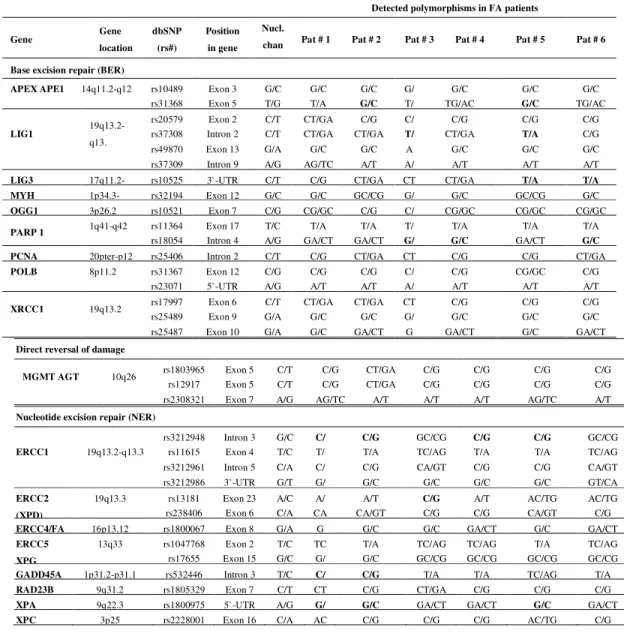
In this study, we have assessed 95 SNPs located in 52 key genes involved in BER, NER, MMR, DSB repair and cell cycle control using an Asper Biotech array. The investigation was performed in five FA-D2 patients and in one FA-A patient. The summary of genes involved in different repair and cell cycle control pathways and corresponding SNPs are presented in Tables 1 - 2. Some genes are involved in more than one pathway, but each is listed only once. All of the SNPs studied are accessible at the NCBI (National Center for Biotechnology Information)
database (http://www.ncbi.nlm.nih.gov/snp).
The polymorphisms studied were synonymous (n=10), nonsynonymous (missense) (n=52) and in non-coding regions of the genome (introns and 5 ‘and 3’ untranslated regions (UTR)) (n=33). Polymorphisms found at the homozygous state (genotypes in bold, see Tables 1-2) are selected for further analysis.
In the BER pathway, the SNPs located in APEX, LIG1, LIG3, MYH, OGG1, PARP1, PCNA, POLB and XRCC1 were evaluated. As shown in Table 1, the most of the studied
polymorphisms were found as heterozygous variants, while the homozygous variants were found in APEX APE1, LIG1, LIG3 and PARP1. The missense variation was found in APEX APE1
(rs3136820) (p.Asp148Glu) in patients #2 and #5. In LIG1, an intronic variation (rs3730849) was
found in patients #3 and #5, while the intronic rs1805403 of PARP1 was detected in patients #3,
#4 and #6. The 3`-UTR rs1052536 of LIG3 was found in patients #5 and #6.
In the NER pathway, we analyzed polymorphisms located in ERCC1, ERCC2 (XPD), ERCC4 (FANCQ), ERCC5 (XPG), GADD45A, RAD23B, XPA and XPC (Table 1). The
homozygous missense variation rs13181 (p.Lys751Gln) of ERCC2 was found inpatient #3. The
intronic rs532446 was detected in patients #1 and #2, while the 5`-UTR variant rs1800975 of XPA was detected in patients #1, #2 and #5.
Table 1. Summary of DNA repair genes screened for polymorphisms
Detected polymorphisms in FA patients
Gene Gene
location dbSNP
(rs#)
Position in gene
Nucl.
chan Pat # 1 Pat # 2 Pat # 3 Pat # 4 Pat # 5 Pat # 6
Base excision repair (BER)
APEX APE1 14q11.2-q12 rs10489 Exon 3 G/C G/C G/C G/ G/C G/C G/C rs31368 Exon 5 T/G T/A G/C T/ TG/AC G/C TG/AC
LIG1 19q13.2-q13.
rs20579 Exon 2 C/T CT/GA C/G C/ C/G C/G C/G rs37308 Intron 2 C/T CT/GA CT/GA T/ CT/GA T/A C/G
rs49870 Exon 13 G/A G/C G/C A G/C G/C G/C
rs37309 Intron 9 A/G AG/TC A/T A/ A/T A/T A/T LIG3 17q11.2- rs10525 3`-UTR C/T C/G CT/GA CT CT/GA T/A T/A MYH 1p34.3- rs32194 Exon 12 G/C G/C GC/CG G/ G/C GC/CG G/C OGG1 3p26.2 rs10521 Exon 7 C/G CG/GC C/G C/ CG/GC CG/GC CG/GC
PARP 1 1q41-q42 rs11364 Exon 17 T/C T/A T/A T/ T/A T/A T/A rs18054 Intron 4 A/G GA/CT GA/CT G/ G/C GA/CT G/C PCNA 20pter-p12 rs25406 Intron 2 C/T C/G CT/GA CT C/G C/G CT/GA POLB 8p11.2 rs31367 Exon 12 C/G C/G C/G C/ C/G CG/GC C/G
rs23071 5`-UTR A/G A/T A/T A/ A/T A/T A/T
XRCC1 19q13.2 rs17997 Exon 6 C/T CT/GA CT/GA CT C/G C/G C/G rs25489 Exon 9 G/A G/C G/C G/ G/C G/C G/C rs25487 Exon 10 G/A G/C GA/CT G GA/CT G/C GA/CT
Direct reversal of damage
MGMT AGT 10q26 rs1803965 Exon 5 C/T C/G CT/GA C/G C/G C/G C/G rs12917 Exon 5 C/T C/G CT/GA C/G C/G C/G C/G rs2308321 Exon 7 A/G AG/TC A/T A/T A/T AG/TC A/T Nucleotide excision repair (NER)
ERCC1 19q13.2-q13.3
rs3212948 Intron 3 G/C C/ C/G GC/CG C/G C/G GC/CG rs11615 Exon 4 T/C T/ T/A TC/AG T/A T/A TC/AG rs3212961 Intron 5 C/A C/ C/G CA/GT C/G C/G CA/GT rs3212986 3`-UTR G/T G/ G/C G/C G/C G/C GT/CA ERCC2
(XPD)
19q13.3 rs13181 Exon 23 A/C A/ A/T C/G A/T AC/TG AC/TG rs238406 Exon 6 C/A CA CA/GT C/G C/G CA/GT C/G ERCC4/FA 16p13.12 rs1800067 Exon 8 G/A G G/C G/C GA/CT G/C GA/CT ERCC5
XPG
Table 1 Continued
Detected polymorphisms in FA patients
Gene Gene location
dbSNP (rs#)
Position in gene
Nucl. chan Pat # 1
Pat #
2 Pat # 3 Pat # 4 Pat # 5 Pat # 6
Mismatch repair (MMR)
MLH1 3p21.3 rs1799977 Exon 8 A/G AG/TC AG/T AG/TC AG/TC A/T AG/TC MSH2 2p22-p21 rs4987188 Exon 6 G/A G/C G/C G/C G/C G/C G/C MSH3 5q11-q12 rs184967 Exon 21 A/G GA/CT G/C GA/CT GA/CT G/C GA/CT
rs26279 Exon 23 G/A AG/TC AG/T G/C G/C A/T G/C MSH6 2p16 rs1800935 Exon 3 T/C T/A CT/G T/A T/A T/A C/G PMS2 7p22.2 rs1805324 Exon 11 G/A G/C G/C G/C G/C G/C GA/CT RECQL 12p12 rs13035 3`-UTR A/C AC/TG AC/T AC/TG A/T A/T A/T Double strand break (DSB) repair
BARD1 2q34-q35 rs2070094 Exon 6 G/A A/T G/C GA/CT GA/CT G/C A/T rs2070093 Exon 6 T/C C/G C/G C/G CT/GA CT/GA C/G
BRCA1 17q21
rs799917
Exon 10
C/T CT/GA C/G CT/GA CT/GA CT/GA C/G
rs4986850 G/A G/C G/C G/C G/C G/C G/C
rs16941 A/G AG/TC A/T AG/TC AG/TC AG/TC A/T
rs4986852 G/A G/C GA/C G/C G/C G/C G/C
rs1799950 A/G A/T A/T A/T A/T AG/TC A/T
BRCA2/
FANCD1 13q12.3
rs1799943 5`-UTR G/A GA/CT A/T GA/CT G/C G/C GA/CT rs144848 Exon 10 A/C A/T A/T A/T A/T A/T A/T rs4987117 Exon 11 C/T C/G C/G CT/GA C/G C/G C/G rs15869 3`-UTR A/C A/T A/T AC/TG AC/TG A/T AC/TG FANCD2 3p26 rs3732974 5`-UTR C/G G/C G/C G/C G/C G/C G/C LIG4 13q33-q34 rs1805388 Exon 2 C/T CT/GA C/G C/G CT/GA CT/GA CT/GA
rs1805389 Exon 2 C/T CT/GA C/G C/G C/G CT/GA C/G NBS1 8q21 rs1063045 Exon 2 G/A G/C G/C GA/CT A/T G/C G/C rs1805794 Exon 6 G/C G/C G/C GC/CG C/G G/C G/C
PARP 4 13q11
rs1050112 Exon 31 C/A C/G CA/G C/G C/G A/T C/G rs13428 Exon 31 G/C G/C CG/G G/C G/C C/G G/C rs4986817 Exon 21 A/T A/T A/T A/T A/T A/T A/T rs4986819 Intron 19 C/G C/G C/G C/G C/G C/G C/G rs7571 Exon 33 G/C G/C CG/G G/C G/C C/G G/C RAD51 15q15.1 rs1801320 5`-UTR G/C G/C G/C GC/CG GC/CG G/C G/C RAD52 12p13- rs11226 3`-UTR C/T C/G C/G CT/GA T/A CT/GA CT/GA RAD54B 8q21.3- rs2291439 Exon 5 T/C C/G TC/A TC/AG TC/AG T/A TC/AG XRCC2 7q36.1 rs3218536 Exon 3 G/A GA/CT G/C G/C G/C G/C G/C
rs718282 3`-UTR C/T C/G C/G C/G C/G C/G C/G
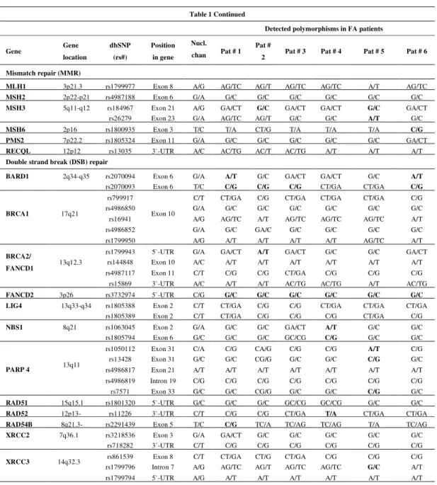
In the MMR pathway, the SNPs located in MLH1, MSH2, MSH3, MSH6, PMS2 and RECQL were evaluated. The homozygous missense variants rs184967 (p.Gln949Arg) and
rs26279 (p.Ala1045Thr) of MSH3 were found in patients #2 and #5, and patient #5, respectively.
The synonymous rs1800935 of MSH6 was detected in patient #6.
In the DSB repair pathway, which consists of both homologous recombination and non-homologous end-joining, SNPs located in BRCA1, BRCA2 (FANCD1), FANCD2, LIG4, NBS1, PARP4, RAD51, RAD52, RAD54B, XRCC2, XRCC3, XRCC4, XRCC5 and XRCC9 (FANCG)
were investigated (Table 1). Similar to the results obtained analyzing the BER, NER and MMR pathways explained above, the most of the nucleotide changes were at the heterozygous state. The
Table 1 Continued
Detected polymorphisms in FA patients
Gene Gen dbSNP Position Nucl. Pat # 1 Pat # Pat # 3 Pat # 4 Pat # 5 Pat # 6
XRCC4 5q13 rs180537 Intron 7 G/A G/C GA/C A/T G/C G/C G/C XRCC5 2q35 rs105167 3`-UTR T/C TC/AG T/A T/A T/A T/A T/A
rs2440 G/A G/C GA/C G/C GA/CT GA/CT G/C
XRCC9/FANCG 9p13 rs223785 Exon 7 C/T C/G C/G C/G C/G C/G C/G
Detected polymorphisms in FA patients
Gene Gene
location
dbSNP (rs#) Position
in gene
Nucl.
change
Pat # 1 Pat # 2 Pat # 3 Pat # 4 Pat # 5 Pat # 6
ATM 11q22-q23
rs664677 Intron 20 T/C TC/A T/A TC/A TC/A C/G TC/A
rs1800057 Exon 22 C/G C/G CG/G C/G C/G C/G C/G
rs1801516 Exon 37 G/A G/C G/C G/C G/C G/C GA/C
rs1801673 Exon 37 A/T G/C G/C G/C G/C G/C GA/C
rs609429 Intron 46 C/G CG/G C/G CG/G CG/G G/C CG/G
CCND1 11q13 rs603965 Exon 4 G/A GA/C GA/C A/T GA/C GA/C GA/C
rs678653 3`-UTR C/G G/C GC/C G/C GC/C GC/C GC/C
CCNH 5q13.3-q14 rs2266690 Exon 7 T/C TC/A T/A T/A T/A TC/A TC/A
p21/CDKN1A 6p21.2 rs1801270 Exon 2 C/A C/G CA/G C/G CA/G C/G C/G
CDKN2A 9p21 rs3731249 rs11515 3`-UTR Exon 2 G/A C/G CG/GG/C G/C C/G G/C C/G C/G G/C C/G G/C G/C C/G
rs3088440 3`-UTR C/T C/G CT/G C/G C/G C/G C/G
CDKN1B 12p13.1-p12 rs34330 Exon 1 T/C CT/G C/G CT/G CT/G T/A C/G
CDK7 5q12.1 - rs nr Exon 10 C/T C/G C/G C/G C/G C/G C/G
CHEK2 22q12.1 - rs nr Exon 13 C/del 1 C/G C/G C/G C/G C/G C/G
GRTH/Ddx25 11q24 rs551373 Intron 6 G/T G/C G/C G/C G/C G/C G/C
rs683155 Exon 10 T/C C/G CT/G CT/G T/A CT/G C/G
RAD9A 11q13.1- rs1064876 3`-UTR G/A G/C G/C G/C G/C G/C G/C
TP53 17p13.1 rs1042522 Exon 4 C/G GC/C GC/C GC/C G/C G/C GC/C
TP53BP1 15q15-q21 rs560191 Exon 9 C/G CG/G CG/G G/C C/G C/G CG/G
rs689647 Exon 11 G/A GA/C G/C G/C G/C G/C GA/C
homozygous variants were found in BARD 1, NBS1, PARP4, RAD54B, RAD52, FANCD2 and XRCC4. The missense rs2070094 (p.Val507Met) of BARD 1 was detected in patients #1 and #6.
The rs1805794 (p.Glu185Gln) of NBS1 was detected in patient #4. The rs1050112
(p.Pro1328Thr) and rs13428 (p.Gly1280Arg) of PARP4 were found in patient #5. The same
patient also carried the rs7571 (p.Ala1656Pro) located in the same gene. Of the synonymous polymorphisms investigated, the rs2070093 of BARD1 was found in patients #1, #2, #3 and #6. The rs1063045 of NBS1 was found in patient #4, while the rs2291439 of RAD54B was found in patient #1. Considering the polymorphisms located in the non-coding regions, the 5`-UTR rs1799943 of BRCA2 was found in patient #2. The 5`-UTR rs3732974 located in FANCD2 was
found in all of the patients. The 3`-UTR rs11226 located in RAD52 was found in patient #4. The
intronic rs1799796 of XRCC3 was found in patient #5, while the intronic rs1805377 of XRCC4
was found in patient #3.
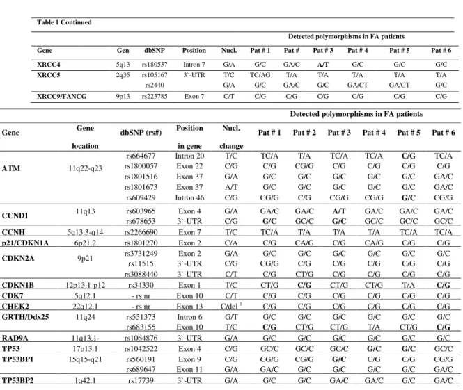
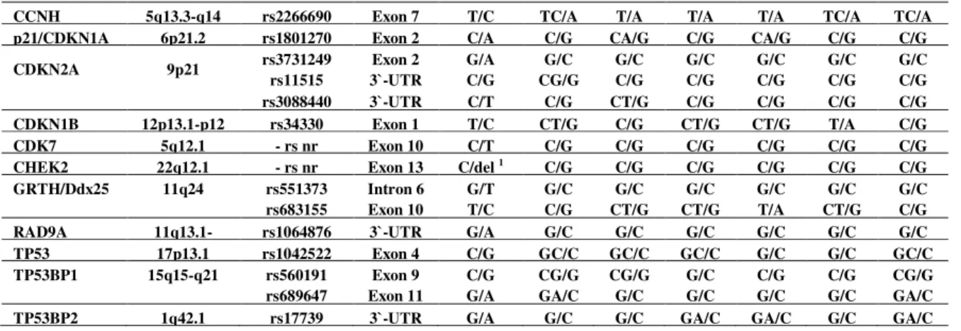
Table 2. Summary of cell cycle control genes screened for polymorphisms
Results of the analysis of SNPs located in the genes involved in cell cycle control and apoptosis are presented in Table 2. We assessed polymorphisms located in ATM, CCND1, CCNH, p21/CDKN1A, CDKN2A, CDKN1B, CDK7, CHEK2, GRTH/Ddx25, RAD9A, TP53, TP53BP1
and TP53BP2. The homozygous variants were found in ATM, CDKN1B, TP53, TP53BP1, CCND1 and GRTH/Ddx25. The missense polymorphism rs34330 (p.Leu332Pro) of CDKN1B
was detected in patients #2 and #6. The rs1042522 (p.Pro72Arg) of TP53 was found in patients
#4 and #5. The rs560191 (p.Asp353Glu) of TP53BP1 was found in patient #3. The synonymous
rs603965 located in CCND1 was detected in patient #3, while the rs683155 of GRTH/Ddx25 was
found in patients #1 and #6. In addition, the intronic variant rs664677 of ATM was identified in
patient #5, while the 3`-UTR rs678653 located in CCND1 was found in patients #1 and #3.
Considering the missense nucleotide substitutions in all of the studied genes, we have observed that among the 52 analyzed SNPs, the 29 (~56%) were identified in our patients as heterozygous variants, while the 12 of them (~23%) were detected as homozygous variants. Of the 10 synonymous SNPs, the six (60%) were found in our patients at the homozygous state. In
CCNH 5q13.3-q14 rs2266690 Exon 7 T/C TC/A T/A T/A T/A TC/A TC/A
p21/CDKN1A 6p21.2 rs1801270 Exon 2 C/A C/G CA/G C/G CA/G C/G C/G
CDKN2A 9p21 rs3731249 Exon 2 G/A G/C G/C G/C G/C G/C G/C
rs11515 3`-UTR C/G CG/G C/G C/G C/G C/G C/G
rs3088440 3`-UTR C/T C/G CT/G C/G C/G C/G C/G
CDKN1B 12p13.1-p12 rs34330 Exon 1 T/C CT/G C/G CT/G CT/G T/A C/G
CDK7 5q12.1 - rs nr Exon 10 C/T C/G C/G C/G C/G C/G C/G
CHEK2 22q12.1 - rs nr Exon 13 C/del 1 C/G C/G C/G C/G C/G C/G
GRTH/Ddx25 11q24 rs551373 Intron 6 G/T G/C G/C G/C G/C G/C G/C
rs683155 Exon 10 T/C C/G CT/G CT/G T/A CT/G C/G
RAD9A 11q13.1- rs1064876 3`-UTR G/A G/C G/C G/C G/C G/C G/C
TP53 17p13.1 rs1042522 Exon 4 C/G GC/C GC/C GC/C G/C G/C GC/C
TP53BP1 15q15-q21 rs560191 Exon 9 C/G CG/G CG/G G/C C/G C/G CG/G
rs689647 Exon 11 G/A GA/C G/C G/C G/C G/C GA/C
view of the 33 SNPs located in the non-coding regions of selected genes, the thirteen (~40%) of them were identified in some of our patients as homozygous variants.
Summarizing the results presented above it could be seen that SNPs identified at the homozygous state were not equally distributed among the patients. Patient #1 carried one missense, three synonymous and five polymorphisms in the non-coding region of the genes. Patient #2 carried three missense, one synonymous and five substitutions in the non-coding regions. Patient #3 carried two missense, two synonymous and five substitutions in the non-coding regions of the genes. Patient #4 carried two missense, one synonymous and four polymorphisms in the non-coding regions. Patient #5 carried seven missense variations, seven variations in the non-coding regions of the genes and no one homozygous synonymous substitution. Finally, patient #6 carried two missense variations, three synonymous and three variations in the non-coding regions of selected genes.
Taken together, the results of our investigation have shown a significant inter-individual variability among patients in the type and the frequency of SNPs.
Extensive analysis of the genotype–phenotype correlations in this cohort was not possible because of the small number of patients.
DISCUSSION
This study was aimed at defining the factors that might influence a DNA repair capacity in FA patients, which may allow for a more rational therapeutic approach. Accumulating evidence suggests that reduced repair capacity is a polymorphic phenotypictrait associated with an increased risk of developing tumors at several sites, including breast, lung, skin, liver, or head/neck (BERWICK and VINEIS, 2000; MOHRENWEISER et al., 2002). Therefore, the data of the
factors of the integrity of DNA repair pathways may be used not only in the development of novel therapeutic agents but also for the understanding of the high degree of phenotypic and clinical heterogeneity in Fanconi anemia which is in evident contrast with the uniform cellular phenotype (HODSON and WALDEN, 2013).
In this study, we have genotyped the known polymorphisms located in the DNA repair and cell cycle control genes in FA patients and observed that most of the polymorphisms are present at the heterozygous state. Considering the polymorphisms with functional consequences i.e., missense polymorphisms, it is possible that heterozygous nucleotide changes in the genes of interest could also have various biological and/or pathophysiological effects on FA patients. Recent report of Alsbeih and coworkers (ALSBEIH et al., 2013) has shown that radiation toxicity in
radiotherapy patients is associated with polymorphisms rs1801516 of ATM and rs25487 of XRCC1. Results of that study suggest that the presence of protective alleles (ATM rs1801516 A
and XRCC1 rs25487 A) at the heterozygous status would increase patients’ follow-up after
radiotherapy by 51 months, while homozygous status would raise this index by 77 months. In our cohort, all of the FA-D2 patients were homozygote carriers of the ATM G allele. These results are
in concordance with our previous investigations reporting the marked radiosensitivity of FA patients (LESKOVAC et al., 2010; JOKSIC et al., 2012; LESKOVAC et al., 2014) and support the use
of SNPs in candidate genes as predictive markers of patients’ radiosensitivity.
In the BER pathway, the homozygous missense nucleotide changes (rs3136820) were found in the APEX. This endonuclease recognizes and begins the process of removing abasic sites
ionizing radiation and an increased risk of colorectal cancer (HU et al., 2001; KASAHARA et al.,
2008). Our previous investigations have shown that exposure to ionizing radiation in vitro
significantly slowed down the proliferation of FANCD2 fibroblasts compared to control fibroblasts (JOKSIC et al., 2012; LESKOVAC et al., 2014).
In the NER pathway, we detected the homozygous missense polymorphism rs13181 (p.Lys751Gln) of ERCC2 which is associated with a risk of acute myeloid leukemia, melanoma, chronic lymphocytic leukemia and squamous head and neck cancer (CHANG-CLAUDE et al., 2009; GANSTER et al., 2009; MITRA et al., 2009; STROM et al., 2010). Of the two homozygous missense
polymorphisms located in MSH3 (MMR pathway), the rs26279 is associated with increased sensitivity to platinum-based chemotherapy in advanced non-small cell lung cancer patients (XU
et al., 2015).
In the DSB repair pathway, the homozygous missense variations were found in BARD 1, NBS1, and PARP4. The rs1805794 (p.Glu185Gln) of NBS1 is associated with increased risk for
urinary system cancer, especially for bladder cancer (ZHANG et al., 2014), while the functional
consequences of the rs1050112 (p.Pro1328Thr), rs13428 (p.Gly1280Arg) and rs7571 of PARP4
as well as of the rs2070094 (p.Val507Met) of BARD1 are still unknown. The significance of these
findings is not known yet but may be important for future testing and improvement of the diagnosis and outcomes in patients with FA.
Considering the genes involved in the cell cycle control and apoptosis, the homozygous missense variations were found in CDKN1B, TP53 and TP53BP1. The p53 protein is essential for regulating different cellular processes such as DNA repair, cell cycle arrest and apoptosis and therefore, has a crucial role in maintaining the genetic integrity of the cells (RILEY et al., 2008). The functional SNPs located in TP53 are confirmed to affect the p53 signaling pathway
(GROCHOLA et al., 2010). The rs1042522 (p.Pro72Arg) of TP53 at homozygous state was found in
two of our patients, while the rest of them were heterozygotes. It has been shown that 72Arg variant has a stronger ability to induce apoptosis then 72Pro variant. In other words, 72Arg is more efficiently translocated to the mitochondria, where it interacts with proapoptotic proteins such as GRP75 and Hsp60, consequently triggering apoptosis (DUMONT et al., 2003). These
results are in accordance with our previous investigation of FA patients describing the elevated level of leukocyte apoptosis either occurring spontaneously or induced by ionizing radiation (PETROVIC et al., 2013). Additionally, the report of Litviakov and coworkers (LITVIAKOV et al., 2010) has shown that 72Arg variant is coupled with higher frequency of aberrant cells and chromatid breaks in comparison to individuals with Pro/Pro genotypes. This polymorphism is also associated with high risk of diabetes II and acute lymphoblast leukemia (GAULTON et al.,
2008; DO et al., 2009). As for the other two found polymorphisms, the rs34330of CDKN1B is
reported to have an association with breast cancer (DRIVER et al., 2008), while the rs560191
(p.Asp353Glu) of TP53BP1 is associated with a risk of lung cancer (TRUONG et al., 2010).
As previously stated, the homozygous synonymous nucleotide changes were found in BARD1, MSH6, NBS1, RAD54B,CCND1 and GRTH/Ddx25. The literature data provide the evidence that
synonymous mutations are “silent” with respect to protein sequence but are not always “silent” regarding protein function (GINGOLD and PILPEL, 2011). Synonymous SNPs could influence protein function affecting mRNA secondary structure and stability consequently reducing protein expression (NACKLEY et al., 2006). In other words, “silent” changes may possibly affect the
(PARMLEY et al., 2006), or, through translational pausing, a protein folding (ZHOU et al., 2013).
Silent mutations are proven to contribute to human cancer (SUPEK et al., 2014).
In this study, the thirteen homozygous variants were found in the non-coding regions of the genes tested. Several studies provided evidence that SNPs located in the non-coding DNA, especially in intronic gene regions near the exon/intron boundaries, could inactivate pre-mRNA splice sites consequently affecting gene expression (LOMELIN et al., 2010; VOGELSTEIN et al., 2013), or could activate cryptic splice sites leading to exonization (WANG and COOPER, 2007). Furthermore, the presence of SNPs in the 3`-UTR of selected genes could modify the binding with specific microRNAs (miRNAs) (NACCARATI et al., 2012). It has been reported that one of
the key mechanisms of gene expression control is a post-transcriptional gene regulation by miRNAs which interact with the 3`-UTR of target mRNAs inducing their degradation or inhibition of their translation, thus silencing gene expression (BARTEK and LUKAS, 2007; FILIPOWICZ et al., 2008). Therefore, the SNPs could alter the miRNA–mRNA interaction
consequently inducing the increase or decrease of protein translation (DUAN et al., 2009). In
addition, the sequence changes in the 5´ -UTR regions of mRNAs can influence translation regulation (WARD and KELLIS, 2012).
There are many association studies providing the linkage between SNPs in non-coding regions of candidate genes and cancer propensity. As for the homozygous polymorphisms detected in our patients, the intronic rs3730849 of LIG1, the 3`-UTR rs1052536 of LIG3 and the intronic rs3212948 of ERCC1 are associated with a risk of lung cancer (LANDI et al., 2006; MA et al., 2007). Similarly, the 5`-UTR variant rs1800975 of XPA is associated with lung cancer risk and also with squamous cell carcinoma (Lou et al, 2014). In GADD45A, an intronic rs532446 was
reported to possess a functional role in acute lung injury (MITRA et al., 2014). The 5`-UTR
rs1799943 of BRCA2 is associated with breast cancer susceptibility (SAPKOTA et al., 2013). The
3`-UTR rs11226 located in RAD52 is reported to has a significant association with
myelodysplastic syndrome (BELICKOVA et al., 2013). The intronic rs1799796 of XRCC3 is
associated with ovarian cancer risk (YUAN et al., 2014), while the intronic rs1805377 of XRCC4 is
associated with an increased risk of glioma development (ZHAO et al., 2013). In addition, the
intronic variant rs664677 of ATM may play an important role in the development of thyroid
cancer (SONG et al., 2015) while the 3`-UTR rs678653 located in CCND1 is associated with oral
cancer (TSAI et al., 2011). Regarding the intronic rs1805403 of PARP1 as well asthe 5`-UTR rs3732974 located in FANCD2, the clinical significance of these substitutions has not yet been elucidated.
Investigation of the relatively large number of SNPs, as in the current report, probably diminishes the significance of findings; therefore, many of the SNPs assessed (e.g. polymorphisms located in ATM, APEX APE 1, XRCC1, ERCC2, MSH3, PARP4, NBS1, BARD1, CDKN1B, TP53, TP53BP1) should be considered as promising candidates for further
investigation in larger cohort. In addition, since Fanconi anemia is a genetically heterogeneous disease, the future investigation should also encompass the SNPs located in other genes that are proven to contribute to FA condition i.e., genes involved in bioenergetic pathways, antioxidant activities, response to stress and metal-chelating proteins, inflammation-related cytokines and also in some selected DNA repair genes such as type II DNA topoisomerase (PAGANO et al., 2013).
ACKNOWLEDGEMENTS
The authors wish to thank Prof. J. Surralles, Prof. H. Hoehn, and Prof. D. Schindler for FA complementation group analysis. This research was supported by the Ministry of Education, Science and Technological Development of the Republic of Serbia (Grant No. 173046).
Received March23th, 2015 Accepted June 29th, 2015
REFERENCES
ALSBEIH, G., M. EL-SEBAIE, N. AL-HARBI, K. AL-HADYAN, M. SHOUKRI and N. AL-RAJHI (2013): SNPs in genes implicated in radiation response are associated with radiotoxicity and evoke roles as predictive and prognostic biomarkers. Radiat Oncol 8: 125.
AMEZIANE, N., D. SIE, S. DENTRO, Y. ARIYUREK, L. KERKHOVEN, H. JOENJE, J. C. DORSMAN, et al. (2012): Diagnosis of fanconi anemia: mutation analysis by next-generation sequencing. Anemia 2012: 132856.
AUERBACH, A. D.(1993): Fanconi anemia diagnosis and the diepoxybutane (DEB) test. Exp. Hematol. 21: 731-733. BARTEK, J. and J. LUKAS (2007): DNA damage checkpoints: from initiation to recovery or adaptation. Curr. Opin. Cell
Biol. 19: 238-245.
BELICKOVA, M., M. D. MERKEROVA, E. STARA, J. VESELA, D. SPONEROVA, D. MIKULENKOVA, R. BRDICKA, et al. (2013): DNA repair gene variants are associated with an increased risk of myelodysplastic syndromes in a Czech population. J Hematol Oncol 6: 9.
BERWICK, M. and P. VINEIS (2000): Markers of DNA repair and susceptibility to cancer in humans: an epidemiologic review. J. Natl. Cancer Inst. 92: 874-897.
BOGLIOLO, M., B. SCHUSTER, C. STOEPKER, B. DERKUNT, Y. SU, A. RAAMS, J. P. TRUJILLO, et al. (2013): Mutations in ERCC4, encoding the DNA-repair endonuclease XPF, cause Fanconi anemia. Am. J. Hum. Genet. 92: 800-806.
BOND, G.L. and A. J. LEVINE (2007): A single nucleotide polymorphism in the p53 pathway interacts with gender, environmental stresses and tumor genetics to influence cancer in humans. Oncogene 26: 1317-1323.
CAPPELLI, E., S. RAVERA, D. VACCARO, P. CUCCAROLO, M. BARTOLUCCI, I. PANFOLI, C. DUFOUR, et al. (2013):
Mitochondrial respiratory complex I defects in Fanconi anemia. Trends Mol Med 19: 513-514.
CHANG-CLAUDE, J., C. B. AMBROSONE, C. LILLA, S. KROPP, I. HELMBOLD, D. VON FOURNIER, W. HAASE,et al. (2009): Genetic polymorphisms in DNA repair and damage response genes and late normal tissue complications of radiotherapy for breast cancer. Br. J. Cancer 100: 1680-1686.
CHEN, H., S. ZHANG and Z. WU (2014): Fanconi anemia pathway defects in inherited and sporadic cancers. Translational Pediatrics 3: 300-304.
DO, T. N., E. UCISIK-AKKAYA, C. F. DAVIS, B. A. MORRISON and M. T. DORAK (2009): TP53 R72P and MDM2 SNP309 polymorphisms in modification of childhood acute lymphoblastic leukemia susceptibility. Cancer Genet. Cytogenet. 195: 31-36.
DRIVER, K. E., H. SONG, F. LESUEUR, S. AHMED, N. L. BARBOSA-MORAIS, J. P. TYRER, B. A. PONDER,et al. (2008): Association of single-nucleotide polymorphisms in the cell cycle genes with breast cancer in the British population. Carcinogenesis 29: 333-341.
DRUMMOND, D. A. and C. O. WILKE (2008): Mistranslation-induced protein misfolding as a dominant constraint on coding-sequence evolution. Cell 134: 341-352.
DUAN, S., S. MI, W. ZHANG and M. E. DOLAN (2009): Comprehensive analysis of the impact of SNPs and CNVs on human microRNAs and their regulatory genes. RNA Biol 6: 412-425.
FILIPOWICZ, W., S. N. BHATTACHARYYA and N. SONENBERG (2008): Mechanisms of post-transcriptional regulation by microRNAs: are the answers in sight? Nat Rev Genet 9: 102-114.
GANSTER, C., J. NEESEN, S. ZEHETMAYER, U. JAGER, H. ESTERBAUER, C. MANNHALTER, B. KLUGE, et al. (2009): DNA repair polymorphisms associated with cytogenetic subgroups in B-cell chronic lymphocytic leukemia. Genes Chromosomes Cancer 48: 760-767.
GAULTON, K. J., C. J. WILLER, Y. LI, L. J. SCOTT, K. N. CONNEELY, A. U. JACKSON, W. L. DUREN, et al. (2008): Comprehensive association study of type 2 diabetes and related quantitative traits with 222 candidate genes. Diabetes 57: 3136-3144.
GINGOLD, H.and Y. PILPEL (2011): Determinants of translation efficiency and accuracy. Mol Syst Biol 7: 481.
GOODMAN, D. B., G. M. CHURCH and S. KOSURI (2013): Causes and effects of N-terminal codon bias in bacterial genes. Science 342: 475-479.
GROCHOLA, L. F., J. ZERON-MEDINA, S. MERIAUX and G. L. BOND (2010): Single-nucleotide polymorphisms in the p53 signaling pathway. Cold Spring Harb Perspect Biol 2: a001032.
HODSON, C. and H. WALDEN (2013): Towards a molecular understanding of the fanconi anemia core complex. Anemia
2012: 926787.
HU, J. J., T. R. SMITH, M. S. MILLER, H. W. MOHRENWEISER, A. GOLDEN and L. D. CASE (2001): Amino acid substitution variants of APE1 and XRCC1 genes associated with ionizing radiation sensitivity. Carcinogenesis 22: 917-922.
JOENJE, H. and K. J. PATEL (2001): The emerging genetic and molecular basis of Fanconi anaemia. Nat Rev Genet 2: 446-457.
JOKSIC, I., D. VUJIC, M. GUC-SCEKIC, A. LESKOVAC, S. PETROVIC, M. OJANI, J. P. TRUJILLO, et al. (2012): Dysfunctional telomeres in primary cells from Fanconi anemia FANCD2 patients. Genome Integrity 3.
KASAHARA, M., K. OSAWA, K. YOSHIDA, A. MIYAISHI, Y. OSAWA, N. INOUE, A. TSUTOU, et al. (2008): Association of MUTYH Gln324His and APEX1 Asp148Glu with colorectal cancer and smoking in a Japanese population. J. Exp. Clin. Cancer Res. 27: 49.
KEE, Y.and A. D. D'ANDREA (2012): Molecular pathogenesis and clinical management of Fanconi anemia. J. Clin. Invest.
122: 3799-3806.
KIM, H. and A. D. D'ANDREA (2012): Regulation of DNA cross-link repair by the Fanconi anemia/BRCA pathway. Genes Dev. 26: 1393-1408.
KUTLER, D. I., A. D. AUERBACH, J. SATAGOPAN, P. F. GIAMPIETROO, S. D. BATISH, A. G. HUVOS, A. GOBERDHAN,et al.
(2003): High incidence of head and neck squamous cell carcinoma in patients with Fanconi anemia. Arch. Otolaryngol. Head Neck Surg. 129: 106-112.
LANDI, S., F. GEMIGNANI, F. CANZIAN, V. GABORIEAU, R. BARALE, D. LANDI, N. SZESZENIA-DABROWSKA, et al. (2006): DNA repair and cell cycle control genes and the risk of young-onset lung cancer. Cancer Res. 66: 11062-11069.
LESKOVAC, A., S. PETROVIC, M. GUC-SCEKIC, D. VUJIC and G. JOKSIC (2014): Radiation-induced mitotic catastrophe in FANCD2 primary fibroblasts. Int. J. Radiat. Biol. 90: 373-381.
LESKOVAC, A., D. VUJIC, M. GUC-SCEKIC, S. PETROVIC, I. JOKSIC, P. SLIJEPCEVIC and G. JOKSIC (2010): Fanconi anemia is characterized by delayed repair kinetics of DNA double-strand breaks. Tohoku J. Exp. Med. 221: 69-76. LITVIAKOV, N. V., E. V. DENISOV, R. M. TAKHAUOV, A. B. KARPOV, E. V. SKOBEL'SKAJA, E. O. VASIL'EVA, O. O. GONCHARIK,
et al. (2010): Association between TP53 gene ARG72PRO polymorphism and chromosome aberrations in
human cancers. Mol. Carcinog. 49: 521-524.
MA, H., L. XU, J. YUAN, M. SHAO, Z. HU, F. WANG, Y. WANG,et al. (2007): Tagging single nucleotide polymorphisms in
excision repair cross-complementing group 1 (ERCC1) and risk of primary lung cancer in a Chinese population. Pharmacogenet Genomics 17: 417-423.
MAGDALENA, N., D. V. PILONETTO, M. A. BITENCOURT, N. F. PEREIRA, R. C. RIBEIRO, M. JENG and R. PASQUINI (2005): Frequency of Fanconi anemia in Brazil and efficacy of screening for the FANCA 3788-3790del mutation. Braz. J. Med. Biol. Res. 38: 669-673.
MATHEW, C. G. (2006): Fanconi anaemia genes and susceptibility to cancer. Oncogene 25: 5875-5884.
MITRA, A. K., N. SINGH, V. K. GARG, R. CHATURVEDI, M. SHARMA and S. K. RATH (2009): Statistically significant association of the single nucleotide polymorphism (SNP) rs13181 (ERCC2) with predisposition to Squamous Cell Carcinomas of the Head and Neck (SCCHN) and Breast cancer in the north Indian population. J. Exp. Clin. Cancer Res. 28: 104.
MITRA, S., M. S. WADE, X. SUN, N. MOLDOBAEVA, C. FLORES, S. F. MA, W. ZHANG, et al. (2014): GADD45a promoter regulation by a functional genetic variant associated with acute lung injury. PLoS One 9: e100169.
MOHRENWEISER, H. W., T. XI, J. VAZQUEZ-MATIAS and I. M. JONES (2002): Identification of 127 amino acid substitution variants in screening 37 DNA repair genes in humans. Cancer Epidemiol. Biomarkers Prev. 11: 1054-1064. NACCARATI, A., B. PARDINI, L. STEFANO, D. LANDI, J. SLYSKOVA, J. NOVOTNY, M. LEVY, et al. (2012): Polymorphisms in
miRNA-binding sites of nucleotide excision repair genes and colorectal cancer risk. Carcinogenesis 33: 1346-1351.
NACKLEY, A. G., S. A. SHABALINA, I. E. TCHIVILEVA, K. SATTERFIELD, O. KORCHYNSKYI, S. S. MAKAROV, W. MAIXNER, et al.
(2006): Human catechol-O-methyltransferase haplotypes modulate protein expression by altering mRNA secondary structure. Science 314: 1930-1933.
PAGANO, G., A. A. TALAMANCA, G. CASTELLO, M. D'ISCHIA, F. V. PALLARDO, S. PETROVIC, B. PORTO, et al. (2013): Bone marrow cell transcripts from Fanconi anaemia patients reveal in vivo alterations in mitochondrial, redox and DNA repair pathways. Eur. J. Haematol. 91: 141-151.
PARMLEY, J. L., J. V. CHAMARY and L. D. HURST (2006): Evidence for purifying selection against synonymous mutations in mammalian exonic splicing enhancers. Mol. Biol. Evol. 23: 301-309.
PETROVIC, S., A. LESKOVAC, I. JOKSIC, J. FILIPOVIC, D. VUJIC, M. GUC-SCEKIC and G. JOKSIC (2013): Leukocyte apoptosis as a predictor of radiosensitivity in fanconi anemia. Curr. Sci. 105: 56-60.
RILEY, T., E. SONTAG, P. CHEN and A. LEVINE (2008): Transcriptional control of human p53-regulated genes. Nat Rev Mol Cell Biol 9: 402-412.
SAPKOTA, Y., J. R. MACKEY, R. LAI, C. FRANCO-VILLALOBOS, S. LUPICHUK, P. J. ROBSON, K. KOPCIUK, et al. (2013): Assessing SNP-SNP Interactions among DNA Repair, Modification and Metabolism Related Pathway Genes in Breast Cancer Susceptibility. PLoS One 8: e64896.
SIGURDSON, A. J., M. HAUPTMANN, N. CHATTERJEE, B. H. ALEXANDER, M. M. DOODY, J. L. RUTTER and J. P. STRUEWING
(2004): Kin-cohort estimates for familial breast cancer risk in relation to variants in DNA base excision repair, BRCA1 interacting and growth factor genes. BMC Cancer 4: 9.
SONG, C. M., T. K. KWON, B. L. PARK, Y. B. JI and K. TAE (2015): Single nucleotide polymorphisms of ataxia telangiectasia mutated and the risk of papillary thyroid carcinoma. Environ. Mol. Mutag. 56: 70-76.
STROM, S. S., E. ESTEY, U. M. OUTSCHOORN and G. GARCIA-MANERO (2010): Acute myeloid leukemia outcome: role of nucleotide excision repair polymorphisms in intermediate risk patients. Leuk. Lymphoma 51: 598-605. SUPEK, F., B. MINANA, J. VALCARCEL, T. GABALDON and B. LEHNER (2014): Synonymous mutations frequently act as driver
mutations in human cancers. Cell 156: 1324-1335.
TSAI, M. H., C. W. TSAI, Y. A. TSOU, C. H. HUA, C. F. HSU and D. T. BAU (2011): Significant association of cyclin D1 single nucleotide polymorphisms with oral cancer in taiwan. Anticancer Res. 31: 227-231.
VOGELSTEIN, B., N. PAPADOPOULOS, V. E. VELCULESCU, S. ZHOU, L. A. DIAZ, JR., K. W. KINZLER (2013): Cancer genome landscapes. Science 339: 1546-1558.
WANG, G. S. and T. A. COOPER (2007): Splicing in disease: disruption of the splicing code and the decoding machinery. Nat Rev Genet 8: 749-761.
WARD, L. D. and M. KELLIS (2012): Interpreting noncoding genetic variation in complex traits and human disease. Nat. Biotechnol. 30: 1095-1106.
WILSON, D. M. and L. H. THOMPSON (1997): Life without DNA repair. Proc. Natl. Acad. Sci. U. S. A. 94: 12754-12757.
XU, X. L., Y. L. YAO, W. Z. XU, J. G. FENG and W. M. MAO (2015): Correlation of MSH3 polymorphisms with response and survival in advanced non-small cell lung cancer patients treated with first-line platinum-based chemotherapy. Genet Mol Res 14: 3525-3533.
YUAN, C., X. LIU, S. YAN, C. WANG and B. KONG (2014): Analyzing association of the XRCC3 gene polymorphism with ovarian cancer risk. Biomed Res Int 2014: 648137.
ZHANG, Y., Y. S. HUANG, W. Q. LIN, S. D. ZHANG, Q. W. LI, Y. Z. HU, R. L. ZHENG, et al. (2014): NBS1 Glu185Gln
polymorphism and susceptibility to urinary system cancer: a meta-analysis. Tumour Biol. 35: 10723-10729. ZHAO, P., P. ZOU, L. ZHAO, W. YAN, C. KANG, T. JIANG and Y. YOU (2013): Genetic polymorphisms of DNA double-strand
break repair pathway genes and glioma susceptibility. BMC Cancer 13: 234.
ISPITIVANJE POJEDINA NIH NUKLEOTIDNIH POLIMORFIZAMA U 52 GENA UKLJU ENA U DNK REPER I KONTROLU ELIJSKOG CIKLUSA KOD
PACIJENATA OBOLELIH OD FANKONIJEVE ANEMIJE
Sandra PETROVI 1, Andreja LESKOVAC1, Ivana JOKSI 1,2, Dragana VUJI 3, 4,
Ana Valenta ŠOBOT1, Jelena FILIPOVI 1 and Gordana JOKSI 1
1 Institut za nuklearne nauke Vin a, Univerzitet u Beogradu, Beograd, Srbija 2Ginekološko akušerska klinika “Narodni front”, Beograd, Srbija
3Medicinski fakultet, Univerzitet u Beogradu, Beograd, Srbija
4Institut za zdravstvenu zaštitu majke i deteta Srbije "Dr. Vukan upi ", Beograd, Srbija
Izvod
Fankonijeva anemija (FA) je geneti ki heterogeno oboljenje koje se manifestuje progresivnom pancitopenijom, razvojnim abnormalnostima i predispozicijom ka razvoju kancera. Nezavisno od mutacija u FANC genima koje su direktni uzro nici bolesti, identifikacija varijacija u DNK, naro ito pojedina nih nukleotidnih polimorfizama u odabranim genima, može omogu iti individualizaciju pristupa svakom pacijentu i poboljšati prognozu bolesti. U ovom istraživanju, ispitali smo 95 pojedina nih nukleotidnih izmena lociranih u 52 klju na gena uklju ena u bazni ekscizioni reper, nukleotidni ekscizioni reper,“ mismatch“ reper, reper dvolan anih prekida DNK, kao i u nekoliko gena odgovornih za kontrolu elijskog ciklusa i apoptoze. Istraživanjem su obuhva ena pet FA-D2 pacijenta i jedan FA-A pacijent, a genotipizacija polimorfizama je izvršena pomo u DNK reper ipa (Asper Biotech). Prou avani su sinonimni (n=10) i nesinonimni (“missense“) polimorfizmi (n=52), kao i polimorfizmi u nekodiraju im regionima genoma (introni i 5‘ i 3’ netranslatorni regioni) (n=33). Rezultati našeg istraživanja pokazuju da se pacijenti me usobno razlikuju po tipu i po broju nukleotidnih polimorfizama i istovremeno ukazuju na potrebu daljeg ispitivanja polimorfizama lociranih u genima ATM, APEX APE 1, XRCC1, ERCC2, MSH3, PARP4, NBS1, BARD1, CDKN1B, TP53 and TP53BP,1 na ve em broju
pacijenata, što bi omogu ilo bolji uvid u klini ku sliku bolesti. Pored toga, pokazano je da
pojedina ni nukleotidni polimorfizmi mogu imati zna ajnu ulogu u predikciji kancera kod FA pacijenata, kao i u proceni efikasnosti i toksi nosti razli itih vidova terapije.
Primljeno 23.III 2015.