This article was downloaded by: [ULPGC. Biblioteca] On: 13 April 2015, At: 13:35
Publisher: Taylor & Francis
Informa Ltd Registered in England and Wales Registered Number: 1072954 Registered office: Mortimer House, 37-41 Mortimer Street, London W1T 3JH, UK
Click for updates
Journal of Aquatic Food Product Technology
Publication details, including instructions for authors and subscription information: http://www.tandfonline.com/loi/wafp20
Microbial Growth Models in Gilthead Sea Bream (Sparus
Aurata) Stored in Ice
Conrado Carrascosaa, Pedro Saavedrab, Rafael Millána, José Raduán Jaberc, Tania Montenegroa, António Raposod & Esther Sanjuána
a
Department of Animal Pathology, Animal Production, Bromatology and Food Technology, Faculty of Veterinary, Universidad de Las Palmas de Gran Canaria, 35413 Arucas, Las Palmas, Spain.
b
Department of Mathematics, Mathematics Building, Campus Universitario de Tafira, 35018 Las Palmas de Gran Canaria, Spain.
c
Department of Morphology, Faculty of Veterinary, Universidad de Las Palmas de Gran, Canaria, 35413 Arucas, Las Palmas, Spain.
d
Centro de Investigação Interdisciplinar Egas Moniz, CiiEM. Instituto Superior de Ciências da Saúde Egas Moniz, ISCSEM. Quinta da Granja. Monte de Caparica. 2829-511 Caparica. Portugal.
Accepted author version posted online: 27 Jan 2015.
To cite this article: Conrado Carrascosa, Pedro Saavedra, Rafael Millán, José Raduán Jaber, Tania Montenegro, António
Raposo & Esther Sanjuán (2015): Microbial Growth Models in Gilthead Sea Bream (Sparus Aurata) Stored in Ice, Journal of Aquatic Food Product Technology, DOI: 10.1080/10498850.2013.848964
To link to this article: http://dx.doi.org/10.1080/10498850.2013.848964
Disclaimer: This is a version of an unedited manuscript that has been accepted for publication. As a service
to authors and researchers we are providing this version of the accepted manuscript (AM). Copyediting, typesetting, and review of the resulting proof will be undertaken on this manuscript before final publication of the Version of Record (VoR). During production and pre-press, errors may be discovered which could affect the content, and all legal disclaimers that apply to the journal relate to this version also.
PLEASE SCROLL DOWN FOR ARTICLE
Taylor & Francis makes every effort to ensure the accuracy of all the information (the “Content”) contained in the publications on our platform. However, Taylor & Francis, our agents, and our licensors make no
representations or warranties whatsoever as to the accuracy, completeness, or suitability for any purpose of the Content. Any opinions and views expressed in this publication are the opinions and views of the authors, and are not the views of or endorsed by Taylor & Francis. The accuracy of the Content should not be relied upon and should be independently verified with primary sources of information. Taylor and Francis shall not be liable for any losses, actions, claims, proceedings, demands, costs, expenses, damages, and other liabilities whatsoever
form to anyone is expressly forbidden. Terms & Conditions of access and use can be found at http:// www.tandfonline.com/page/terms-and-conditions
Accepted Manuscript
Running title: Microbiological growth in sea bream
MICROBIAL GROWTH MODELS IN GILTHEAD
SEA BREAM (SPARUS AURATA) STORED IN ICE
Authors: Conrado Carrascosaa*, Pedro Saavedrab, Rafael Millána, José Raduán Jaberc, Tania Montenegroa, António Raposod and Esther Sanjuána
a Department of Animal Pathology, Animal Production, Bromatology and Food Technology, Faculty of Veterinary, Universidad de Las Palmas de Gran Canaria, 35413 Arucas, Las Palmas, Spain.
b Department of Mathematics, Mathematics Building, Campus Universitario de Tafira, 35018 Las Palmas de Gran Canaria, Spain.
c Department of Morphology, Faculty of Veterinary, Universidad de Las Palmas de Gran
Canaria, 35413 Arucas, Las Palmas, Spain.
d Centro de Investigação Interdisciplinar Egas Moniz, CiiEM. Instituto Superior de Ciências da Saúde Egas Moniz, ISCSEM. Quinta da Granja. Monte de Caparica. 2829-511 Caparica. Portugal.
* Corresponding author: Conrado Carrascosa E-mail address: [email protected] Phone: (+34) 928457434
Fax: (+34) 928451142
Accepted Manuscript
Abstract
This study analyzes microbiological changes in whole, ungutted farmed gilthead sea bream (Sparus aurata) stored for an 18-day period in ice using traditional methods for mesophilic aerobic bacteria, psychrotrophic, Pseudomonas spp., Aeromonas spp., Shewanella putrefaciens, Enterobacteriaceae, sulphide-reducing Clostridium (Clostridia), and Photobacterium
phosphoreum in muscle, skin, and gills, evaluating their seasonal differentiation. Two different
statistical models were used to analyze microbiological growth. Simultaneously, physicochemical parameters such as the temperature, pH, biological oxygen demand (BOD5), total dissolved solids, salinity, ammonia nitrogen, and total phosphorus content of growing waters were analyzed. The results showed that by the end of the storage time, specific spoilage bacteria (SSB) such as Pseudomonas spp., Aeromonas spp. and S. putrefaciens as H2S-producing bacteria were dominant in sea bream harvested in temperate water in the Canary Islands. Muscle tissue had least contamination, followed by skin and gills. The values of the analyzed seawater parameters were constant during the four seasons, except that the temperature showed a small difference between winter and summer. Seasonal effects were observed among the fish analyzed, suggesting that the lower levels of contamination detected in winter may have been due to the slight difference observed in water temperature in that season.
Keywords: gilthead sea bream (Sparus aurata); ice storage; microbiological evolution; tissues;
seasons and statistical models; seawater.
Accepted Manuscript
Introduction
The determination of microbial growth and, thereby, fish shelf life with traditional microbiological tests is expensive and time-consuming (Bruckner et al., 2013). An alternative is the concept of predictive microbiology, which uses mathematical models to predict microbiological growth and, thus, to estimate the shelf life (McMeekin et al., 1993; Whiting, 1995). Predictive microbiology involves the development of mathematical models to describe the effect of the most important environmental factors controlling the responses of microorganisms in foods (Ross et al., 2000). The majority of these models are based on experimental data derived from laboratory media, and limited information is available for assessing individual cell variability in foods (François et al., 2006; Manios et al., 2011). The models have immediate practical application to improve microbial food safety and quality and also can provide quantitative understanding of the microbial ecology of foods (Ross et al., 2000).
In Spain, the farming of gilthead sea bream (Sparus aurata) has increased, with overall output rising from 127 tonnes in 1985 to 19430 tonnes in 2012. Although production has fallen by 15.9% over the previous year (2011), the Canary Islands are the third most important region in Spain for the production of gilthead sea bream (2740 tonnes in 2012), (APROMAR, 2013), mainly due to the ideal conditions of the ocean, namely temperature, salinity, nutrients, currents, and the morphology and nature of the seabeds (Pérez-Sánchez and Moreno-Batet, 1991). Water surface temperature in these islands ranges from 18ºC in winter to 22-23ºC in summer, an aspect that is important to take into consideration when studying the microbiology of farmed fish.
Accepted Manuscript
Nevertheless, no studies have evaluated microbiology during the handling, distribution, and storage of this species harvested in the Canary Islands.
The growing production of this species of fish has increased the importance of maintaining their good quality during storage. Fish quality declines due to a complex process involving physical, chemical, and microbiological forms of deterioration. Enzymatic and chemical reactions are normally responsible for the initial loss of freshness, whereas microbial activity accounts for the obvious spoilage and thereby establishes product shelf-life (Guillén-Velasco et al., 2004; De Koning, 2004). Many factors can influence the rate of the microbial spoilage of fish, such as the bacterial flora present, storage conditions, handling, and temperature (Ward and Baj, 1988). Some bacterial groups are particularly associated with this spoilage, such as Shewanella
putrefaciens and Pseudomonas spp., which are the specific spoilage bacteria (SSB) of iced fresh
fish regardless of the origin of the fish (Gram and Huss, 1996). At room temperature, motile aeromonads are the specific spoilers of aerobically stored freshwater fish (Gorczyca and Pek Poh Len, 1985; Gram et al., 1990). Moreover, representatives of the families Vibrionaceae (Huss et al., 1995) and Enterobacteriaceae, lactic acid bacteria and yeasts (Koutsoumanis and Nychas, 1999; Koutsoumanis and Nychas, 2000) should also be considered. Modified atmosphere stored marine fish from temperate waters are spoiled by the CO2 resistant Photobacterium phosphoreum, whereas Gram-positive bacteria are likely spoilers of CO2 packed fish from fresh or tropical waters (Gram and Huss, 1996).
Clostridia are an important gastrointestinal pathogen associated with diarrheal disease of both humans and animals (Arroyo et al., 2005; Keel et al., 2007). Isolates that are indistinguishable
Accepted Manuscript
from strains implicated in human disease can often be isolated from varying percentages of food and food producing animals. The presence of important Clostridium difficile strains in food sources has raised concerns about the potential for food-borne transmission (Rupnik, 2007). Impedance is a microbiological method that can be used to determine bacterial counts within a short period of time (Koutsoumanis et al., 1999a). However, other methods such as polymerase chain reaction (PCR) have been used for the enumeration and identification of bacterial species (Boulares et al., 2011; Metcalf et al., 2011). In addition, traditional microbiological techniques have also been widely used for the identification and enumeration of different bacterial species in fish (Katikou et al., 2006; Özden et al., 2007; Baixas-Nogueras et al., 2009), including S.
putrefaciens (Özden et al., 2007) and P. phosphoreum (Dalgaard et al., 1993; Dalgaard, 1995).
In recent years, several models have been developed to predict the growth of the different bacteria in fresh fish and fishery products (Dalgaard, 1995; Ross et al., 2000). The majority of the developed predictive mathematical models for chilled products describe the growth of SSB and pathogens based on the temperature, because this is the most important factor influencing shelf life (Zwietering et al., 1991; McMeekin et al., 1992; Gunvig et al., 2013). A two-step approach is employed: primary models are used to describe the growth of the SSB as a function of time, such as the modified Gompertz, Baranyi, and Roberts models or the Logistic model (Zwietering et al., 1990; Whiting, 1995; McDonald and Sun, 1999). Secondary models [e.g. the Arrhenius model, the Square root model (SRM), or response surface equations] are used to describe the temperature dependency of the growth of the SSB (Labuza and Riboh, 1982; Bratchell et al., 1990; Ross and McMeekin, 1994; Whiting, 1995, McDonald and Sun, 1999).
Accepted Manuscript
But it is well-known that the prerequisite for the development of a successful predictive shelf life model requires sufficient knowledge of the spoilage process, the SSB itself, and the spoilage level, which determines the end of shelf life (Dalgaard et al., 1997, Koutsoumanis and Nychas, 2001).
The aim of this study was to develop a mathematical model that predicts growth of eight different bacteria in relation to seawater temperature in four different seasons and, moreover, to improve microbiological knowledge of gilthead sea bream (Sparus aurata) by determining the evolution of microorganisms responsible for spoilage and their counts in different tissues such as muscle, skin, and gills during iced storage.
Materials and methods
Gilthead sea breams, with an initial average weight of 490.8 g (420 g – 580 g), were obtained from an aquaculture farm located in Gran Canaria (Canary Islands, Spain; Atlantic Ocean, 27° 57′ 31″N, 15° 35′ 33″W). The fish were cultivated in floating cages, and four samplings were carried out: the first in winter (January), when average water temperature was 18 ºC (batch 1); the second in spring (April), 19.30 ºC (batch 2); the third in summer (June), 22 ºC (batch 3); and the fourth and last sampling in autumn (November), 21.30 ºC (batch 4); similar temperatures were described by Ginés et al. (2004). Fourteen fish per batch were sacrificed by immersion in iced fresh water (hypothermia), delivered to the laboratory within 1 hour of harvesting, and packed in polystyrene boxes with ice.
Accepted Manuscript
On the day of slaughter (day 0 of the study), one whole ungutted fish was analyzed, while the other fish were kept in ice in polystyrene boxes with drainage holes. The ice was produced under hygienic conditions in an ice machine (ITV model IQ 135) and replenished when necessary. The fish were kept in a refrigerator with controlled temperature at 2 ± 1ºC. Further microbiological analyses were performed on days 2, 4, 7, 10, 14, and 18. Another fish was examined randomly in each analysis. Each sample was analyzed in duplicate; the results are the mean from both determinations.
Seawater samples were collected for four months, one sample per week, from two floating cages. In every unit, temperature, pH, salinity, total dissolved solids, and BOD5 (APHA, 1992) were recorded using a Horiba U 22XD (Kyoto, Japan), which was placed in a depth of 1 m and at a distance of 8 m away from the cages, in order to avoid the direct influence of the fish discharges and the food residues. Seawater samples were also taken and analyzed for ammonia nitrogen and total phosphorus using the Agilent G1369A Spectrophotometer (Waldbronn, Germany).
Sample Preparation and Microbiological Analysis
Sea bream flesh (25 g/fish) was obtained aseptically from the dorsal anterior region of the right side of each fish, using the technique described by Slattery (1998). Sea bream skin (25 g) was removed aseptically from the central dorsal region of both sites of the fish. The samples were transferred to a Stomacher bag (Seward Medical, London, UK) containing 225 ml of 0.1% peptone water (Cultimed 413795) with salt (NaCl 0.85% w/v) (Drosinos and Nychas, 1996) and homogenized for 60 seconds using a Stomacher Lab Blender 400 at high speed (Stomacher, IUL
Accepted Manuscript
Instruments, Barcelona, Spain). From these microbiological extracts, 9 decimal dilutions were prepared.
In addition to skin and flesh, gills were also analyzed. Gills from each fish were removed using sterilized scalpels and forceps (Cakli et al., 2006) and weighed (they had a weight approaching 10 g/fish), and the resulting value was multiplied by 9 to obtain the millilitres of the first serial dilution (Pascual and Calderón, 2002). No more than 30 minutes passed between dilution preparation and spreading on the appropriate media to avoid changes in the microbial population. Then, 1 ml of each one of the ten serial dilutions of three different tissues (skin, muscle, gills) was spread on the surface of each dry media except for the Enterobacteriaceae and Clostridia counts where the dilutions were introduced into molten agar.
Mesophilic bacteria were determined using Plate Count Agar (PCA Cultimed, 413799) incubated at 31 ºC for 72 h (Pascual and Calderón, 2002; ISO 4833:2003; Álvarez, et al., 2008; Corbo et al., 2008; Calanche et al., 2013; Genç et al., 2013). PCA was also used for psychrotrophic bacteria and incubated at 6.5 ºC for 7-10 days (technique proposed by ISO 17410, 2001; Broekaert et al., 2011). All colonies were counted on plates of countable dilutions.
Pseudomonas spp. were determined by spreading on Pseudomonas F agar (Cultimed, 413796)
incubated at 31ºC for 48 h and cream, fluorescent, or greenish colonies were counted.
Aeromonas spp. were determined on BD Yersinia Aeromonas agar (BD, PA-25405605), after
incubation at 31ºC for 48 h: pale colonies with a rose to red center and oxidase positive were counted.
Accepted Manuscript
The counts of Shewanella putrefaciens (H2S producing bacteria) were determined on Iron Agar Lyngby (IAL, prepared following indications and ingredients provided by OXOID). Iron agar plates were incubated at 20ºC for 48-72 h to count the black colonies formed by the production of H2S (Dalgaard, 1995).
Photobacterium phosphoreum were also counted on Iron Agar Lyngby, with 0.1 ml being spread
on a dried surface and incubated at 5ºC for 14 days. P. phosphoreum colonies appeared on the plates as transparent drops of dew (Dalgaard, 1995).\
Enterobacteriaceae were determined using Violet Red Bile Glucose Agar (VRBG) (Cultimed, 413745). From each dilution, 1 ml was inoculated into 10 ml of molten agar (45°C). After setting, a 10 ml overlay of molten medium was pour-platedapplied. Incubation was carried out at 37°C for 24 h and Enterobacteriaceaethese bacteria appeared as large colonies with purple haloes (Pascual and Calderon, 2002).
Clostridia (spores and vegetative cells) were determined on sulfite polymyxin sulfadiazine (SPS) (Cultimed, 414125); 1 ml of the dilutions was inoculated into tubes (15 ml) with molten medium (45ºC) and incubated at 46ºC for 24-48 hours. The black colonies observed in the tubes were multiplied by a dilution factor to obtain the number of CFU/g (Pascual and Calderón, 2002).
Counts were performed in duplicate and examined visually for typical colony types and morphology characteristics associated with each growth medium. The data were reported as colony forming units (log CFU/g). Conventional biochemical tests were carried out to ensure the
Accepted Manuscript
final identification, and the strains were identified according to Barrow and Feltham (2004) (Table 1) and Smith Svanevik and Tore Lunestad (2011).
Statistical Analysis
For each bacteria,
Ni, j,k,t indicates the count (CFU/g) corresponding to ith fish, jth tissue
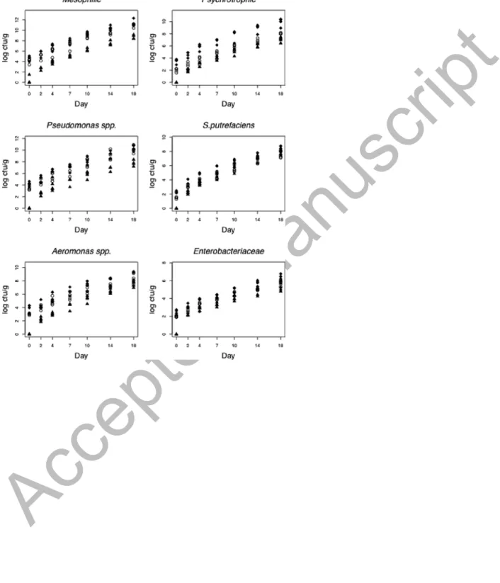
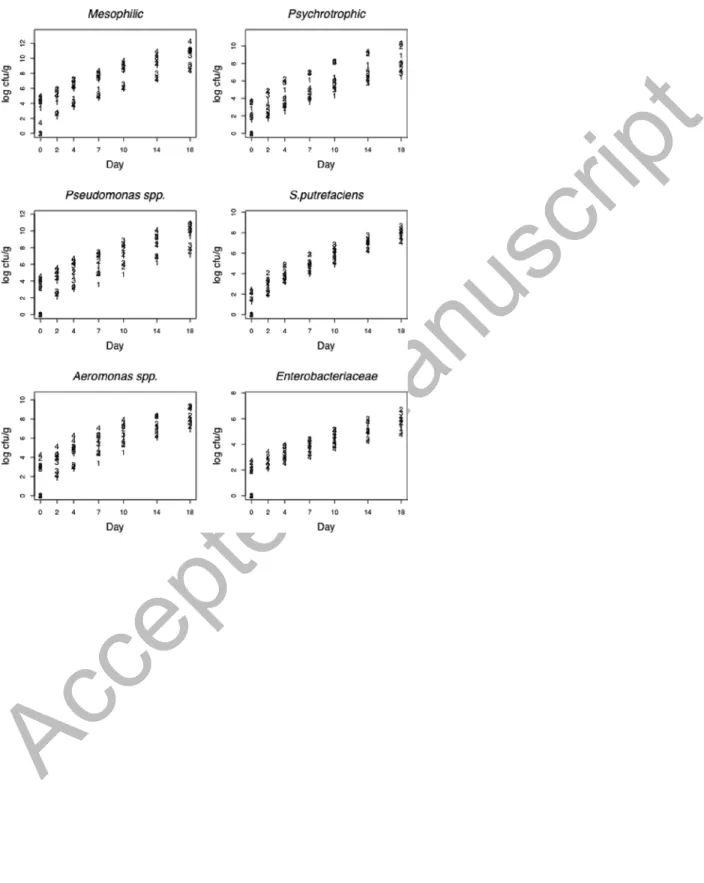
(muscle, gill, skin), kth batch (seasons), and at the t observation day. In order to explore bacterial growth on the observation days, the log10 Ni, j,k,t was plotted against the day and by tissue (Figure 1) and batch (Figure 2). The plots for mesophiles, psychrotrophic, Pseudomonas spp., S.
putrefaciens, Aeromonas spp., and Enterobacteriaceae suggested that the linear mixed-effects
model should be used (Laird and Ware, 1982):
(Model 1) log10Ni, j,k ,t =θ+ fishi +αj +λk +Pr
()
t +ei, j,k ,twhere fishi indicated the random effect of the fish (i = 1,...56),αj the fixed tissue effect ( j = muscle,gill,skin ), λk the batch effect ( k = 1,2,3,4 ),Pr
( )
t is a polynomial of degree r (trend),and ei, j,k,t random error. The following formula was used: αmuscle =λ1=0 (muscle and batch 1
were the reference categories). It was assumed that the random effects fishi were independent random variables distributed according to
N 0,
( )
σf and the error ei, j,k,t, independent randomvariables N 0,
( )
σe and independent from the random effects fishi. In order to determinate the optimum degree for the polynomial Pr( )
t (trend), we considered the degrees r = 1, 2, 3, and thenAccepted Manuscript
the one that optimized the Akaike criterion (AIC) was selected. For all the bacteria, the optimum degree obtained was r = 2, and, thus, P2
()
t =β1⋅t +β2⋅t2 (the intercept is subsumed in θ). Thegoodness of fit of the model was evaluated by the coefficient R2, which measures the proportion of the variability corresponding to the fixed effects of the model.
To account for excess zeros for the Clostridia and P. phosphoreum (44.6% for Clostridia and 46.4% for P. phosphoreum), the zero-inflated Poisson (ZIP) model was defined as (Hall, 2000):
This model consists of a combination of two distributions to incorporate extra zeros, where ln µj,k,t =θ + αj+λk+β ⋅t and πt was the probability of being an extra zero. The logistic model logitπt =γ +η⋅t (the zeros only depended on day t) was considered. Here, the vector
xj,k,t summarized the covariate tissue (j), batch (k), and day (t). Note that the effects of tissue,
batch, and day are expressed with the same parameters for both models. All of the regression models were fitted to our data using the R packages nlme (LME procedure) and PSCL (Zeroinfl procedure).
Accepted Manuscript
Results and Discussion
The investigation showed the general possibility to predict the expected growth values of eight microorganisms studied in sea bream stored in ice, in four different batches.
Models for the analysis of microbiological growth
Model 1
For all bacteria represented by model 1, the optimal polynomial trend obtained was the quadratic being all the coefficients β2 negatives, which means that the growth has a linear deceleration. Table 2 shows the coefficients R2, all comparisons among tissues and among batches, and, finally, the coefficients β1 and β2 corresponding to the quadratic trends.
Figures 1 and 2 show the expected growth values of log10Ni, j,k,t mesophilic aerobic bacteria, psychrotrophic bacteria, Aeromonas spp., Pseudomonas spp., Enterobacteriaceae, and
S. putrefaciens plotted against each observation day and according to tissue and batch,
respectively. The Table 3 shows the mean of bacterial counts of four batches (P < 0.001). The linear trend for all log-counts over time justified the use of this model (1). Table 2 shows the estimates of the model parameters (SE) and all paired comparisons. For these microorganisms, spoilage levels in gills were higher than in skin and muscle (P < 0.001), with muscle being the least contaminated tissue. Almost all estimates ofλ2−λ1, λ3−λ1 and λ4−λ1were significantly
Accepted Manuscript
greater than zero, with lower levels of spoilage corresponding to batch 1 (winter). Batch 4 was the most contaminated, except for Enterobacteriaceae and S. putrefaciens.
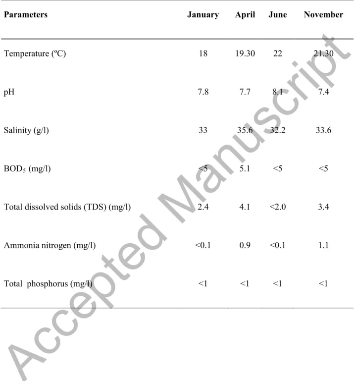
An effect over the batches was observed, supposedly due to the lower water temperature in winter (4 ºC lower) compared to summer, while the rest of the physicochemical values stood constant during the four studied seasons (Table 4). This was the determining factor for the growth of different types of microflora present in fish. According to Ward and Baj (1988), the microbiological condition of fish muscle is directly related to fishing ground and environmental factors. These results coincide with those published by Iliopoulou-Georgudaki et al. (2009), who observed that temperature, dissolved oxygen, and conductivity had a greater influence on microbiological populations in aquacultured fish. Other authors (Grigorakis et al., 2003, 2004) detected differences between microbial counts from December (water temperature, 14ºC) and July (water temperature, 25ºC), with results showing that summer fish presented higher rates of autolytic activity but lower rates of microbial spoilage.
Model 2
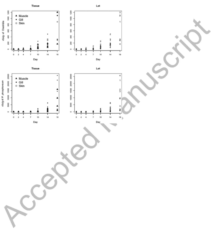
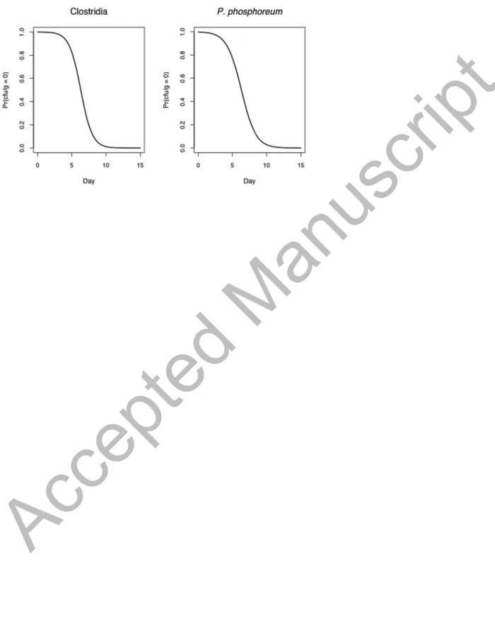
Figure 3 shows the counts Ni, j,k,t of Clostridia and P. Phosphoreum over time according to tissue and per batch. Note that the excess of zeros for both counts suggested the use of model 2. The estimated values
µj,k,t (expected growth) corresponding to this model and the estimated
probabilities of zeros πt are summarized in Table 5. The negative coefficients of the parameter
η indicated that the probability of zeros for Clostridia and P. phosphoreum decreased over the
Accepted Manuscript
The plot of these probabilities versus days (Figure 4) showed that the probability of zeros (no counts) decreased over time; from day 10, the probability of spoilage was near one.
Table 5 shows that the contamination levels for Clostridia and P. phosphoreum displayed significant differences (P < 0.001) between the three tissues sampled. Muscle was the least contaminated tissue, followed by skin and gills for Clostridia; skin was the least contaminated tissue for P. phosphoreum.
Significant differences (P < 0.001) were observed for the microorganisms among the four batches studied, except between batches 2-1 for P. phosphoreum. These results suggested that the irregular growth observed for the two bacteria was independent of parameters analyzed in this study. Growth delay may be due to the fact that Clostridia and P. phosphoreum can be the dominant cultivable bacterium in the gut tracts of some fish, such as hatchetfish, and it is a normal member of the gut flora of cod (Gadus morua) (Dalgaard et al., 1997).
Descriptive microbiological analysis
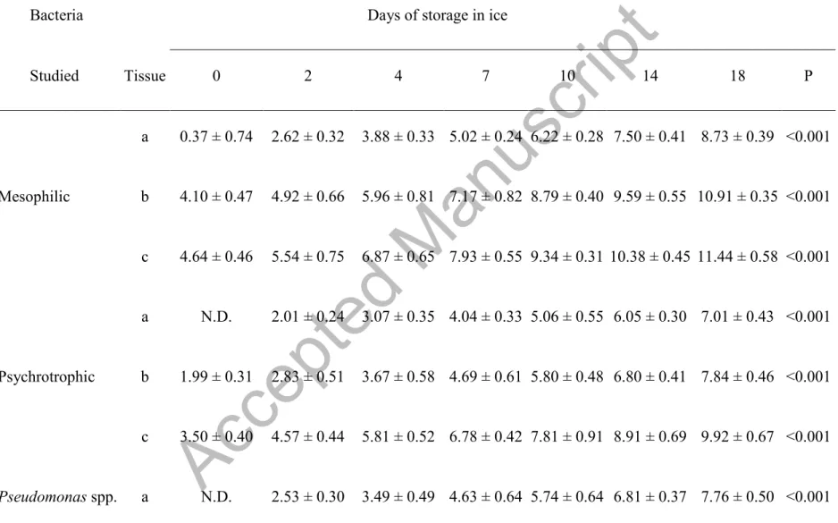
The data for microbial flora (log CFU/g) of aquacultured ungutted sea bream stored aerobically in ice at 2 ± 1 ºC by tissue are shown in Table 3. The results are expressed as averages of the four batches sampled, allowing the comparison of these results with other similar studies.
All analyzed bacteria in the present study increased gradually throughout the storage period from day 0, except in the case of Clostridia and P. phosphoreum, which started to grow from days 4 and 7 in muscle, respectively. Mesophilic and psychrotrophic bacteria on the initial day (day 0)
Accepted Manuscript
were 0.37 and not detected in muscle, 4.10 and 1.99 log CFU/g in skin, and 4.64 and 3.50 log CFU/g in gills, respectively. Similar results were obtained in muscle on days 0 and 3 (Grigorakis et al., 2003) or in skin on day 1 (Drosinos and Nychas, 1996) in ungutted sea bream stored in ice. However, other authors have reported higher results in the initial values of mesophilic and psychrotrophic bacteria in muscle of ungutted sea bream (Tejada and Huidobro, 2002; Lougovois et al., 2003; Kilinc et al., 2007; Özden et al., 2007), sea bream fillets (Erkan and Ueretener, 2010), and ungutted sea bass (Papadopoulos et al., 2003) or in skin of whole sea bream (Cakli et al., 2007; Erkan, 2007). The differences observed in mesophilic and psychrotrophic bacteria between the different studies may have been due to the microbiological conditions of fish muscle in ungutted sea bream, directly related to fishing ground, sanitary conditions of the slaughterhouse, and environmental factors (Ward and Baj, 1988).
Mesophilic and psychrotrophic bacteria counts reached 7 log CFU/g on days 10 and 18 in muscle, days 7 and 14 in skin, and days 4 and 7 in gill, respectively. This value was considered to be the maximum acceptability limit for fresh water and marine species as defined by the ICMSF (1986). Other authors studying sea bream muscle reported mesophilic and psychrotrophic values of 7.81 and 7.11 log CFU/g, respectively, after 21 days of storage (Álvarez et al., 2008) and 6.7 and 7 log CFU/g, after 16 and 13 days, respectively (Erkan and Ueretener, 2010), and 7 log CFU/g after 11 and 14 days, respectively, in different culture conditions (López-Caballero et al., 2002). Similar results were reported in skin with mesophilic and psychrotrophic counts of 7.20 and 7.35 log CFU/g after 15 days, respectively, and 6.6 and 6.8 log CFU/cm2 after 13 days of storage, respectively (Cakli et al., 2007; Erkan, 2007).
Accepted Manuscript
the psychrotrophic counts (8.73 and 7.01 log CFU/g in muscle, 10.91 and 7.84 log CFU/g in skin, and 11.44 and 9.92 log CFU/g in gills, respectively).
Initial counts of SSB (specific spoilage bacteria: Pseudomonas spp., S. putrefaciens, as H2 S-producing bacteria, and Aeromonas spp.) were below the detection threshold (<1 log CFU/g) in muscle: 3.51, 1.10, and 3 log CFU/g in skin; 4.14, 2.31, and 3.65 log CFU/g in gills, respectively. Low counts of Pseudomonas spp. for ungutted European hake stored in ice were also reported by Baixas-Nogueras et al. (2009). Other authors described higher initial counts of
Pseudomonas spp. in muscle, with 3.9 log CFU/g for sea bream (Özden et al., 2007) and 3.0 log
CFU/g for sea bass (Papadopoulos et al., 2003; Paleologos et al., 2004), as well as 3.3 log CFU/g in gutted sardine samples (Erkan, 2008) and 2.88 log CFU/g in horse mackerel (Tzikas et al., 2007). Initial S. putrefaciens counts accounted for a large proportion of the microflora in muscle of several species such as: sea bream, with values of 4.4 log CFU/g (Özden et al., 2007); sea bass, with values of 2.2 log CFU/g (Paleologos et al., 2004); sardines, 3.3 log CFU/g (Erkan, 2008); and sea bream skin, 3.3 log CFU/g (Erkan, 2007). However, these values differed from those reported in our study, which coincided with those described by López-Caballero et al. (2002), Lougovois et al. (2003), and Baixas-Nogueras et al. (2009).
In general, Pseudomonas spp. was the dominant population on day 18 of storage, followed by
Aeromonas spp. and S. putrefaciens, with values of 7.76, 7.49, and 8.05 log CFU/g in muscle;
10.11, 8.24, and 7.49 log CFU/g in skin; and 10.40, 9.02, and 8.05 log CFU/g in gills, respectively. Others authors have reported similar results in terms of Pseudomonas spp. and S.
putrefaciens counts in muscle of sea bream, ranging from 6-7.8 log CFU/g (López-Caballero et
Accepted Manuscript
al., 2002; Lougovois et al., 2003; Özden et al., 2007), sea bass, with 7-7.2 log CFU/g (Pseudomonas spp.), and 6.6 and 7 log CFU/g (S. putrefaciens) (Papadopoulos et al., 2003; Paleologos et al., 2004), as well as in sardines with values of 4 and 4.9 log CFU/g, respectively, after 9 days of storage (Erkan, 2008), horse mackerel with values of 6.42 and 5.12 log CFU/g, respectively, after 12 days of ice storage (Tzikas et al., 2007), and sea bream skin with 6.7 log CFU/g after 13 days of ice storage for H2S-producing bacteria counts (Erkan, 2007).
Similar counts for Pseudomonas spp. and S. putrefaciens have been reported as for SSB in fish from temperate and tropical waters (Gillespie, 1981; Lima Dos Santos et al., 1981; Gram and Huss, 1996) and in fresh Mediterranean fish stored aerobically under refrigeration (Koutsoumanis and Nychas, 1999) or ice storage (Gennari and Tomaselli, 1988; Gennari et al., 1999; Sant’Ana et al., 2011). The values for S. putrefaciens reported in this study were lower than those observed for Pseudomonas spp. at the end of the storage period, possibly due to the fact that Pseudomonas spp. and S. putrefaciens have specific iron chelating systems (siderophores), and when these are grown in co-culture on fish samples, siderophore-producing
Pseudomonas spp. inhibits the growth of S. putrefaciens (Gram and Dalgaard, 2002; Olafsdóttir
et al., 2006).
Regarding Aeromonas spp., Nagar and Bandekar (2011) showed a few counts in Rohu (Labeo
rohita) of 5.24 to 6.64 log CFU/g on day 0 and 7, respectively; both results were higher than
those obtained in our study. However, most of the literature refers to the prevalence of this microorganism in the fish, not counts (CFU/g) (Galindo and Chopra, 2007), highlighting the importance of A. hydrophila as a pathogen conveyed through water (Daskalov, 2006).
Accepted Manuscript
In this study, Enterobacteriaceae counts were lower than those of SSB at the end of storage, coinciding with the results reported for different fresh Mediterranean fish at the end of shelf life (Koutsoumanis et al., 1999b; Gennari et al., 1999; Tejada and Huidobro, 2002). Initial Enterobacteriaceae counts were not detected in muscle, 1.99 log CFU/g in skin, and 2.48 log CFU/g in gills, increasing to 5.19, 5.75, and 6 .35 log CFU/g, respectively, after 18 days of ice storage. Initial Enterobacteriaceae counts in fresh fish muscle were similar to those reported for ungutted European hake (Baixas-Nogueras et al., 2009). Nevertheless, other authors have described higher initial counts, although these same authors have reported similar values to those described here after the period of ice storage in different species, such as sea bass with counts of 2 and 4.2 log CFU/g (initial and final counts, respectively) (Papadopoulos et al., 2003), sea bream with 3.9 and 5.6 log CFU/g (initial and final counts, respectively) (Özden et al., 2007), and sardines with 3.5 and 5.08 log CFU/g (initial and final counts, respectively) (Erkan, 2008). The contribution of Enterobacteriaceae to the microflora of fish and its spoilage potential must be taken into consideration especially in the case of polluted water or delay in chilling after catch (Chouliara et al., 2004), as well as in the filleting process (Moini et al., 2009). Although this group can grow at low temperatures, their abundance decreases during iced storage, possibly because their growth rate is lower than that of other Gram-negative psychrotrophic spoilers, making them poor competitors (Bahmani et al., 2011).
Initial counts of Clostridia and P. phosphoreum were not detected in all the tissues analyzed. However, these counts increased after 18 days of iced storage to 3.02 and 3.70 log CFU/g in muscle, 3.03 and 3.15 log CFU/g in skin, and 2.78 and 4.23 log CFU/g in gills, respectively. The
Accepted Manuscript
study. Similar results were found in boque fish stored aerobically, where the contribution of P.
phosphoreum was very small and relatively unimportant (Koutsoumanis et al., 1999b).
Regarding Clostridia, the source of contamination could be a result of carriage by the fish, water contamination, or cross-contamination during processing and sale (Metcalf et al., 2011). Thus, in research carried out by Lalitha and Surendran (2002), the presence of Clostridia in fresh fish was described in 19% of the samples.
Conclusions
The mathematical models applied in the present study revealed that the lowest contamination was detected in muscle during winter. In addition, the final counts of SSB were similar to those obtained in fish from temperate and tropical waters in other studies. Since registered physicochemical parameters (except the temperature) remained relatively constant throughout the whole year, this could be the main factor for the observed seasonal difference. Future studies would offer more data about the influence of other physicochemical parameters over the microbiological evolution in ungutted sea bream in order to confirm whether the temperature is the determining factor.
Acknowledgements
We are very grateful to our families for their support during the preparation of this manuscript. We also want to thank to Dr. Pablo Lupiola and Alevines and Doradas, S.A. for their help in this study.
Accepted Manuscript
References
Álvarez, A., García, B., Garrido, M.D. and Hernández, M.D. 2008. The influence of starvation time prior to slaughter on the quality of commercial-sized gilthead sea bream (Sparus aurata) during ice storage. Aquaculture. 284: 106–114.
APHA. 1992. Standard methods for examination of water and wastewater. 18th Edition, American Public Health Association. Washington D.C.
APROMAR. 2013. La Acuicultura Marina de Peces en España.
https://docs.google.com/file/d/0B4_4E-v9oqL_X1ZjQUVPOFphUDA/edit?pli=1.
Arroyo, L., Kruth, S., Willey, B., Staempfli, H., Low, D., Weese, J.S. 2005. PCR ribotyping of
Clostridium difficile isolates originating from human and animal sources. J. Med. Microbiol. 54
(Pt 2): 163-166.
Bahmani, Z.A., Rezai, M., Hosseini, S.V., Regenstein, J.M., Böhme, K., Alishahi, A. and Yadollahi, F. 2011. Chilled storage of golden gray mullet (Liza aurata). LWT - Food Sci. Tech. 44: 1894-1900.
Baixas-Nogueras, S., Bover-Cid, S., Veciana-Nogué, M.T. and Vidal-Carou, M.C. 2009. Effect of gutting on microbial loads, sensory properties, and volatile and biogenic amine contents of European Hake (Merluccius merluccius var. mediterraneus) stored in ice. J. Food Prot. 72: 1671–1676.
Barrow, G.I. and Feltham, R.K. 2004. Cowan and Steel's manual for the identification of medical bacteria. 3rd Edition, Cambridge University, UK. Cambridge.
Accepted Manuscript
Boulares, M., Mejri, L. and Hassouna, M. 2011. Study of the microbial ecology of wild and aquacultured tunisian fresh fish. J. Food Prot. 10: 1762-1768.
Bratchell, N., McClure, P.J., Kelly, T.M. and Roberts, T.A. 1990. Predicting microbial growth: graphical methods for comparing models. Int. J. Food Microbiol. 11: 279–287.
Broekaert, K., Heyndrickx, M., Herman, L., Devlieghere, L. and Vlaemynck, G. 2011. Seafood quality analysis: Molecular identification of dominant microbiota after ice storage on several general growth media. Food Microbiol. 28: 1162-1169.
Bruckner, S., Albrecht, A., Petersen, B. and Kreyenschmidt, J. 2013. A predictive shelf life model as a tool for the improvement of quality management in pork and poultry chains. Food Control. 29: 451-460.
Cakli, S., Kilinc, B., Cadun, A., Dincer, T. and Tolasa, S. 2007. Quality differences of whole ungutted sea bream (Sparus aurata) and sea bass (Dicentrarchus labrax) while stored in ice. Food Control 18: 391-397.
Calanche., J., Samayoa, S., Alonso, V., Provincial, L., Roncales, P. and. Beltran, J.A. 2013. Assessing the effectiveness of a cold chain for fresh fish salmon (Salmo salar) sardine (Sardina
pilchardus) in a food processing plant. Food Control, 33, 126-135.
Chouliara, I., Sawaidis, L.N., Riganakos, K. and Kontaminas, M.G. 2004. Preservation of salted, vacuum-packaged, refrigerated sea bream (Sparus aurata) fillets by irradiation: microbiological, chemical and sensory attributes. Food Microbiol 21: 351–359.
Accepted Manuscript
Corbo, M.R., Speranza, B., Filippone, A., Granatiero, S., Conte, A., Sinigaglia, M. and Del Nobile, M.A. 2008. Study on the synergic effect of natural compounds on the microbial quality decay of packed fish hamburger. Int J Food Microbiol 127, 261–267.
Dalgaard, P., Gram, L. and Huss, H.H. 1993. Spoilage and shelf-life of cod fillets packed in vacuum or modified atmospheres. Int. J. Food Microbiol. 19: 283-294.
Dalgaard, P. 1995. Qualitative and quantitative characterisation of spoilage bacteria from packed fish. Int. Journal of Food Microbiology, 26: 319-334.
Dalgaard, P., Mejlholm, O., Christiansen, T.J. and Huss, H.H. 1997. Importance of
Photobacterium phosphoreum in relation to spoilage of modified atmosphere-packed fish
products. Lett Appl Microbiol 24: 373–378.
Daskalov, H. 2006. The importance of Aeromonas hydrophila in food safety. Food Control 17: 474-483.
De Koning, A.J. 2004. Rates of Cholesterol Ester Formation During Storage of Anchovy (Engraulis capensis) at Various Temperatures. Int J Food Prop 7: 321–327.
Drosinos, E.H. and Nychas, G.J.E. 1996. Brochothrix thermosphacta, a dominant microorganism in Mediterranean fresh fish (Sparus aurata) stored under modified atmosphere. It J Food Sci 8: 323-329.
Erkan, N. 2007. Sensory, chemical, and microbiological attributes of sea bream (Sparus aurata): effect of washing and ice storage. Int J Food Prop 10: 421-434.
Accepted Manuscript
Erkan, N. 2008. Quality assessment of whole and gutted sardines (Sardina pilchardus) stored in ice. Int J Food Sci Tech 43: 1549-1559.
Erkan, N. and Ueretener, G. 2010. The effect of high hydrostatic pressure on the microbiological, chemical and sensory quality of fresh gilthead sea bream (Sparus aurata). Eur Food Res Tech 230: 533-542.
François, K., Devlieghere, F., Uyttendaele, M., Standaert, A.R., Geeraerd, A.H., Nadal, P. et al. 2006. Single cell variability of L. monocytogenes grown on liver pâté and cooked ham at 7ºC: comparing challenge test data to predictive simulations. J Appl Microbiol 100: 800-812. Galindo, C.L. and Chopra, A.K. 2007. Aeromonas and Plesiomonas species. In: Food Microbiology: Fundamentals and Frontiers, 3rd ed. Doyle, M.P., Beuchat, L.R. (Eds.). ASM Press, Washington, D.C., pp. 381–400.
Genç, I. Y., Esteves, E, Anibal, J. and Diler, A. 2013. Effects of chilled storage on quality of vacuum packed meagre fillets. J Food Eng 115: 486–494.
Gennari, M. and Tomaselli, S. 1988. Changes in aerobic microflora of skin and gills of
Mediterranean sardines (Sardina Pilchardus) during storage in ice. Int J Food Microbiol 6: 341-347.
Gennari, M., Tomasselli, S. and Cotrona, V. 1999. The microflora of fresh and spoiled sardines (Sardina pilchardus) caught in Adriatic (Mediterranean) sea and stored in ice. Food Microbiol 16: 15–28.
Accepted Manuscript
Gillespie, N.C. 1981. A numerical taxonomic study of Pseudomonas-like bacteria isolated from fish in Southeastern Queensland and their association with spoilage. J Applied Microbiol 50: 29– 44.
Ginés, R., Afonso, J.M., Argüello, A., Zamorano, M.J. and López, J.L. 2004. The effects of long-day photoperiod on growth, body composition and skin colour in immature gilthead sea bream (Sparus aurata L.). Aquaculture Res 35: 1207-1212.
Gorczyca, E. and Pek Poh Len. 1985. Mesophilic spoilage of bay trout (Arripis trurta), bream (Acanthropagrus butchri) and mullet (Aldrichetta forsteri). In: Spoilage of Tropical Fish and Product Development. FAO Fish. Rep. 317 Suppl. FAO Reilly, A. (Ed). Rome, Italy, pp. 123-132.
Gram, L., Wedell-Neergaard, C. and Huss, H.H. 1990. The bacteriology of fresh and spoiling Lake Victorian Nile perch (Lates niloticus). Int J Food Microbiol 10: 303-316.
Gram, L. and Huss, H. 1996. Microbiological spoilage of fish and fish products. Int J Food Microbiol 33: 589–595.
Gram, L. and Dalgaard, P. 2002. Fish spoilage bacteria – problems and solutions. Curr Opinion Biotech 13: 262–266
Grigorakis, K., Taylor, K.D.A. and Alexis, M.N. 2003. Seasonal pattern of spoilage of ice stored cultured gilthead sea bream (Sparus aurata). Food Chem 81: 263–268.
Grigorakis, K., Alexis, A., Gialamas, I. and Nikolopoulou, D. 2004. Sensory, microbiological, and chemical spoilage of cultured common sea bass (Dicentrarchus labrax) stored in ice: a
Accepted Manuscript
Guillén-Velasco, S., Ponce-Alquicira, E., Farrés-González Saravia, A. and Guerrero-Legarreta, I. 2004. Histamine Production by Two Enterobacteriaceae Strains Isolated from Tuna (Thunnus thynnus) and Jack Mackerel (Trachurus murphyii ). Int J Food Prop 7: 91-103.
Gunvig, A., Hansen, F. and Borggaard, C. 2013. A mathematical model for predicting
growth/no-growth of psychrotrophic C. botulinum in meat products with five variables. Food Control 29: 309–317.
Hall, D.B. 2000. Zero-inflated Poisson and binomial regression with random effects: a case study. Biometrics 56: 1030-1039.
Huss, H.H., Dalgaard, P. and Gram, L. 1995. Microbiology of fish and fish products. In Seafood from Producer to Consumer, Integrated approach to Quality. Proceedings of the International Seafood Conference on the occasion of the 25th anniversary of the WEFTA, held in
Noordwijkerhout, The Netherlands, 13-16 November, 1995.
ICMSF (International Commission on Microbiological Specifications for Foods). 1986.
Sampling plans for fish and shellfish in microorganisms in foods. Sampling for microbiological analysis: principles and scientific applications, 2nd. Edt, vol 2. University of Toronto Press, Toronto, 181–196.
Iliopoulou-Georgudaki, J., Theodoropoulos, C., Venieri, D. and Lagkadinou, M. 2009. A model predicting the microbiological quality of aquacultured sea bream (Sparus aurata) according to physicochemical data: an application in western Greece fish aquaculture. Int J Bio Life Sci 5: 1-8.
Accepted Manuscript
ISO, 2001. ISO 17410: Microbiology of Food and Animal Feeding Stuffs – Horizontal Method for the Enumeration of Psychrotrophic Microorganisms. International Organization for
Standardization, Geneve, Switzerland, 7 pp.
ISO, 2003. ISO 4833: Microbiology of Food and Animal Feeding Stuffs – Horizontal Method for the Enumeration of Microorganisms – Colony-Count Technique at 30 ºC. International
Organization for Standardization, Geneve, Switzerland, 9 pp.
Katikou, P., Georgantelis, D., Paleologos, E.K., Ambrosiadis, I. and Kontominas, M.G. 2006. Relation of biogenic amines’ formation with microbiological and sensory attributes in
Lactobacillus-inoculated vacuum-packed rainbow trout (Oncorhynchus mykiss) fillets. J Agri
Food Chem 54: 4277–4283.
Keel, K., Brazier, J.S., Post, K.W., Weese, J.S. and Songer, J.G. 2007. Prevalence of PCR ribotypes among Clostridium difficile isolates from pigs, calves, and other species. J Clin Microbiol 45: 1963-1964.
Kilinc, B., Cakli, S., Cadun, A., Dincer, T. and Tolasa, S. 2007. Comparison of effects of slurry ice and flake ice pretreatments on the quality of aquacultured sea bream (Sparus aurata) and sea bass (Dicentrarchus labrax) stored at 4 degrees ºC. Food Chemistry 104: 1611-1617.
Koutsoumanis, K., Tassou, C., Taoukis, P. and Nychas, G-J.E., 1999a. The use of impedance in predictive microbiology. In: Predictive Modelling of Microbial Growth and Survival in Foods, COST914, Proceedings of Workshop 5. Instrumental Methods For Data Capture For Advanced Predictive Modelling, 15–16 May 1998, Bologna, Italy, Eur19103 En, Roberts, T.A (Ed.) pp. 295–303.
Accepted Manuscript
Koutsoumanis, K., Lampropoulou, K. and Nychas, G.J.E. 1999b. Biogenic Amines and Sensory Changes Associated with the Microbial Flora of Mediterranean Gilt-head Sea Bream (Sparus
aurata) Stored Aerobically at 0.8 and 15°C. J Food Prot 62: 398–402.
Koutsoumanis, K. and Nychas, G. 1999. Chemical and sensory changes associated with
microbial flora of Mediterranean boque (Boops boops) stored aerobically at 0, 3, 7 y 10ºC. Appl Env Microbiol 65: 698-706.
Koutsoumanis, K. and Nychas, G.J.E. 2000. Application of a systematic procedure to develop a microbial model for rapid fish shelf life predictions. Int J Food Microbiol 60: 171–184.
Koutsoumanis, K. and Nychas, G.J.E. 2001. Application of a systematic experimental procedure to develop a microbial model for rapid fish shelf life prediction. Int J Food Microbiol 60: 174– 184.
Labuza, T.P. and Riboh, D. 1982. Theory and application of Arrhenius kinetics to the prediction of nutrient losses in foods. Food Tech 36: 66–74.
Laird, N.M. and Ware, J.H. 1982. Random-Effects models for longitudinal data. Biometrics 38: 963-974.
Lalitha, K.V. and Surendran, P.K. 2002. Occurrence of Clostridium botulinum in fresh and cured fish in retail trade in Cochin (India). Int J Food Microbiol 72: 169-174.
Lima Dos Santos, C., James, D. and Teutscher, F. 1981 Guidelines for chilled fish storage experiments. FAO Fish Tech. Pap., 210 p.
Accepted Manuscript
López-Caballero, M.A., Huidobro, A., Pastor, A. and Tejada, M. 2002. Microflora of gilthead sea bream (Sparus aurata) stored in ice. Effect of washing. Eur Food Res Tech 215: 396-400. Lougovois, V.P., Kyranas, E.R. and Kyrana, V.R. 2003. Comparison of selected methods of assessing freshness quality and remaining storage life of iced gilthead sea bream (Sparus
aurata). Food Res Int 36: 551–560.
Manios, S.G., Konstantinidis, N., Gounadaki, A. and Skandamis, P.N. 2011 Single cell
variability and population dynamics of Listeria monocytogenes and Salmonella Typhimurium in fresh-cut salads and their sterile liquid or solidified extracts. In: Seventh international conference on predictive modelling in foods. Conference proceedings (pp. 49–52). E. Cummins, J. M. Frias, & V. P. Valdramidis (Eds.) Dublin, Ireland: UCD, DIT, Teagasc.
McDonald, K. and Sun, D.W. 1999. Predictive food microbiology for the meat industry: a review. Int J Food Microbiol 52: 1–27.
McMeekin, T.A., Ross, T. and Olley, J. 1992. Application of predictive microbiology to assure the quality and safety of fish and fish products. Int J Food Microbiol 15: 13–32.
McMeekin, T.A., Olley, J., Ross, T. and Ratkowsky, D.A. 1993. Predictive microbiology: Theory and application. Innovation in microbiology. Taunton, UK: Research Studies Press and John Wiley and Sons.
Metcalf, D., Avery, B.P., Janecko, N., Matic, N., Reid-Smith, R. and Weese, J.S. 2011.
Clostridium difficile in seafood and fish. Anaerobe 17 (2): 85-86.
Accepted Manuscript
Moini, S., Tahergorabi, R., Hosseini, S.V., Rabbani, M., Tahergorabi, Z., Feàs, X. and Aflaki, F. 2009. Effect of gamma radiation on the quality and shelf life of refrigerated rainbow trout (Oncorhynchus mykiss) fillets. J Food Prot 72: 1419–1426.
Nagar, V. and Bandekar, J. R. 2011. Effectiveness of radiation processing in elimination of
Aeromonas from food. Radiation Physics Chem 80: 911-916.
Olafsdóttir, G., Lauzon, H.L., Martinsdóttir, E. and Kristbergsson, K. 2006. Influence of storage temperature on microbial spoilage characteristics of haddock fillets (Melanogram musaeglefinus) evaluated by multivariate quality prediction. International J Food Microbiol 111: 112–125. Özden, O., Inugur, M. and Erkan, N. 2007. Preservation of iced refrigerated sea bream (Sparus
aurata) by irradiation: microbiological, chemical and sensory attributes. Eur Food Res Tech 225:
797–805.
Paleologos, E.K., Savvaidis, I.N. and Kontominas, M.G. 2004. Biogenic amines formation and its relation to microbiological and sensory attributes in ice-stored whole, gutted and filleted Mediterranean sea bass (Dicentrarchus labrax), Food Microbiol 21: 549-557.
Papadopoulos, V., Chouliara, I., Badeka, A., Savvaidis, I. and Kontominas, M. 2003. Effect of gutting on microbiological, chemical, and sensory properties of aquacultured sea bass
(Dicentrarchus labrax) stored in ice. Food Microbiol 20: 411–420.
Pascual, M.R. and Calderón V. 2002. Microbiología alimentaria: metodología analítica para alimentos y bebidas. 2nd Edition. Edt. Díaz de Santos. Madrid. Spain.
Pérez-Sánchez, J.M. and Moreno-Batet, E. 1991. Invertebrados marinos de Canarias. (Edited by
Accepted Manuscript
R Core Team 2013. R: A language and environment for statistical computing. R Foundation for Statistical Computing, Vienna, Austria. URL http://www.R-project.org/.
Ross, T. and McMeekin T.A. 1994. Predictive microbiology. Int J Food Microbiol 23: 241–264. Ross, T., Dalgaard, P. and Tienungoon, S. 2000. Predictive modelling of the growth and survival of Listeria in fishery products. Int J Food Microbiol 62: 231–245.
Rupnik, M. 2007. Is Clostridium difficile-associated infection a potentially zoonotic and foodborne disease?. Clin Microbiol Infect 13: 457-459.
Sant’Ana, L.S., Soares, S.and Vaz-Pires, P. 2011. Development of a quality index method (QIM) sensory scheme and study of shelf-life of ice-stored blackspot sea bream (Pagellus bogaraveo). LWT- Food Sci Tech 44: 2253–2259.
Slattery, S.L. 1998. Shelf-life of Spanish mackerel (Scomberomorus commerson) from northern Australian waters. J Aquat Food Prod Tech 7: 63–79.
Smith Svanevik, C and Tore Lunestad, B. 2011. Characterisation of the microbiota of Atlantic mackerel (Scomber scombrus). Int J Food Microbiol 151: 164–170.
Tejada, M. and Huidobro, A. 2002. Quality of farmed gilthead sea bream (Sparus aurata) during ice storage related to the slaughter method and gutting. Eur Food Res Tech 215: 1–7.
Tzikas, Z., Ambrosiadis, I., Soultos, N. and Georgakis, S. 2007. Quality assessment of Mediterranean horse mackerel (Trachurus mediterraneus) & blue jack mackerel (Trachurus
picturatus) during storage in ice. Food Control 18: 1172–1179
Accepted Manuscript
Ward, D.R. and Baj, N.J. 1988. Factors affecting microbiological quality of seafood. Food Tech 42: 85–89.
Whiting, R.C. 1995. Microbial modelling in foods. Crit Rev Food Sci Nutr 35: 467–494. Zwietering, M.H., Jongenburger, I., Rombouts, F.M. and van’t Riet, K. 1990. Modeling of the bacterial growth curve. Appl Env Microbiol 56: 1875–1881.
Zwietering, M.H., de Koos, J.T., Hasenack, B.E., de Wit, J.C.and van’t Riet, K. 1991. Modeling of bacterial growth as a function of temperature. Appl Env Microbiol 57: 1094–1101.
Accepted Manuscript
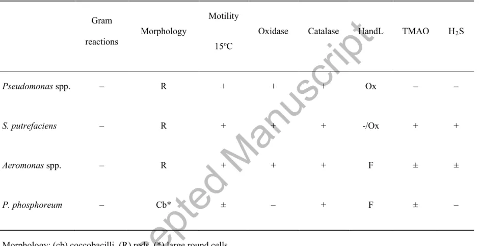
Table 1. Provisional identification of strains isolated from sea bream (Sparus aurata) stored in ice.Gram
reactions Morphology
Motility 15ºC
Oxidase Catalase HandL TMAO H2S
Pseudomonas spp. – R + + + Ox – –
S. putrefaciens – R + + + -/Ox + +
Aeromonas spp. – R + + + F ± ±
P. phosphoreum – Cb* ± – + F ± –
Morphology: (cb) coccobacilli, (R) rods, (*) large round cells.
HandL: Oxidative or fermentative metabolism of glucose was performed in the medium of Hugh and Leifson (Hugh and Leifson, 1953).
TMAO: trimethylamine oxide (TMAO) reduction.
Accepted Manuscript
Table 2. Mixed model for microbial growth in sea bream for model (1).Mesophilic Psychrotrophic Pseudomonas
spp.
S. putrfaciens Aeromonas spp.
Entero-bacteriaceae 0.976 0.985 0.975 0.956 0.964 0.938 3.12 (0.13)** 2.87 (0.08)** 2.78 (0.11)** 0.87 (0.13)** 2.22 (0.12)** 1.22 (0.11)** 2.45 (0.13)** 0.91 (0.08) 2.30 (0.11)** 0.17 (0.14) 1.63 (0.12)** 0.73 (0.11)** 0.67 (0.13)** 1.95 (0.08)** 0.49 (0.11)** 0.71 (0.14)** 0.59 (0.12)** 0.49 (0.11)** 0.59 (0.15)** 0.89 (0.10)** 0.62 (0.14)** 0.39 (0.16) 0.57 (0.14)** 0.27 (0.12) 0.56 (0.15)** 0.69 (0.10)** 0.98 (0.14)** 0.71 (0.15)** 0.46 (0.14)** 0.11 (0.13) R2 αgill−αmuscle αskin−αmuscle αgill−αskin λ2−λ1 λ3−λ1
Accepted Manuscript
0.81 (0.15)** 0.94 (0.10)** 1.04 (0.14)** 0.13 (0.15) 0.98 (0.14)** 0.04 (0.12) 0.03 (0.15) 0.20 (0.10) -0.36 (0.14) -0.32 (0.16) 0.11 (0.14) 0.15 (0.13) -0.22 (0.15) -0.05 (0.10) -0.42 (0.14)* 0.27 (0.16) -0.41 (0.14)* 0.23 (0.12) -0.25 (0.15) -0.25 (0.10) -0.06 (0.14) 0.58 (0.15)** -0.52 (0.14)** 0.07 (0.13) 0.60 (0.03)** 0.53 (0.02)** 0.56 (0.03)** 0.57 (0.03)** 0.52 (0.03)** 0.37 (0.03)** -0.01 (0.002)** -0.01 (0.001)** -0.01 (0.001)** -0.01 (0.002)** -0.01 (0.002)** -0.008 (0.001)**(*) P < 0.05; (**) P < 0.001; all p-values correspond to a multiple linear comparison.
λ4−λ1 λ2−λ3 λ2−λ4 λ3−λ4 β1 β2
Accepted Manuscript
Table 3. Changes in the bacterial count (log CFU/g) in the muscle (a), skin (b), and gills (c) of sea bream (Sparus aurata) stored in ice.
Bacteria Days of storage in ice
Studied Tissue 0 2 4 7 10 14 18 P Mesophilic a 0.37 ± 0.74 2.62 ± 0.32 3.88 ± 0.33 5.02 ± 0.24 6.22 ± 0.28 7.50 ± 0.41 8.73 ± 0.39 <0.001 b 4.10 ± 0.47 4.92 ± 0.66 5.96 ± 0.81 7.17 ± 0.82 8.79 ± 0.40 9.59 ± 0.55 10.91 ± 0.35 <0.001 c 4.64 ± 0.46 5.54 ± 0.75 6.87 ± 0.65 7.93 ± 0.55 9.34 ± 0.31 10.38 ± 0.45 11.44 ± 0.58 <0.001 Psychrotrophic a N.D. 2.01 ± 0.24 3.07 ± 0.35 4.04 ± 0.33 5.06 ± 0.55 6.05 ± 0.30 7.01 ± 0.43 <0.001 b 1.99 ± 0.31 2.83 ± 0.51 3.67 ± 0.58 4.69 ± 0.61 5.80 ± 0.48 6.80 ± 0.41 7.84 ± 0.46 <0.001 c 3.50 ± 0.40 4.57 ± 0.44 5.81 ± 0.52 6.78 ± 0.42 7.81 ± 0.91 8.91 ± 0.69 9.92 ± 0.67 <0.001 Pseudomonas spp. a N.D. 2.53 ± 0.30 3.49 ± 0.49 4.63 ± 0.64 5.74 ± 0.64 6.81 ± 0.37 7.76 ± 0.50 <0.001
Accepted Manuscript
b 3.51 ± 0.48 4.50 ±0.43 5.48 ± 0.72 6.55 ± 0.81 7.82 ± 0.76 9.05 ± 0.85 10.11 ± 0.63 <0.001 c 4.14 ± 0.39 5.17 ± 0.51 6.19 ± 0.59 7.06 ± 0.67 8.16 ± 0.63 9.30 ± 0.55 10.40 ± 0.51 <0.001 S. putrefaciens a N.D. 2.28 ± 0.26 3.40 ± 0.23 4.57 ± 0.47 5.82 ± 0.68 7.03 ± 0.48 8.05 ± 0.45 <0.001 b 1.10 ± 0.74 2.83 ± 0.51 3.72 ± 0.33 4.60 ± 0.54 5.56 ± 0.55 6.68 ± 0.39 7.49 ± 0.44 <0.001 c 2.31 ± 2.31 3.53 ± 0.40 4.45 ± 0.52 5.34 ± 0.74 6.33 ± 0.56 7.28 ± 0.38 8.05 ± 0.50 <0.001 Aeromonas spp. a N.D. 2.23 ± 0.34 3.04 ± 0.19 4.24 ± 0.51 5.28 ± 0.53 6.42 ± 0.33 7.49 ± 0.43 <0.001 b 3.00 ± 0.12 4.00 ± 0.35 4.94 ± 0.59 5.71 ± 0.56 6.72 ± 0.58 7.52 ± 0.60 8.24 ± 0.72 <0.001 c 3.65 ± 0.58 4.37 ± 0.56 5.39 ± 0.62 6.39 ± 0.56 7.33 ± 0.66 8.10 ± 0.54 9.02 ± 0.57 <0.001 Enterobacteriaceae a N.D. 2.22 ± 0.14 2.85 ± 0.18 3.30 ± 0.22 3.99 ± 0.27 4.67 ± 0.39 5.19 ± 0.38 <0.001 b 1.99 ± 0.08 2.72 ± 0.22 3.34 ± 0.21 3.95 ± 0.12 4.50 ± 0.25 5.03 ± 0.10 5.75 ± 0.29 <0.001Accepted Manuscript
c 2.48 ± 0.25 3.17 ± 0.27 3.59 ± 0.69 4.34 ± 0.16 5.34 ± 0.26 5.75 ± 0.29 6.35 ± 0.34 <0.001 Clostridia a N.D. N.D. N.D. 0.25 ± 0.50 1.74 ± 0.38 2.38 ± 0.77 3.02 ± 0.59 <0.001 b N.D. N.D. N.D. 0.83 ± 0.57 1.76 ± 0.32 2.31 ± 0.21 3.03 ± 0.46 <0.001 c N.D. N.D. 0.40 ± 0.80 1.55 ± 0.33 2.03 ± 0.26 2.34 ± 0.24 2.78 ± 0.34 <0.001 P. phosphoreum a N.D. N.D. N.D. N.D. 2.05 ± 0.51 2.93 ± 0.53 3.70 ± 0.53 <0.001 b N.D. N.D. N.D. 1.38 ± 0.92 2.32 ± 0.05 2.64 ± 0.09 3.15 ± 0.11 <0.001 c N.D. N.D. 0.58 ± 0.68 2.51 ± 0.27 3.16 ± 0.18 3.64 ± 0.19 4.23 ± 0.20 <0.001 Mean of four batches ± standard deviation; P < 0.001N.D. = not detected.
Accepted Manuscript
Table 4. Mean values of seawater properties.
Parameters January April June November
Temperature (ºC) 18 19.30 22 21.30
pH 7.8 7.7 8.1 7.4
Salinity (g/l) 33 35.6 32.2 33.6
BOD5 (mg/l) <5 5.1 <5 <5
Total dissolved solids (TDS) (mg/l) 2.4 4.1 <2.0 3.4
Ammonia nitrogen (mg/l) <0.1 0.9 <0.1 1.1
Total phosphorus (mg/l) <1 <1 <1 <1
Accepted Manuscript
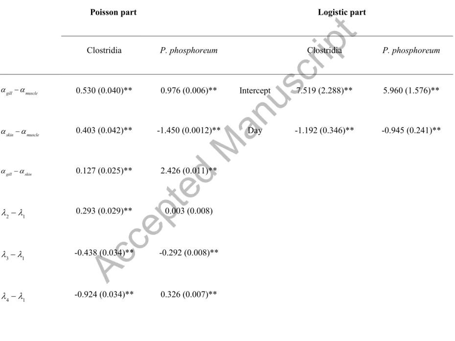
Table 5. Zero-inflated models with its Poisson and logistic parts for Clostridia and P. phosphoreum in sea bream stored in ice.
Poisson part Logistic part
Clostridia P. phosphoreum Clostridia P. phosphoreum
αgill−αmuscle 0.530 (0.040)** 0.976 (0.006)** Intercept 7.519 (2.288)** 5.960 (1.576)**
αskin−αmuscle 0.403 (0.042)** -1.450 (0.0012)** Day -1.192 (0.346)** -0.945 (0.241)**
αgill−αskin 0.127 (0.025)** 2.426 (0.011)**
λ2−λ1 0.293 (0.029)** 0.003 (0.008) λ3−λ1 -0.438 (0.034)** -0.292 (0.008)** λ −λ -0.924 (0.034)** 0.326 (0.007)**
Accepted Manuscript
λ2−λ3 0.731 (0.037)** 0.295 (0.008)**λ2 −λ4 1.217 (0.037)** -0.323 (0.007)** λ3−λ4 0.486 (0.040)** -0.618 (0.008)** β (slope) 0.262 (0.004)** 0.350 (0.001)**
(*) P < 0.05; (**) P < 0.001; all p-values correspond to multiple linear comparison.
Accepted Manuscript
Figure 1. Log counts of microorganisms (four batches) over time and according to tissue: = Muscle; = Gill; = Skin.
Accepted Manuscript
Figure 2. Log counts of microorganisms (four batches) over time and per batch.
Accepted Manuscript
Figure 3. Counts of Clostridia and P. phosphoreum according to tissue and batch. The zero rates were 44.6% for Clostridia and 46.4% for P. phosphoreum.
Accepted Manuscript
Figure 4. Probabilities of zeros for Clostridia and P. phosphoreum by observation day.