ORIGINAL ARTICLE
Autologous fat transfer with SEFFI (superficial enhanced fluid fat injection)
technique in periocular reconstruction
Benjamin Riescoa, Cristina Abascalb, Ana Duartec, Rocio Mariel Floresd, Guillermina Rouauxd, Raul Sampayod, Francesco Bernardinie, and Martin Devotod
aOculoplástica, MIRA Clínica Oftalmológica, IOPA Clínica Oftalmológica, Hospital del Salvador, Santiago, Chile;bOftalmología, Complejo
Hospitalario de Navarra, Pamplona, Spain;cOculoplástico, Centro Hospitalar Lisboa Central and Hospital Cuf Descobertas, Lisboa, Portugal;
dOculoplástica, Consultores Oftalmológicos, Buenos Aires, Argentina;eOculoplastics, Oculoplastica Bernardini, Genova, Italy
ABSTRACT
Purpose: To evaluate the aesthetic and functional outcomes of autologous fat transfer using the SEFFI (superficial enhanced fluid fat injection) technique for reconstruction of the periocular area.
Methods: Autologous fat injections prepared with the 0.5 mL and 0.8 mL SEFFI technique were used in four patients for periocular rehabilitation.
Results: Case 1 (C1): A patient with left-sided progressive facial hemiatrophy underwent ipsilateral volumizing with 0.8 SEFFI in the superior, temporal, and inferior periorbital areas, and 0.5 SEFFI in both eyelids. C2: A 21-year-old female with a post trauma frontal scar, left ptosis, and lower eyelid retraction was treated with 0.5 SEFFI applied in the scar area associated with an upper eyelid conjunctivomullerectomy and resection of the lower eyelid retractors. C3: A patient with previous left-eye evisceration and orbital floor and medial wall fractures underwent socket reconstruction with buccal mucosal graft in the lower fornix and 0.5 SEFFI injections in both superior and inferior eyelids. SEFFI was also applied in the intraorbital space for correction of the enophthalmos. C4: A patient with lower lid retraction post blepharoplasty was treated with 0.8 SEFFI injections in lower eyelids and malar areas, complemented with a bilateral lateral cantopexy.
Conclusions: Autologous fat transfer with SEFFI technique is an effective and safe procedure in cases of periocular rehabilitation.
ARTICLE HISTORY
Received 19 January 2017 Accepted 19 September 2017
KEYWORDS
Superficial enhanced fluid fat injection; periocular reconstruction; autologous fat transfer
Introduction
Restoring a normal and healthy facial appearance after trauma, progressive disfiguring diseases, surgical, and non surgical oncologic treatments and unsuccessful cosmetic surgery present a true challenge for the oculoplastic sur-geon. More than 120 years ago, Nueber published the first results with autologous fat transfer for scar improvement.1Since then, a therapeutic arsenal including autologous fat, hyaluronic acid fillers, flaps, grafts, and more recently mesenchymal stem cells have been devel-oped trying to overcome the arduous purpose of restoring a near-to-normal volume, symmetry, skin quality, and function during periocular reconstruction. Prominent names of plastic surgery such as Coleman2and Rohrich
3, among others, have provided a significant amount of
literature demonstrating the potential of fat grafting for customized volume increase and facial rejuvenation. In 2015, Bernardini proposed the injection of autologous fat rich in stem cells at a superficial level pointing out its
regenerative, anti-inflammatory, and immunomodulat-ing benefits. This technique, SEFFI (superficial enhanced fluid fat injection), uses 0.5 and 0.8 mL micro-cannulas in order to collect fine fat, rich in pluripotent cells.4We have expanded the use of SEFFI for periocular rehabilitation with good results.
Materials and methods
A seven-step standardized protocol, currently used in our practice, was followed (Table 1). Scar tissue and the orbicular muscle were injected with 0.5 mL SEFFI, and 0.8 mL SEFFI was used when a more pronounced volumizing effect was needed. The volume of fat injected was between 12 and 35 mL (Video).
The selection of the donor site was performed during the pre-surgical examination. Previous lipo-suctioned areas were avoided. We prefer the internal side of the knees and, as a second choice, the abdo-men, as both provide an easy access.
CONTACTBenjamin Riesco benjaminriesco@gmail.com Oculoplástica, MIRA Clínica Oftalmológica, Av. Kennedy 7500, Santiago, Chile Color versions of one or more of the figures in the article can be found online atwww.tandfonline.com/IORB.
https://doi.org/10.1080/01676830.2017.1383470
© 2017 Taylor & Francis
In the periocular area, fat filling was performed using a retrograde injection technique (to prevent real risk of fat embolism) exerting the least possible pres-sure. Five cc 0.5 mL SEFFI syringes with 23G needles and 5 cc 0.8 mL SEFFI syringes with 20G needles were used4
The results were subjectively evaluated both by sur-geons and patients using standardized photographic exams (Canon® EOS Rebel T3i with 60 mm lens, Tokyo, Japan) pre- and post- intervention with a mini-mum follow-up of 6 months. Written consent was obtained from all identifiable patients.
Results
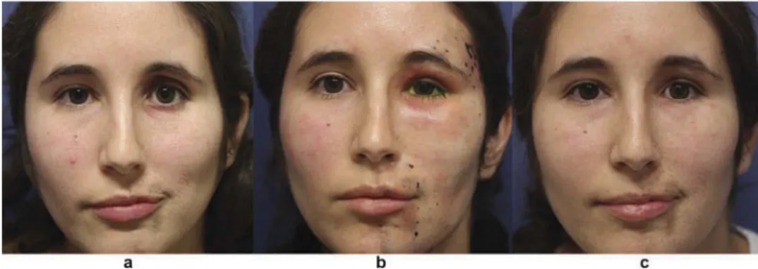
C1: A 29-year-old female patient with Parry Romberg Syndrome (Figure 1a). A total of 24 mL of 0.8 SEFFI was used in the left superior, temporal,
and inferior periorbital areas. The eyelids were trea-ted with 6 mL of 0.5 SEFFI (Figure 1b). A satisfac-tory improvement was obtained at 1 year of follow-up (Figure 1c).
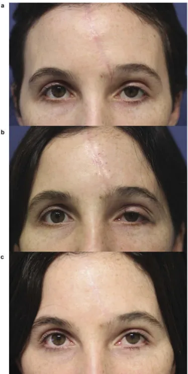
C2: A 21-year-old female patient with left ptosis, lower lid retraction, and a prominent frontal scar resulting from a previous motor vehicle accident was referred to our clinic (Figure 2a). The scar area was treated with 6 cc of 0.5 SEFFI, and a left con-junctivomullerectomy combined with left lower eye-lid retractors resection were used to treat the ptosis and lower eyelid retraction, respectively (Figure 2b). A good result was obtained 6 months after surgery (Figure 2c).
C3: A 36-year-old female patient with previous left-eye evisceration and left orbital floor and medial wall fractures was evaluated due to enophthalmos (Figure 3a). Socket reconstruction was performed
Figure 1.Parry Romberg Syndrome. Table 1.Autologous fat transfer technique.
(1) Anesthesia
Under sterile technique, at the same time as facial anesthesia Donor site marking
Local anesthesia: 500 mL saline solution + 20 mL 2% lidocaine + 1 mL 1:1000 epinephrine Stab incision with 11 blade. Inject local with 20 mL syringe and 2 mm diameter cannula 20 mL in deep fat, 20 mL in superficial fat. Wait 15 min for vasoconstriction
(2) Harvesting
Fat is suctioned with SEFFI cannulas (0.5 and 0.8 mm side ports) using 10 mL syringes Not exceed 2 cc of negative pressure to suck
The volume of fat harvested is twice the amount needed for injection Donor site does not need suturing
(3) Centrifugation
Each syringe is placed on a sterile tube in the centrifuge Fat is centrifuged 3 min at 3000 rpm
(4) Processing
The infranatant (blood + local) is discarded Supernatant (oil) is also discarded (5) Platelet rich plasma (PRP)
20 mL of blood is withdrawn in 2 citrated tubes and centrifuged 2 min at 1000 rpm PRP is mixed with the processed fat in a volume of 20% of the volume of the fat (6) Injection
Fat + PRP is transferred to 5 mL syringes
Injection is performed in a superficial level using 21 G needles or intraorbital with cannula (7) Donor site care
A small patch is applied to the entry point Compressive bandages are applied
Figure 2.Scar from a previous motor vehicle accident.
Figure 3.Anophthalmic socket.
using a buccal mucosal graft on the lower fornix, and 4 mL of 0.5 SEFFI were injected into the upper lid, 3 mL to the lower lid, and 5 mL into the intraorbital space using an inferotemporal transpalpebral approach. At 1 year follow-up, a good improvement was noticed (Figure 3b).
Case 4: A 43-year-old male patient was evaluated for lower lid retraction post blepharoplasty (Figure 4a). We used 10 mL of 0.8 SEFFI in each lower eyelid and malar region, associated with a bilateral lateral cantopexy. A satisfactory result was obtained after 1 year of follow-up (Figure 4b).
Discussion
In this report, we describe four illustrative cases of our experience with SEFFI from a total of 20 reconstructive cases. The use of autologous fat transfer for facial rejuvenation is a well-accepted and largely widespread procedure. 2–4 Recent advances in cell therapy have highlighted the adipose tissue-derived stem cells (ASCs) potential, leading to the concept of enriched lipotransfer, containing higher quantities of these plur-ipotent cells, with proven benefits.5
We have started to expand the SEFFI technique to non-purely aesthetic indications with encouraging results. As the management of such cases should first take into account the primary need of each situation, the quantity and place of injection are individually adjustable. In the treatment of disfiguring scars, the transfer of micro-fat rich in ASCs, such as SEFFI, offers great advantages, since the main purpose is tissue regeneration.6 When the loss of substance is a major concern (such as in cases 1,3,4), the goal should be double, and volume restoration should be complemen-ted with elements that might improve the microenvir-onment around the fat graft. This is more easily
achieved with a micro-transfer technique such as SEFFI, rich in trophic and immunomodulatory factors, than with previous macro-transfer procedures.
In our clinical practice, SEFFI has proven to be effec-tive in sculpturing target areas in a homogeneous and long-term predictable way, addressing volume concerns and also improving skin appearance. We can therefore highlight several advantages of this method compared to Coleman’s harvesting technique using 2-mm side-port cannulae and other macro-grafting procedures. In addi-tion to being enriched with ASCs as menaddi-tioned, SEFFI harvesting is made with“micro” side-port cannulae that obviate other fat processing; it is enhanced with platelet-rich plasma and is easily manipulated and injected using syringe needles. All these features make SEFFI an excel-lent alternative in reconstructive cases.
Platelet-rich plasma is an autologous concentration of platelets in a small volume of plasma. A pilot study demonstrated that mixing platelet-rich plasma with fat improves the fat’s survival when injected in rats, increases vascularization, and produces fewer cysts, vacuoles, and fibrosis.7 Besides improving the survival and quality of the fat graft, it helps the graft to be more fluid, therefore avoiding the obstruction of the syringe at the time of injection.4In our experience fat injection without PRP is much more difficult due to repeated needle blockage. In all cases, we used a centrifugation velocity of 1,000 rpm during 2 minutes, as a slower centrifugation better preserves the components.8
We found difficult to manage cases of post surgical unequal distribution and irregularity (formation of nodules), mainly observed after 0.5 mL SEFFI injec-tions in the eyelids, where the skin is typically thinner. We believe that the recently published micro-superficial enhanced fluid fat injection (M-SEFFI) technique, using the 0.3 mL multi-perforated cannula, will signifi-cantly avoid this complication.9 We did not have any
Figure 4.Lower lid retraction post blepharoplasty.
case of fat embolism after injecting the periocular area; however, we are aware of this potential risk.
We hope this report will open a new insight on autologous fat transfer use. Even though the use of fat in reconstructive surgery is not a new concept, we are now attempting to expand all the proven SEFFI benefits to the reconstructive field. We emphasize, however, that a higher number of cases, longer follow-up and the use of objective volumetric measures are still needed in order to definitely improve our understanding of the long-term results and potential of this procedure.
Conclusion
Lipotransfer with the SEFFI technique is an effective and safe procedure in cases of periocular and orbital rehabilitation.
Disclosure statement
The authors report no conflicts of interest. The authors alone are responsible for the content and writing of the article.
References
1. Neuber F. Fettransplantation Bericht uber die Verhandlungen der Deutscht Gesellsch Chir. Zentralblatt Fur Chirurgie. 1893;22:66.
2. Coleman SR. Facial recontouring with lipostructure. Clin Plast Surg. 1997;24:347–367.
3. Rohrich RJ, Ghavami A, Constantine FC, et al. Lift-and-fill face lift: integrating the fat compartments. Plast Reconstr Surg. 2014;133:756e–767e. doi:10.1097/01. prs.0000436817.96214.7e.
4. Bernardini FP, Gennai A, Izzo L, et al. Superficial enhanced fluid fat injection (SEFFI) to correct volume defects and skin aging of the face and periocular region. Aesthet Surg J. 2015;35:504–515. doi:10.1093/ asj/sjv001.
5. Zhou Y, Wang J, Li H, et al. Efficacy and safety of cell-assisted lipotransfer: a systematic review and meta-analysis. Plast Reconstr Surg. 2016;137:44e–57e. doi:10.1097/PRS.0000000000001981.
6. Bertheuil N, Chaput B, M_Enard C, et al. Adipose derived stromal cells: history, isolation, immunomodula-tory properties and clinical perspectives. Ann Chir Plast Esthet. 2015;60:94e102. doi:10.1016/j.anplas.2014.09.014. 7. Oh DS, Cheon YW, Jeon YR, Lew DH. Activated
platelet-rich plasma improves fat graft survival in nude mice: a pilot study. Dermatol Surg. 2011 May;37 (5):619–625. doi:10.1111/j.1524-4725.2011.01953.x. 8. Sabarish R, Lavu V, Rao SR. A comparison of
plate-let count and enrichment percentages in the plateplate-let rich plasma obtained following preparation by three different methods. J Clin Diagn Res. 2015 Feb;9(2): ZC10–2.
9. Gennai A, Zambelli A, Repaci E, et al. Skin rejuvena-tion and volume enhancement with the Micro super-ficial enhanced fluid fat injection (M-SEFFI) for skin aging of the periocular and perioral regions. Aesthet Surg J. 2017 Jan;37(1):14–23.