UNIVERSIDADE DE LISBOA
Faculdade de Medicina Veterinária
PRELIMINARY INVESTIGATION INTO FIXATION OF THE DISTAL TIBIAL TUBEROSITY IN THE MODIFIED MAQUET PROCEDURE
PEDRO ALEXANDRE PALHAIS ALVES
CONSTITUIÇÃO DO JÚRI ORIENTADOR
Doutor António José de Almeida Ferreira Dr. Malcolm Graham Ness Doutor João José Martins Afonso
Doutor Luís Miguel Alves Carreira CO-ORIENTADOR
Doutor Luís Miguel Alves Carreira
2014 LISBOA
UNIVERSIDADE DE LISBOA
Faculdade de Medicina Veterinária
PRELIMINARY INVESTIGATION INTO FIXATION OF THE DISTAL TIBIAL TUBEROSITY IN THE MODIFIED MAQUET PROCEDURE
PEDRO ALEXANDRE PALHAIS ALVES
DISSERTAÇÃO DE MESTRADO INTEGRADO EM MEDICINA VETERINÁRIA
CONSTITUIÇÃO DO JÚRI ORIENTADOR
Doutor António José de Almeida Ferreira Dr. Malcolm Graham Ness Doutor João José Martins Afonso
Doutor Luís Miguel Alves Carreira CO-ORIENTADOR
Doutor Luís Miguel Alves Carreira
2014 LISBOA
i
iii
Acknowledgements
Firstly, I would like to thank Malcolm Ness, my supervisor, for accepting me as a trainee, for his enthusiasm and dedication towards the profession, for the constant availability and support, and his help and efforts to carry this project out successfully. A mentor and an inspiration, without whom this project would not have happened.
I would also like to thank Prof. Dr. Miguel Carreira, my co-supervisor, for his support and availability throughout the last part of the degree, including his help with this thesis. His attitude, dedication, and professionalism make him an example to follow.
To Orthomed UK Ltd. for supplying the blocks, wires, and staples, without which this study would have been impossible.
To Prof. Dr. Leigh Fleming and the University of Huddersfield for the testing facility, and Mr. Chris Dawson for the assistance with the testing machine.
To the amazing staff at Croft. I cannot be more grateful to Malcolm Ness and Judith Joyce for accepting me as a trainee at Croft, allowing me to be part of the Croft team and have one the most important experiences of my life. To Joanne Jobling for her constant availability and support. To Carol Thompson and the nurse team for their help and support during my stay; their skills, dedication, and professionalism would make many vets blush. To the outstanding vet team for their remarkable help and untiring patience with me, with a special reference to Dani McCready, Mark Gosling, Rieke Hettrick, and Sean Henney.
To Hugo Martins, faculty colleague, co-worker, housemate, and above all, friend, to whom I am truly grateful for all the help and advice before, during,andafter my stay in the UK. To all my friends from FMV. Especially to Nuno Milharadas, Pedro Silveira, and Rafael Gomes, my friends and companions during the course of the degree, for the support during the good and bad times throughout this chapter of my life, I cannot imagine going through it without you by my side.
To all my friends from my hometown Amadora, with a special reference to João Cheira and André Sousa, for their valuable friendship and incessant support for the last decade.
Finally, to my parents, for a humble education and for giving me the opportunities they never had, even with all the obstacles that came up in the way. I am proud of you, Ihope to make youproudofme.
v
Abstract
PRELIMINARY INVESTIGATION INTO FIXATION OF THE DISTAL TIBIAL TUBEROSITY IN THE MODIFIED MAQUET PROCEDURE
Cranial cruciate ligament rupture is one of the most common causes of lameness in the dog, having a significant impact in Veterinary Medicine. From the plethora of available options, it has yet to be found the perfect treatment for this condition. The Modified Maquet Procedure (MMP) for the advancement of the tibial tuberosity (TT) provides a surgical alternative to existing techniques. In the MMP, stabilization of the distal TT is supported by the placement of an orthopaedic staple or orthopaedic wire in a figure-of-eight pattern. In this in vitro mechanical study, we tested the behaviour of different types of implants used in the stabilization of the distal TT, when submitted to an acute monotonic unidirectional axial load. Three sizes of wire (0.8, 1.0, 1.2 mm diameter) and two sizes of staple (1.6 mm, 2.0 mm width) were used. A specimen consisted of two rigid foam polyurethane blocks, linked up by an orthopaedic staple or orthopaedic wire in a figure-of-eight pattern. There were 50 samples in total, organized in 10-sample groups according to implant type. Testing was performed in a universal materials testing machine, with each sample submitted to 20 N preload and distracted at 5 mm/min until failure of the construct. The recorded parameters were: displacement at 100 N (D100), 200 N (D200), and failure (DFAIL), load to failure (LTF), stiffness
(STIF), yield load (YL), and mode of failure. Mean D100 was highest in group 0.8, and no
significant differences were shown between groups 1.2, 1.6 and 2.0. The highest mean D200
was seen in group 0.8, with no significant differences between groups 1.6 and 2.0. Regarding DFAIL, all groups were significantly different from each other (p < 0.05), with group 1.0
showing the highest mean. Results failed to show a significant difference in mean LTF between groups 1.0, 1.6, and 2.0, with the highest values being observed in group 1.2. Mean STIF was highest for the 2.0 group, and no significant differences were seen between groups 0.8, 1.0 and 1.2. Results failed to show a significant difference in mean YL between groups 1.6 and 2.0, with group 1.2 showing the highest YL values. All the specimens failed by knot untwisting in groups 0.8 and 1.0, and by block breakage in the remaining groups. Based on our results the 2.0 width orthopaedic staple proved to be the most advantageous option. Given the poorer performance we would not recommend using the 0.8 mm and 1.0 mm wire.
Keywords: Orthopaedic wire, Orthopaedic staple, Modified Maquet Procedure, fixation,
vi
Resumo
ESTUDO PRELIMINAR SOBRE A FIXAÇÃO DA TUBEROSIDADE TIBIAL DISTAL NO MODIFIED MAQUET PROCEDURE
A ruptura do ligamento cruzado cranial é uma das causas mais frequentes de claudicação em cães, tendo um impacto significativo em Medicina Veterinária. Apesar de haver uma miríade de tratamentos, ainda não há um superior aos restantes. O Modified Maquet Procedure (MMP) para o avanço da tuberosidade tibial (TT) é uma alternativa cirúrgica às técnicas já existentes. No MMP utiliza-se arame ortopédico em figura-de-oito ou um agrafo ortopédico como suporte à estabilização da TT distal. Neste estudo mecânico in vitro, testou-se o comportamento de diferentes tipos de implantes usados na estabilização da TT distal, quando submetidos a uma carga axial, unidireccional e monotónica. Usou-se arame de três diâmetros diferentes (0.8, 1.0, e 1.2 mm) e agrafos de duas espessuras diferentes (1.6 e 2.0 mm). Cada espécimen foi consituído por dois blocos de poliuretano de espuma rígida unidos por um agrafo ortopédico ou arame ortopédico em figura-de-oito. No total testaram-se 50 amostras, organizadas em grupos de 10, de acordo com o tipo de implante. As amostras foram testadas numa máquina de teste de materiais universal, e cada uma delas submetida a 20 N de pré-carga e a uma velocidade de distracção de 5 mm/min ate colapsarem. Os parâmetros registados foram: deformação aos 100 N (D100), 200 N (D200), e à ruptura (DFAIL), tensão à
ruptura (LTF), rigidez (STIF), tensão de limite elástico (YL) e modo de ruptura. D100 médio
foi mais alto no grupo 0.8, sem se observar diferenças significativas (DF) entre os grupos 1.2, 1.6 e 2.0. No grupo 0.8 observou-se o D200 médio mais elevado e ausência de DF entre os
grupos 1.6 e 2.0. No que toca ao DFAIL, todos os grupos foram significativamente diferentes (p
< 0.05), com o grupo 1.0 a obter a média mais alta. Não se registaram DF entre os grupos 1.0, 1.6 e 2.0 no que toca a LTF, tendo o grupo 1.2 a média mais alta. O grupo 2.0 registou a STIF média mais elevada, sem se observar DF entre os grupos 0.8, 1.0 e 1.2. Não se observou DF na YL média entre os grupos 1.6 e 2.0, tendo-se observado os valores mais altos no grupo 1.2. Nos grupos 0.8 e 1.0 todas as amostras colapsaram devido ao desenrolamento do arame. Nos restantes grupos as amostras colapsaram todas por quebra dos blocos. Com base nestes resultados, o agrafo ortopédico com 2.0 mm de espessura parece ser o tipo de implante mais vantajoso. Dada a pior performance das amostras com arame de 0.8 e 1.0 mm de espessura, não se recomenda o seu uso.
Palavras-chave: Arame ortopédico, agrafo ortopédico, Modified Maquet Procedure, fixação,
viii
Table of contents
Internship report ... 1
I. Introduction ... 3
1. Cranial cruciate ligament morphology and function... 3
1.1. General anatomy ... 3
1.2. Microanatomy and neurovascular supply ... 6
1.3. Functional anatomy ... 7
1.4. Biomechanics of the cranial cruciate ligament-intact stifle ... 7
2. Cranial cruciate ligament failure ... 9
2.1. Pathogenesis ... 9
2.2. Epidemiology ... 11
2.3. Biomechanics of the cranial cruciate ligament-deficient stifle ... 12
2.4. History and clinical signs ... 14
2.5. Diagnostic imaging ... 16
2.6. Arthroscopy ... 18
3. Treatment of cranial cruciate ligament rupture ... 19
3.1. Conservative management ... 19
3.2. Surgical management ... 19
3.3. Tibial Plateau Levelling Osteotomy and Tibial Tuberosity Advancement... 22
3.4. Modified Maquet Procedure ... 25
4. Objectives... 29
II. Materials and Methods ... 30
1. Samples ... 30 2. Testing ... 35 3. Statistical analysis ... 36 III. Results... 38 1. Displacement at 100 N ... 40 2. Displacement at 200 N ... 40 3. Displacement at Failure ... 41 4. Load to Failure ... 42 5. Stiffness ... 42 6. Yield Load... 43 7. Mode of failure... 44 IV. Discussion ... 45 V. Conclusion ... 58 References ... 60 Annex... 82
Annex 1: Descriptive statistics, one-way ANOVA, and Bonferroni adjustments for the “Displacement at 100 N (mm)” variable ... 82
Annex 2: Descriptive statistics, one-way ANOVA, and Bonferroni adjustments for the “Displacement at 200 N (mm)” variable ... 82
Annex 3: Descriptive statistics, one-way ANOVA, and Bonferroni adjustments for the “Displacement at Failure (mm)” variable ... 83
Annex 4: Descriptive statistics, one-way ANOVA, and Bonferroni adjustments for the “Load to Failure (N)” variable ... 84
Annex 5: Descriptive statistics, one-way ANOVA, and Bonferroni adjustments for the “Stiffness (N/mm)” variable ... 85
ix
Annex 6: Descriptive statistics, one-way ANOVA, and Bonferroni adjustments for the “Yield Load (N)” variable ... 85 Annex 7: Samples after testing ... 86 Annex 8: Load to failure values, respective wire diameters, and knot features reported in several biomechanical studies using unbent twist knots. ... 87 Annex 9: Yield load values, respective wire diameters, and knot features reported in several biomechanical studies using unbent twist knots. ... 87
x
List of Figures
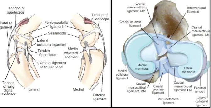
Figure 1 - Ligaments and menisci of the stifle joint ... 4
Figure 2 - Mediolateral radiographic views of a normal stifle and one with partial rupture of the cranial cruciate ligament ... 16
Figure 3 - Slocum’s model of the stifle joint ... 23
Figure 4 - Tepic’s model of the stifle joint ... 24
Figure 5 - Two techniques for advancement of the tibial tuberosity ... 26
Figure 6 - MMP with wire support ... 28
Figure 7 - MMP with staple support ... 29
Figure 8 - Components of the samples... 30
Figure 9 - Wire sample assembling... 32
Figure 10 - Staple sample assembling ... 33
Figure 11 - The 50 samples labelled and ready for testing ... 34
Figure 12 - Samples ready for testing ... 35
Figure 13 - Example of a load-displacement curve and the mechanical variables interpreted in this study ... 36
Figure 14 - Load-displacement curves of each group obtained by polynomial regression . 38 Figure 15 - Examples of a typical load-displacement curve for each group ... 39
Figure 16 - Comparison of mean and SD for Displacement at 100 N between groups ... 40
Figure 17 - Comparison of mean and SD for Displacement at 200 N between groups ... 41
Figure 18 - Comparison of mean and SD for Displacement at Failure between groups ... 41
Figure 19 - Comparison of mean and SD for Load to Failure between groups ... 42
Figure 20 - Comparison of mean and SD for Stiffness between groups... 43
Figure 21 - Comparison of mean and SD for Yield Load between groups ... 43
Figure 22 - Examples of implant failure by knot untwisting, in a 1.0 mm diameter wire sample and 0.8 mm sample ... 44
Figure 23 - Examples of block failure, in a 1.2 mm diameter wire sample, and 2.0 mm width staple ... .44
xi
List of Abbreviations and Symbols
CLB – Caudolateral bandCMB – Craniomedial band CT – Computed tomography
CTWO – Cranial tibial wedge osteotomy CaCL – Caudal cruciate ligament
CrCL – Cranial cruciate ligament D100 – Displacement at 100 N
D200 – Displacement at 200 N
DFAIL – Displacement at failure
ECM – Extracellular matrix
g/cm3 – Grams per cubic centimetre
ICN – Intercondylar notch Kg – Kilogram
LCL – Lateral collateral ligament LTF – Load to failure
MCL – Medial collateral ligament MMP – Modified Maquet Procedure MMT – Modified Maquet technique MRI – Magnetic resonance imaging mm – Millimeters
N – Newton
NSAID – Nonsteroidal anti-inflammatory drug p – p-value
SD – Standard deviation STIF – Stiffness variable TPA – Tibial plateau angle
TPLO – Tibial plateau levelling osteotomy TT – Tibial tuberosity
TTA – Tibial tuberosity advancement UK – United Kingdom
USA – United States of America YL – Yield load
YP – Yield point
1
Internship report
To fulfill the requirements of the Integrated Masters in Veterinary Medicine from the Faculty of Veterinary Medicine, University of Lisbon, I completed a 6-month training at Croft Veterinary Surgeons A&E Hospital, UK, between 7th October 2013 and 11th April 2014, in a total of approximately 1200 hours.
During that time I was deeply involved in the Hospital’s routine, assisting and participating, under supervision, in several procedures englobing different areas of referral and first opinion small animal Veterinary Medicine. Overall I was able to put my knowledge to practice and to develop my clinical case solving skills. In terms of Anaesthesia, I was able to assist and practice procedures such as induction, intubation, and general anaesthetic monitoring. In terms of Surgery, I was able to assist the surgeons during soft-tissue, orthopaedic, and spinal surgeries. Some of these included total ear canal ablation, portosystemic shunt resolution, lung lobectomy, Modified Maquet Procedure for tibial tuberosity advancement, total hip replacement, shoulder arthroscopy, hemilaminectomy, and ventral slot. With respect to Internal Medicine, I was mainly involved in clinical solving and participating in diagnostic tests. I was able to collect blood, catheterize and collect urine, and run several laboratory tests such as haematology, biochemistry, and urinalysis. In terms of Diagnostic Imaging, I was able to assist and practice patient positioning during radiographies and computerized tomography scans. In addition, I assisted the responsible surgeon during ultrasonography, endoscopy and MRI scans. After these procedures I was taught to interpret the results and relate them with the clinical case. I was also able to practice, under supervision of senior surgeons, basic surgical procedures such as castrations (dogs, cats, and rabbits), ovariohisterectomies (cats), and dentistry procedures such as teeth scaling.
In addition, I have participated in other veterinary-related activities. During my stay I attended several in-house lectures given by the senior surgeons, on subjects such as Orthopaedics, Dermatology, and Rabbit Medicine. In the beginning of April 2014 I have also attended all days of the BSAVA congress in Birmingham. Furthermore, I have also attended Orthomed UK’s Modified Maquet Procedure course given by Malcolm Ness, being a fundamental starting point for the study from which this thesis resulted.
From February 2014 to April 2014 the study was designed and developed. However, it was only possible to successfully perform the mechanical testing in June 2014.
3
I. Introduction
Since it was first described in 1926, canine cranial cruciate ligament (CrCL) rupture has been a focus of scientific research and generated a plenitude of literature. Albeit rare in the feline species, CrCL rupture is one of the most common causes of lameness in the dog, therefore, this dissertation shall focus mainly on canine CrCL failure. CrCL rupture has a marked clinical and economic relevance in small animal practice. A decade ago, pet owners in the USA spent approximately 1.32 billion dollars for treatment of this condition (Wilke, Robinson, Evans, Rothschild, & Conzemius, 2005). As research moves forward, the complex physiopathology is gradually clarified and the continuous pursuit for a superior treatment is far from its ending. At the time, proximal tibial osteotomies seem to be the most popular treatment, either by levelling of the tibial plateau or advancement of the tibial tuberosity (TT). Thus far, no technique has proven to surpass the other and further investigation is invariably warranted. The Modified Maquet Procedure is an alternative method of advancing the TT with its origins in human medicine, and offers a different surgical option for veterinary patients. However, the literature on this procedure is but scarce and further research becomes necessary.
1. Cranial cruciate ligament morphology and function
1.1. General anatomy
The canine stifle is a complex condylar synovial joint involving the femur, patella, and tibia (Evans & Lahunta, 2013a). It has two interdependent articulating components, the femoropatellar and femorotibial joints. The space between the convex femoral and tibial articulating surfaces is occupied by two wedge-shaped fibrocartilaginous structures, the medial and lateral menisci. Distally to the patella, within the fibrous layer of the capsule, lies the infrapatellar fat pad (Evans & Lahunta, 2013a). In addition to the patella, there are three more sesamoid bones in the stifle. The lateral and medial fabellae, caudal to the stifle on the lateral and medial femoral condyle, respectively, and a sesamoid in the tendon of origin of the popliteus muscle (Evans & Lahunta, 2013b). The joint capsule of the stifle joint is the largest in the body, forming three intercommunicable sacs: medial and lateral femorotibial, and femoropatellar sacs. The femoropatellar is considerably larger than the femorotibial sacs (Evans & Lahunta, 2013a).
4
Within each femorotibial sac lies a meniscus. The menisci are developments of the fibrous layer of the joint capsule and are covered by synovial membrane. However, the lateral meniscus has lost his attachment to the joint capsule (Evans & Lahunta, 2013a). Each meniscus is attached to the tibia by a cranial and a caudal meniscotibial ligament, and to each other by the transverse ligament (Fig. 1). The lateral meniscus is connected to the femur by the meniscofemoral ligament (Evans & Lahunta, 2013a). The lateral meniscus is smaller and more circular than the medial one (Pozzi & Cook, 2011). They are semilunar, wedge-shaped fibrocartilaginous discs, with a sharp concave axial, and thick convex abaxial borders (Evans & Lahunta, 2013a). The menisci are highly efficient shock absorbers that contribute to joint stability and stifle kinematics by enhancing congruity between the tibial plateau and the femoral condyles. In addition, by being consecutively submitted to loading-unloading cycles, they induce synovial fluid circulation, promoting joint nutrition and lubrication (Pozzi & Cook, 2011).
Figure 1: Ligaments and menisci of the stifle joint. (Left) Ligaments of the left stifle. (From:
Evans, H. E., & Lahunta, A. de. Arthrology. Miller’s Anatomy of the Dog, 4th ed., 2013, p 178. Saint Louis: Elsevier Health Sciences). (Right) Dorsal view of the ligaments and menisci of the right stifle. MM, Medial meniscus; LM, lateral meniscus. (From: Kowaleski, M. P., Boudrieau, R. J., & Pozzi, A. Stifle Joint. In Tobias, K. M. & Johnston, S. A., editors: Veterinary Surgery: Small Animal, 2012, p. 907. Saint Louis: Elsevier Saunders.)
5
Apart from the meniscal ligaments, there are four other main ligaments in the stifle joint: the medial and lateral collateral ligaments, and the caudal and cranial cruciate ligaments (Fig. 1). However, the portion of the tendon of the quadriceps femoris muscle between the patella and the tibial tuberosity is also commonly referred to as “patellar ligament” (Evans & Lahunta, 2013a). The medial collateral ligament (MCL) is proximally attached to an oval area on the medial epicondyle of the femur and distally to a rectangular area on the medial proximal tibia (Fig. 1). As the MCL passes along the joint capsule it blends with it and fuses with the medial meniscus (Vasseur & Arnoczky, 1981). The lateral collateral ligament (LCL) attaches proximally to an oval area on the lateral femoral epicondyle, courses caudodistally, and inserts on the fibular head (Fig. 1). The LCL has a superficial band that arises from the lateral femorofabellar ligament area, merges with the major component as it crosses the joint surface, and dispersedly inserts on the fascia of the fibularis longus muscle. Contrarily to what happens on the medial side of the joint, the LCL is loosely attached to the joint capsule and does not fuse with the lateral meniscus (Vasseur & Arnoczky, 1981).
The caudal cruciate ligament (CaCL) is attached proximally to a fossa in the ventral aspect of the lateral side of the medial femoral condyle, courses caudodistally, and inserts on the medial aspect of the popliteal notch (Fig. 1). It has two components that function independently of one another in flexion and extension, the cranial and caudal bands. Although it is not attached to the menisci, in some cases its femoral attachment may contain fibers of the femoromeniscal ligament (Arnoczky & Marshall, 1977). The CaCL crosses the CrCL medially at their proximal ends in the intercondylar fossa, and is slightly longer and broader than the CrCL (Arnoczky & Marshall, 1977; Heffron & Campbell, 1978; Evans & Lahunta, 2013a).
The CrCL is attached proximally to a fossa on the caudal portion of the medial side of the lateral femoral condyle, courses cranio-medio-distally, and attaches distally on the cranial intercondyloid area of the tibia, caudally to the cranial meniscotibial ligament of the medial meniscus and cranially to the cranial meniscotibial ligament of the lateral meniscus (Fig. 1; Arnoczky & Marshall, 1977; Heffron & Campbell, 1978). Due to the orientation of its femoral and tibial attachments it also presents a proximal-to-distal outward spiral of approximately 90 degrees (Arnoczky & Marshall, 1977). The CrCL has two components, the craniomedial (CMB) and caudolateral (CLB) bands, which are more individualized than those of the CaCL, and named according to their relative tibial attachment (Arnoczky & Marshall, 1977; Heffron & Campbell, 1978). The CLB is shorter and straighter than the spiral CMB (Heffron & Campbell, 1978). The CrCL has its narrowest portion at its middle and fans out
6
proximally and distally, and its length is directly proportional with body weight (Heffron & Campbell, 1978; Vasseur, Pool, Arnoczky, & Lau, 1985).
1.2. Microanatomy and neurovascular supply
The CrCL is mainly constituted by mostly parallel bundles of longitudinally oriented collagen fibers intercalated with fibroblasts (Heffron & Campbell, 1978). These collagen fibers are organized in sub-fascicles, which, in turn, are organized in fascicles (Yahia & Drouin, 1989). These fascicles have variable diameters as they can be composed of 1 to 10 sub-fascicles (Yahia & Drouin, 1989). Blood vessels, adipocytes, and elastin occupy the space between these subfascicles (Yahia & Drouin, 1989).
The cruciate ligaments are covered by a fold of synovial membrane that incompletely divides the joint in the sagittal plane, being absent only on the surface of contact between CrCL and CaCL (Arnoczky, Rubin, & Marshall, 1979; Vasseur et al., 1985). That synovial membrane is constituted by dense connective tissue and fibroblasts, being more cellular than the rest of the ligaments (Heffron & Campbell, 1978), and has many small holes that are thought to play a role in ligament nutrition (Kobayashi et al., 2006).
The major vascular contribution to the center of the stifle joint occurs from the genicular branches, which arise from the popliteal artery (Evans & Lahunta, 2013c). The blood supply to both cruciate ligaments is predominantly of soft tissue origin (Arnoczky et al., 1979), with the most important sources of vessels being the infrapatellar fat pad and the richly vascularized synovial membranes that ensheat the cruciate ligaments (Arnoczky et al., 1979; Kobayashi et al., 2006). The intraligamentous vessels are less abundant in the central part of the mid portion of both cruciates (Arnoczky et al., 1979; Vasseur et al., 1985). The CaCL appears to be better vascularized than the CrCL, having a greater density of periligamentous and synovial vessels than the CrCL (Tirgari, 1978; Arnoczky et al., 1979).
The main articular nerves of the stifle are the medial articular nerve, which branches from the saphenous nerve, the lateral articular nerve, which branches from the common peroneal nerve, and the caudal articular nerve, which branches directly from the tibial nerve or from one of its muscular branches (O’Connor & Woodbury, 1982). The first is the largest supplier to the stifle joint and the third is sometimes absent in dogs (O’Connor & Woodbury, 1982). There are axons that penetrate the centre of the cruciate ligaments branching from the nerves in the enveloping ligament synovium (Yahia, Newman, & St-Georges, 1992). Within the CrCL
7
there are proprioceptors and mechanoreceptors that can induce quadriceps and hamstring muscle activity (Yahia et al., 1992; Miyatsu, Atsuta, & Watakabe, 1993). Their number is highest in the proximal third of the ligament (Arcand, Rhalmi, & Rivard, 2000), but there are fewer than in the CaCL (Yahia et al., 1992).
1.3. Functional anatomy
In general, the bulk of the CrCL remains taut in extension and becomes relaxed in flexion, as opposed to the bulk of the CaCL which remains relaxed in extension and becomes taut in flexion (Arnoczky & Marshall, 1977). Each component of a ligament functions independently of the other during flexion and extension (Arnoczky & Marshall, 1977). In extension, the CMB and the CLB of the CrCL are taut; with flexion, the CMB curves and twists around the CLB, and there is a shift in tension from the CLB, which becomes relatively relaxed, to the CMB, which remains taut (Arnoczky & Marshall, 1977; Heffron & Campbell, 1978). With flexion, both the CrCL and the CaCL become twisted, albeit to a lesser extent in the latter (Arnoczky & Marshall, 1977). In the CaCL, the cranial part is loose in extension and becomes taut in flexion, with the opposite occurring on the caudal part (Arnoczky & Marshall, 1977).
1.4. Biomechanics of the cranial cruciate ligament-intact stifle
The stifle allows motion in three planes, which is characterized by a combination of three rotations during the swing phase and a pure flexion-extension motion during the stance phase of gait (Korvick, Pijanowski, & Schaeffer, 1994; Pozzi & Kim, 2011). In Labrador Retrievers, the flexion-extension range-of-motion varies between approximately 160° in full extension and 40° in full flexion (Jaegger, Marcellin-Little, & Levine, 2002; Mostafa, Griffon, Thomas, & Constable, 2010). However, the stifle does not work as a pure hinge joint, as the flexion-extension movement results from a combination of rolling and gliding of the femur on the tibia (Pozzi & Kim, 2011). With flexion, LCL becomes more relaxed and allows the lateral condyle to move further caudally, resulting in internal rotation of the tibia (Vasseur & Arnoczky, 1981). This rotation occurring over a range-of-motion has been named the “screw-home” mechanism (Pozzi & Kim, 2011). Albeit to a much lesser extent, there is also internal-external and varus-valgus rotational movement occurring at the stifle joint during the swing phase of the walking gait (Korvick et al., 1994). Translation motion is absent during the stance phase (Korvick et al., 1994).
Given the peculiar congruence between the femoral condyles and the tibial plateau, the stifle relies on dynamic and passive stabilizers to provide adequate joint stability (Pozzi & Kim, 2011; Hayes, Granger, Langley-Hobbs, & Jeffery, 2013). Dynamic stabilization of the stifle is
8
mainly provided by the quadriceps, hamstrings, and gastrocnemius muscles (Hayes et al., 2013). It is a result of simultaneous contraction (co-contraction) of different muscles (Pozzi & Kim, 2011). Their activation and accurate coordination is mediated by the nervous system, and is fundamental for joint stability (Miyatsu et al., 1993; Hayes et al., 2013). Passive stabilization is provided by the soft tissues surrounding or within the joint (Pozzi & Kim, 2011). The femorotibial ligaments, the menisci, and the joint capsule all contribute for passive joint stabilization (Pozzi & Kim, 2011; Hayes et al., 2013). The menisci generally act as secondary stabilizers, with their role depending on the integrity of the primary stabilizers, particularly the CrCL (Pozzi et al., 2006). In extension, both bands of the CrCL are in tension and prevent cranial tibial translation relative to the femur (Arnoczky & Marshall, 1977; Heffron & Campbell, 1978). However, the primary restrain against cranial tibial translation is the CMB, with the CLB being the secondary (Arnoczky & Marshall, 1977). The CaCL, on the other hand, is the primary check against caudal tibial translation (Arnoczky & Marshall, 1977). Because they twist on themselves, the CrCL and the CaCL are the primary check against internal rotation of the tibia (Arnoczky & Marshall, 1977; Vasseur & Arnoczky, 1981). However, in extension, work as the secondary restraint, as the MCL and the LCL constitute the primary restraint against internal rotation of the tibia (Vasseur & Arnoczky, 1981). The collateral ligaments are also responsible for prevention of external rotation of the tibia during both extension and flexion (Vasseur & Arnoczky, 1981). The CrCL is also the primary check against hyperextension, with the CaCL being the secondary (Arnoczky & Marshall, 1977). Regarding flexion, the role of the cruciates is not that well defined, but it is believed that they contribute to limiting flexion (Arnoczky & Marshall, 1977). The collateral ligaments are the primary check against varus or valgus angulation, but, should they fail, stability is provided by the cruciate ligaments, as they constitute the secondary restraint against that type of movement (Vasseur & Arnoczky, 1981).
9
2. Cranial cruciate ligament failure
2.1. Pathogenesis
Failure of the CrCL can occur by avulsion or rupture of the ligament. The former is rare, usually traumatic, and occurs in skeletally immature dogs as a result of the higher strength of the ligament compared with that of the bone itself (Gielen, Saunders, Ryssen, & Bree, 2011; Kowaleski, Boudrieau, & Pozzi, 2012). Acute rupture is also rare and is commonly characterized by tearing of the mid-portion of the ligament secondary to a traumatic event (Kowaleski et al., 2012). The majority of CrCL ruptures occur as a result of chronic degenerative changes within the ligament (Vasseur et al., 1985; Griffon, 2010). These changes include a decrease in ligament fibroblast cellularity, chondroid metaplasia of remaining ligament fibroblasts, loss of the normal architecture of the extracellular matrix (ECM) of collagen, and proliferation of the epiligamentous tissue (Vasseur et al., 1985; Hayashi et al., 2003). In spite of the proliferative epiligamentous repair response, there is no effective spontaneous bridging scar formation (Vasseur et al., 1985; Hayashi et al., 2003). These changes progress with age and are associated with the progressive partial or complete failure of the CrCL (Vasseur et al., 1985; Hayashi et al., 2003).
Despite the plenitude of published studies, the exact aetiopathogenesis of CrCL rupture remains incompletely understood (Cook, 2010; Griffon, 2010; Comerford, Smith, & Hayashi, 2011). It is believed to be influenced by multiple factors that lead to a vicious cycle of abnormal mechanics and abnormal biology, osteoarthritis progression, and overall failure of the stifle joint (Cook, 2010; Griffon, 2010; Comerford et al., 2011). Thus far, it is not known if the cascade of pathological processes is initiated by abnormal biomechanics (leading to biological changes), by abnormal biology (resulting in altered biomechanics), or by a mixture of both, but it is generally agreed that both factors play an important role in the progression of the disease (Cook, 2010; Griffon, 2010; Comerford et al., 2011).
Because antibodies to type I and type II collagen were found in the serum and synovial fluid of dogs with spontaneous CrCL rupture, it has been suggested that there may be an immune-mediated component in CrCL rupture (Niebauer, Wolf, Bashey, & Newton, 1987). However, further studies have showed that the synovial anti-collagen autoantibodies are not specific for CrCL disease and are unlikely to play a role in the initiation of CrCL damage (de Rooster, Cox, & Bree, 2000; de Bruin, de Rooster, van Bree, & Cox, 2007).
10
Causal factors related to the CrCL itself include cellular apoptosis and ECM metabolism. Some studies have suggested apoptosis may have a role in CrCL rupture rather than being simply a consequence of it (Gyger et al., 2007; Krayer et al., 2008). Comerford and colleagues (2005) observed that higher ECM turnover in the CrCL of dogs predisposed to rupture was related to greater stifle laxity and lower ultimate tensile strength. It has also been suggested that rupture may be influenced by the development of cellular ischemia resulting from the poor CrCL blood supply, particularly in the mid-portion of the ligament (Vasseur et al., 1985; Hayashi et al., 2003).
Hindlimb conformational abnormalities have been a focus of research. Distal femoral conformation has been investigated as a risk factor for CrCL, particularly the intercondylar notch (ICN; Comerford, Tarlton, Avery, Bailey, & Innes, 2006; Lewis, Allen, Henrikson, & Lehenbauer, 2008). It has been observed that dogs with uni or bilateral CrCL rupture have a narrower ICN when compared with normal dogs, and that dogs of high-risk breeds (such as Labrador and Golden Retrievers) have a narrower ICN than dogs of a low-risk breed (Greyhounds; Comerford et al., 2006; Lewis et al., 2008). These findings suggest that the CrCL impingement by the narrower ICN may cause biochemical changes within the ECM of the ligament, leading to a reduced CrCL structural integrity and predisposing it to increased laxity, thus leading to CrCL degeneration (Comerford et al., 2006; Comerford, 2011). Medial patellar luxation, with the associated genu varum and misalignment of the quadriceps mechanism, has been suggested to contribute to CrCL rupture by increasing stress on the ligament (Comerford, 2011). An increased tibial plateau angle (TPA) has also been suggested to predispose to CrCL rupture by leading to higher stresses loading to the ligament (Morris & Lipowitz, 2001). Although Morris and Lipowitz (2001) have observed that dogs with CrCL injuries has significantly greater TPA than normal dogs, the effect of TPA on CrCL is still controversial as further studies have failed to substantiate those findings (Wilke, Conzemius, Besancon, Evans, & Ritter, 2002 ; Reif & Probst, 2003; Guastella, Fox, & Cook, 2008). The angle between the patellar tendon and the tibial plateau as also been suggested to influence CrCL and was found to be marginally greater in stifles affected by partial rupture than in intact joints (Dennler, Kipfer, Tepic, Hassig, & Montavon, 2006; Schwandt et al., 2006). Cranial angulation of the proximal tibia has been described and suggested to be a risk factor for CrCL rupture (Read & Robins, 1982). In a study with Labrador Retrievers, this angulation has been shown to be greater in affected and predisposed (contralateral) limbs than in normal ones (Mostafa, Griffon, Thomas, & Constable, 2009). Inauen, Koch, Bass, and Haessig (2009) have identified TT width as a risk factor for CrCl rupture. In that study they suggested
11
that smaller TT widths would lead to a larger cranial tibial thrust, resulting in faster CrCL degeneration and therefore rupture in a younger population of dogs. In a study analyzing the morphometric characteristics of Labrador retrievers, Mostafa et al. (2009) observed that cranial angulation of the proximal tibia, excessive TPA, and distal femoral torsion appear more likely to contribute to the pathogenesis of CrCL disease than femoral angulation, increased inclination of the patellar ligament, ICN stenosis, and tibial torsion.
Hayes and colleagues (2013) have used electromyography to compare hamstring reflex of normal and CrCL-deficient dogs. They observed that the response of one of the components of the hamstring reflex is delayed in dogs with naturally occurring CrCL rupture. In dogs with unilateral rupture of the CrCL the response was altered in the contralateral stifle as well, suggesting that a delayed response could be a sign of chronic impairment of the dynamic stabilizers of stifle, which would then predispose to CrCL rupture (Hayes et al., 2013).
Obesity is considered to be a factor implicated in CrCL as higher bodyweight increases the loading forces occurring on the joint and, consequently, the strain on the ligament (Vasseur et al., 1985; Griffon, 2010). In addition, it can exacerbate dynamic imbalance caused by conformational abnormalities (Griffon, 2010). It also appears to exist a link between CrCL rupture and breed or neutering status, with some breeds and neutered dogs (regardless of gender) having a higher prevalence of disease (Whitehair, Vasseur, & Willits, 1993; Witsberger, Villamil, Schultz, Hahn, & Cook, 2008). Epidemiology is further discussed in “I.2.2. Epidemiology”. The higher prevalence of CrCL rupture in certain breeds may be due to genetic predisposition. In fact, a study with Newfoundlands has shown a possibly recessive mode of inheritance and heritability of 0.27, suggesting that, in that group, CrCL rupture could be attributed to genetics to a certain extent (Wilke et al., 2006).
2.2. Epidemiology
Rupture of the CrCL can affect dogs of any age or breed, and its prevalence has been increasing since the mid-sixties (Witsberger et al., 2008). Nevertheless, it appears that dogs older than 4 years of age are more likely to be diagnosed with CrCL rupture, with the highest prevalence occurring between 7 and 10 years (Whitehair et al., 1993; Witsberger et al., 2008). In addition, a study has identified Newfoundlands, Rottweilers, and Labrador Retrievers as breeds with higher odds of having CrCL disease (Witsberger et al., 2008). On the contrary, Miniature Dachshunds, Dachshunds, and Greyhounds appear to have a lower risk of suffering from CrCL disease (Witsberger et al., 2008). Whitehair and colleagues (1993) observed
12
higher prevalence of CrCL rupture in Rottweilers, Newfoundlands, and Staffordshire Terriers; and lower prevalence in Dachshunds, Basset Hounds, and Old English Sheepdogs. It has been shown that Rottweilers have less stifle stability and inferior CrCL structural and material properties than Greyhounds, which may explain to some extent the relative prevalence within each breed (Wingfield, Amis, Stead, & Law, 2000a; Wingfield, Amis, Stead, & Law, 2000b). Heavier dogs are more likely to suffer CrCL rupture, with higher prevalence being observed in patients weighting over 22 Kg (Whitehair et al., 1993; Duval, Budsberg, Flo, & Sammarco, 1999). In addition, Whitehair and colleagues (1993) observed that larger dogs ruptured their CrCL earlier in life, compared with smaller dogs. The results of these studies are consistent with the findings of Vasseur et al. (1985). In that study, they observed that in larger dogs, CrCL degeneration is more severe and has an earlier onset and faster progression. In one study, female dogs had higher prevalence of CrCL rupture than males (Whitehair et al., 1993). Neutered dogs have been shown to be more likely to have CrCL rupture than sexually intact dogs, regardless of gender (Whitehair et al., 1993; Duval et al., 1999; Witsberger et al., 2008). Bilateral rupture may be present on admission in approximately 11% to 17% of cases (Cabrera, Owen, Mueller, & Kass, 2008; Buote, Fusco, & Radasch, 2009). Moreover, in cases with unilateral CrCL loss, contralateral rupture may happen in 22% to 48% of patients (Moore & Read, 1995; de Bruin et al., 2007; Cabrera et al., 2008; Buote et al., 2009).
2.3. Biomechanics of the cranial cruciate ligament-deficient stifle
It is believed that the instability at the CrCL-deficient stifle joint, which translates in an increase in tangential shear forces and abnormal contact mechanics (Pozzi et al., 2006; Anderst & Tashman, 2009), has an important role in osteoarthritis progression (Pozzi & Kim, 2011). It has been shown that dogs with a CrCL-deficient stifle adapt to joint instability by decreasing the load on the affected limb and carrying it more flexed while walking and trotting (Korvick et al., 1994; DeCamp et al., 1996; Tashman, Anderst, Kolowich, Havstad, & Arnoczky, 2004; Ragetly, Griffon, Mostafa, Thomas, & Hsiao-Wecksler, 2010). These adapting mechanisms are believed to be a result of a neuromuscular response to the pain level induced by joint instability (Korvick et al., 1994; DeCamp et al., 1996; Ragetly et al., 2010). The majority of changes occurring after experimental transection of the CrCL are observed during the stance phase (Korvick et al., 1994; Tashman et al., 2004).
In vivo studies have identified that the loss of the CrCL consistently leads to a dramatic
13
1994; Tashman et al., 2004). However, tibial translation pattern changes in the long term, with cranial tibial translation decreasing at 2 years post-transection, as a result of a more persistent cranial tibial subluxation throughout the gait cycle rather than a return to normal kinematics (Tashman et al., 2004). Korvick et al. (1994) and Tashman et al. (2004) have suggested that quadriceps contraction is one of several factors that lead to cranial tibial translation during the stance phase. Because the flexion angles during the swing phase do not allow the quadriceps to induce cranial tibial luxation, it also suggested that the swing phase is CrCL-independent (Korvick et al., 1994; Tashman et al., 2004). As the findings of Korvick et al. (1994) and Tashman et al. (2004) are a result of experimental sectioning of the CrCL, Pozzi and Kim (2011) have suggested that cranial tibial translation in clinically occurring CrCL rupture may be less pronounced, as a result of periarticular fibrosis.
The existing concept of tibial instability in the CrCL-deficient stifle has been questioned. In a study by Böttcher and Rey (2010) using biplanar fluoroscopy, during the stance phase, caudal translation of the femur relative to the tibia was observed. In that study, the same motion pattern was observed even in chronic cases with periarticular fibrosis and apparent macroscopical stability. More recently, using uniplanar fluoroscopy, Rey, Fischer, and Böttcher (2014) assessed sagittal motion pattern of CrCL-deficient stifles, and their findings were consistent with those of Böttcher and Rey (2010). They observed that the femoro-tibial motion pattern was consistently characterized by a sudden caudal translation of the femur at early stance phase, with spontaneous repositioning at the end of the stance phase. In addition, no translational movement of the tibia was apparent during the stance phase (Rey et al., 2014). These findings appear to question the pre-existing concept of tibial instability in CrCL-deficient stifles and the validity of previous in vitro CrCL rupture models (Rey et al., 2014). Although it has been shown to occur in vitro (Warzee, Dejardin, Arnoczky, & Perry, 2001; Kim, Pozzi, Banks, Conrad, & Lewis, 2009), Tashman and colleagues (2004) have failed to show internal tibial rotation after CrCL loss in vivo. They have suggested that gait does not generate internal torques high enough for the CrCL to act as a restraint, and therefore, it would only be the secondary check against that motion after bone geometry, muscle forces, and other soft tissues. In addition, it has been shown that the abduction-adduction range-of-motion is altered after CrCL transection, increasing over the first year and being kept at high values at least until 2 years post-transection (Tashman et al., 2004). In CrCL-deficient stifles, medio-lateral translation was higher only during the first year, suggesting that gradual stabilization was achieved, possibly due to joint capsule thickening and osteophyte formation (Tashman et al., 2004).
14
2.4. History and clinical signs
In the majority of cases, although patient history is suggestive of trauma, further analysis reveals that the onset of hindlimb lameness is either insidious or occurs after a minor trauma during an everyday activity (Muir, 2011). Lameness in affected dogs is usually more severe following exercise or after periods of rest, and its duration is highly variable (Muir, 2011; Kowaleski et al., 2012). Although it is frequently weight-bearing, its severity usually depends on the extent of ligament disruption (Muir, 2011; Kowaleski et al., 2012). In most cases of complete CrCL rupture, there is a period of several days of non-weight-bearing lameness, followed by moderate to severe weight-bearing lameness (Kowaleski et al., 2012). Stiffness after rest is frequently associated, especially after periods of exercise, and on occasion, audible clicking during walking may be present (Kowaleski et al., 2012). In cases of relatively stable partial tears, lameness is more subtle, commonly bilateral, and more easily identifiable following strenuous activity (Muir, 2011; Kowaleski et al., 2012). In these cases, lameness is generally continuous and relatively refractory to nonsteroidal anti-inflammatory drugs (NSAID; Muir, 2011). Less frequently, patients can be presented with a history of major trauma (e.g. road traffic accident) which is usually associated with traumatic avulsion fracture of a CrCL attachment site (Muir, 2011).
Physical examination of affected dogs typically reveals uni or bilateral weight-bearing hindlimb lameness, stifle pain upon flexion and extension, variable crepitus, and possible audible clicking during walking associated with a meniscal tear (Muir, 2011; Kowaleski et al., 2012). Physical examination is of great importance in cases where bilateral rupture occurs as clinical signs may mimic those of a neurological disease (Muir, 2011). When lameness is bilateral, dogs may also alter their stance by leaning forward, in an attempt to unload the pelvic limbs (Muir, 2011). When unilaterally affected, dogs will present external rotation of the affected limb while sitting (abnormal “sit test”) and during walking (Muir, 2011; Kowaleski et al., 2012). In chronic cases, atrophy of pelvic limb musculature is evident, and periarticular fibrosis is most easily palpated on the medial side of the stifle (Muir, 2011; Kowaleski et al., 2012). This medial firm thickness is also referred to as “medial buttress” and is almost always indicative of CrCL rupture (Muir, 2011; Kowaleski et al., 2012). When a partial tear is present, full extension of the stifle usually elicits a pain response (Scavelli, Schrader, Matthiesen, & Skorup, 1990; Kowaleski et al., 2012). Joint effusion is typically found during examination of the stifle and is characterized by loss of definition of the medial and lateral borders of the patellar tendon (Muir, 2011; Kowaleski et al., 2012). In fact, it has
15
been shown that patellar tendon palpation presents the same sensitivity and specificity than radiography for CrCL disease detection (Carobbi & Ness, 2009).
Cranio-caudal tibiofemoral instability can be tested with the cranial drawer test or the tibial compression test. In the cranial drawer test the operator creates cranio-caudal tibial translation by applying a force to the tibia, while holding the femur stable (Kowaleski et al., 2012). In general, any resulting motion is considered abnormal; however, in immature dogs, a small degree of physiologic cranio-caudal instability of 1-3 millimeters may be present (Muir, 2011; Kowaleski et al., 2012). It is termed “puppy drawer” and is characterized by a sudden stop in tibiofemoral translation during the cranial drawer test, as opposed to the soft or spongy stop occurring with CrCL rupture (Muir, 2011; Kowaleski et al., 2012). Tibiofemoral sagittal instability should be assessed from nearly full extension to flexion (Kowaleski et al., 2012). In full extension the collateral ligaments become taut and partially or completely prevent cranial drawer (Kowaleski et al., 2012). In dogs affected with partial CrCL rupture, cranial drawer could not be elicited in half of the cases and the other half it was usually evident only when the joint was in flexion (Scavelli et al., 1990). When only the CMB is ruptured, cranial drawer is present in flexion only because the CLB is taut in extension; when rupture happened in the CLB, no cranial drawer is present because the CMB is taut throughout the whole range of motion (Arnoczky & Marshall, 1977). If CrCL rupture is suspected but cranial drawer cannot be elicited in a conscious dog even with the stifle in flexion, the test should be repeated under general anaesthesia (Scavelli et al., 1990).
In the tibial compression test (Henderson & Milton, 1978) the operator creates stifle joint compression by flexing and extending the tarsocrural joint, simulating the contraction of the gastrocnemius muscle (Kowaleski et al., 2012). Any cranial motion detected by monitoring the TT is considered abnormal and reflects CrCL impairment (Kowaleski et al., 2012).
While performing these tests, it is important to ensure appropriate placement of the examining fingers on the bony prominences, as failure to do so may allow interpretation of skin and soft tissue movement as tibiofemoral translation (Muir, 2011; Kowaleski et al., 2012). In dogs that are nervous or suffer from chronic arthritis with periarticular fibrosis, it may be particularly important to repeat these tests under sedation or general anaesthesia to ensure that even subtle instability is identified (Muir, 2011). Indeed, it has been shown that the sensitivity and specificity of these testes are far from ideal but greatly increased in anaesthetized patients (Carobbi & Ness, 2009).
16
2.5. Diagnostic imaging
Radiographic examination of the stifle should be performed in all cases to confirm stifle pathology, to verify osteoarthritis in routine cases, and to discard other differentials such as fracture or neoplasia (Kowaleski et al., 2012). Both stifles should be included for comparison (Vasseur, 2003). Stifle effusion is one of the earliest and most consistent findings, characterized by partial or complete replacement of the infrapatellar fat opacity by a soft tissue opacity in the lateral view (Kowaleski et al., 2012). Usually the fat-to-soft tissue opacity transition occurs at the cranial margin of the tibial condyle and courses proximo-caudally until it reaches the femoral condyle (Fig. 2). Any alteration to these limits is consistent with stifle effusion or infrapatellar fad pad oedema (Kowaleski et al., 2012). Other typical radiographic findings in CrCL disease are consistent with stifle osteoarthritis, including enthesiophytosis, osteophytosis, and subchondral sclerosis, and will depend on its degree (Kowaleski et al., 2012). Tibial compression stress radiography can be used to confirm or discard CrCL damage, particularly when cranial drawer is absent during physical examination (Bree, Rooster, & Gielen, 2011). It consists in performing two lateral views of the stifle in 90° of flexion, one in neutral position (no compression), followed by one while performing the tibial compression test (compression position) (Bree et al., 2011). Similar to what happens with a regular tibial compression test, cranial translation of the tibia in the second radiograph is considered abnormal. This is an easy, reliable, cheap technique that can detect partial and complete ruptures of the CrCL with high sensitivity and perfect specificity (Bree et al., 2011). In young dogs affected by avulsion of the CrCL, radiography may also help identify the avulsed bone fragment (Vasseur, 2003).
Figure 2: Mediolateral radiographic views of a normal stifle (Left) and one with partial
rupture of the cranial cruciate ligament (Right). (From: Kowaleski, M. P., Boudrieau, R. J., & Pozzi, A. Stifle Joint. In Tobias, K. M. & Johnston, S. A., editors: Veterinary Surgery: Small Animal, 2012, p. 919. Saint Louis: Elsevier Saunders.)
17
Computed tomography (CT) allows three-dimensional radiographic examination. It appears to be helpful when bone overlapping is to be avoided and detection of small bone fragments is important (Gielen et al., 2011). CT is, therefore, particularly useful for the confirmation and diagnosis of CrCL avulsion in young dogs (Gielen et al., 2011). However, it is not very useful to evaluate integrity of the cruciate ligaments or menisci (Gielen et al., 2011).
Single-plane fluoroscopy has been shown to be a highly repeatable and highly accurate non-invasive method of assessing stifle joint stability (Jones et al., 2014). It allows the analysis of dynamic motions that occur in conscious patients during regular daily activities (Jones et al., 2014). In patients with CrCL rupture, single-plane fluoroscopic analysis shows abnormal cranio-caudal tibiofemoral sagittal instability (Rey et al., 2014). Additionally, it can be used to evaluate the post-surgical stability and, consequently, the efficacy of dynamic stabilizing surgical procedures (Rey et al., 2014; Jones et al., 2014).
Ultrasonography is a reliable non-invasive imaging method that allows evaluation of intra and extra-articular soft tissues structures of the stifle joint (Cook, 2011). It can be particularly useful in the assessment of the menisci, quadriceps and long digital extensor tendons, collateral and patellar ligaments, and for the detection of osteochondritis dissecans and osteoarthritic changes, including joint effusion and synovitis (Gnudi & Bertoni, 2001; Arnault et al., 2009). However, its use as diagnostic method in CrCL rupture is limited, as it has been shown to identify CrCL rupture in approximately 15% to 20% of the cases only (Gnudi & Bertoni, 2001; Arnault et al., 2009). Rupture may be more difficult to identify if it is closer to the midsection or femoral attachment of the ligament (Cook, 2011). If it occurs nearer the tibial attachment it may be easier to identify, with the ligament appearing as a hypoechogenic structure surrounded by echogenic fat (Arnault et al., 2009; Cook, 2011). Effusion is usually mild to severe in acute cases (Cook, 2011). In chronic cases where identification of the ligament is possible, it may appear thickened, irregular, and with retracted rupture ends (Cook, 2011). Interstitial tears can be identified on occasion (Cook, 2011). Usage of ultrasound for the interpretation and diagnosis of stifle conditions is essentially dependent on the training and experience of the operator (Arnault et al., 2009; Cook, 2011).
18
Although the interest in magnetic resonance imaging (MRI) has been growing in Veterinary Medicine, it has yet to be shown that its cost-benefit ratio is superior to that of other existing diagnostic techniques (Scrivani, 2011). MRI is a non-invasive method that can be used to identify ligament rupture, but it appears to be most useful to diagnose pathologic changes in the menisci, subchondral bone, articular cartilage, or to identify a cause of lameness that may have passed undiagnosed by other methods (Scrivani, 2011).
2.6. Arthroscopy
Arthroscopy is presently considered the gold standard of joint evaluation (Kowaleski et al., 2012). It is a minimally invasive, highly accurate surgical technique with low intra and postoperative morbidity that provides a thorough evaluation and direct probing of the intra-articular structures, allowing the treatment of any existing lesions (Beale & Hulse, 2011; Kowaleski et al., 2012). Magnification of the cruciate ligaments, menisci, synovium, joint pouches, and tibial, femoral, and patellar cartilages allows a more accurate diagnosis of lesions and higher treatment precision (Beale & Hulse, 2011; Kowaleski et al., 2012). Arthroscopy is commonly used in the assessment and treatment of CrCL tears. The findings will depend on the stage of the disease and, in early partial tearing, include loss of the normal fiber pattern, ligament oedema, loss of ligament tension, and tearing of a portion of fibers (Kowaleski et al., 2012). The observed lesions will depend on the stage of the disease, with the proportion of torn fibers and ligament laxity gradually increasing (Kowaleski et al., 2012). In addition, with the progression of the disease, further osteoarthritis lesions can be found, such as synovitis, cartilage fibrillation and eburnation, or osteophytosis (Kowaleski et al., 2012). Arthroscopy is also very useful in the evaluation, diagnosis, and treatment of meniscal tears (Beale & Hulse, 2011; Kowaleski et al., 2012).
Arthroscopy-assisted arthrotomy consists in inserting an arthroscope in an arthrotomy incision. The advantages when compared with normal arthroscopy include the ability to use the arthrotomy incision for the placement of the arthroscopy instruments, a dramatic shortening of the arthroscopy learning curve, and a lesser probability of fluid extravasation within the surrounding soft tissues (Beale & Hulse, 2011). On the other hand, when compared with traditional arthrotomy, arthroscopy-assisted arthrotomy dramatically enhances visibility of the intra-articular structures, precision and accuracy of treatment, decreases pain, and allows an earlier return to function (Beale & Hulse, 2011).
19
3. Treatment of cranial cruciate ligament rupture
3.1. Conservative management
The main goals of conservative management are to minimize the clinical signs of osteoarthritis (particularly the associated pain), maintain or improve limb use, and, if possible, to slow disease progression (Jaegger & Budsberg, 2011). The use of a multimodal therapy allows a synergic effect of treatment acting in noncompeting modes of action (Jaegger & Budsberg, 2011). Multimodal therapy for the treatment of osteoarthritis incorporates the use of nonsteroidal anti-inflammatory drugs (NSAIDs), weight loss, and exercise modification (Jaegger & Budsberg, 2011; Vasseur, 2003). Other adjunctive analgesics, chondromodulating agents, nutraceuticals, and dietary supplements may also be added to therapy (Jaegger & Budsberg, 2011). In spite of the similar efficacies between different types of NSAIDs, some patients may show a different analgesic or adverse response to a determinate NSAID, thus it may be necessary to change NSAIDs until acceptable analgesia is achieved or the patient experiences an adverse response (Jaegger & Budsberg, 2011). Exercise modification passes by an initial exercise restriction and a controlled increase in activity (Vasseur, 2003). The rehabilitation program may include proprioceptive training, exercises of range-of-motion, and swimming (Vasseur, 2003; Arnoldy, 2011). Conservative management appears to be most suitable for dogs weighing less than 15 to 20 Kg (Pond & Campbell, 1972; Vasseur, 1984). Nevertheless, some authors still recommend surgical management in the majority of cases as a mean to minimize joint instability and disease progression (Vasseur, 1984; Piermattei, Flo, DeCamp, & Brinker, 2006a). Because osteoarthritis appears to have a different clinical impact between different dogs, and each patient responds differently to treatment, it is important to create and adjust the multimodal treatment according to a determinate patient’s needs and response to treatment (Jaegger & Budsberg, 2011).
3.2. Surgical management
Numerous treatments have been described for the treatment of stifle joint instability with the aim of resolving the lameness caused by joint instability and provide adequate long-term function of the injured limb (Kim, Pozzi, Kowaleski, & Lewis, 2008). Overall they can be divided in three categories: intra-articular, extracapsular, and tibial osteotomies.
Intra-articular reconstruction of the ligament can be obtained by ligament repair or ligament replacement. Ligament repair by apposition of the free ends of the ligament has been shown
20
to be a rather unsuccessful technique due to the intrinsic poor healing mechanisms of the ligament and the influence of the constant loads to which it is submitted (O’donoghue, Charles a. Rockwood, Frank, Jack, & Kenyon, 1966). Many techniques for ligament replacement have been described using different types of materials: autografts, allografts, and prosthetics (Manley, 2011). The first surgical procedure for the management of CrCL deficiency was described by Paatsama in 1952 and consists in an intra-articular fascia lata autograft placement (Paatsama, 1988). The free end of the lateral fascial strip was passed through bone tunnels drilled in the femur and the tibia to simulate normal CrCL attachments and orientation (Paatsama, 1988). From then on, plenty of intra-articular autograft techniques were described. Dickinson and Nunamaker (1977) described a modification of Paatsama’s technique in which only a femoral tunnel was drilled. In 1979, Arnoczky and colleagues described the over-the-top procedure, which consisted in passing the medial third of the patellar tendon proximally through the joint space, passing it over the top of the lateral femoral condyle, and securing it to the tissues on the lateral femoral condyle (Arnoczky, Tarvin, & Marshall, 1982). Later, Hulse, Michaelson, Johnson, and Abdelbaki (1980) described a modification of the over-the-top procedure using an autograft comprised of fascia lata, lateral retinacular fascia and the lateral third of the patellar tendon. Disadvantages of using these autografts include the need to undergo revascularization and the inability to replicate the mechanical properties of the CrCL (Arnoczky et al., 1982; Butler et al., 1983). More recently a study has shown that intra-articular stabilization with an autograft yields inferior results than extracapsular stabilization or levelling of the tibial plateau (Conzemius et al., 2005). In that study, dogs treated with the over-the-top procedure had worse limb function and a reduced chance of achieving a clinically substantial improvement than those treated with lateral suture stabilization or tibial plateau levelling osteotomy (Conzemius et al., 2005). The tissues more frequently used as allografts for CrCL replacement are the common calcaneal tendon and CrCL (Manley, 2011). Their use is not popular as they are associated with an increased immune-directed inflammatory response, difficulty of preservation, sub-optimal mechanical properties, and potential disease transmission (Manley, 2011). Prosthetics are synthetic grafts that can be used as primary replacement for the CrCL or as augmentation device for biologic graft protection, but, in spite of their apparent short-term success, long-term performance is impaired by wear and deterioration (Manley, 2011). Recently, the use of intra-articular scaffolds has gained some interest as they would have the advantage of performing like a prosthesis initially, but gradually allowing and promoting infiltration by soft tissue and undergo neoligamentization (Kowaleski et al., 2012). Despite the variety of
intra-21
articular techniques, the anatomical configuration and attachments of the CrCL are difficult to replicate with any type of graft, therefore, failure is frequently attributed to the inability to recreate these conditions rather than intrinsic graft mechanical properties (Manley, 2011). Due to the premature biological or mechanical failure of intra-articular grafts, extra-articular techniques are often the first choice of treatment (Manley, 2011).
There is a wide range of extracapsular stabilization techniques for the treatment of CrCL deficiency (Tonks, Lewis, & Pozzi, 2011). Extracapsular techniques are relatively simple procedures that do not require expensive highly technical equipment, and are associated with good outcomes, in terms of safety and efficiency, in CrCL-deficient patients (Cook, 2011). These are passive stabilizing techniques that rely on periarticular fibrosis for long-term stability, therefore, their goal is to provide adequate initial stability until adequate fibrosis is formed (Cook, 2011; Kowaleski et al., 2012). One of these procedures it the fibular head transposition that was first described by Smith and Torg (1985). It relies on the LCL to prevent cranial tibial translation and to minimize internal tibial rotation, by surgically moving the fibular head cranially (Smith & Torg, 1985). This technique appears to have become less popular due to the early and continued joint instability resulting from LCL elongation, and the frequent need for implant removal (Kowaleski et al., 2012). The lateral fabellotibial suture is a modification of the technique described by DeAngelis and Lau and is one of the most frequently used extracapsular procedures (DeAngelis & Lau, 1970; Cook, 2011; Kowaleski et al., 2012). It consists in passing a non-absorbable suture proximally around the lateral fabella and through a tunnel in the proximal tibial methaphysis (Kowaleski et al., 2012). Most of the complications associated with this technique result from failure of the stabilizing material due to inferior mechanical properties, inadequate placement, or fixation failure (Cook, 2011). However, this technique is appears to be superior to the fibular head transposition with regards to joint stability and limb function (Moore & Read, 1995). More recently a minimally invasive modification of the lateral fabellotibial suture has been described (TightRope CCL, Arthrex Inc., Naples, FL, USA) using a synthetic braided tape (FiberTape, Arthrex Inc., Naples, FL, USA) that is passed by a femoral and a tibial tunnel (Cook, Luther, Beetem, Karnes, & Cook, 2010). The tape is kept in place by two toggle buttons in a quasi-isometric position (Cook, Luther, et al., 2010). This technique is a viable alternative associated with good outcomes and a higher safety‐to‐efficacy ratio than tibial osteotomies (Christopher, Beetem, & Cook, 2013).
In 1984, Slocum and Devine described the cranial tibial wedge osteotomy (CTWO; Slocum & Devine, 1984). It was the first dynamic stabilizing technique to be described, as its main