Directory of Open Access Journals ©2008-2014. IJARNP-HS Publication
Original Research
Characterization and inhibitory activity of chitosan on
hyphae growth and morphology of Botrytis cinerea
plant pathogen
Silva Júnior S
1, Stamford NP
1*, Lima MAB
1, Arnaud, TMS
1, Pintado, MM
2, Sarmento, BF
3 1Department of Agronomy and Biology, Federal Rural University of Pernambuco, Recife - PE, Brazil2Centro de Biotecnologia e Química Fina-Laboratório Associado, Escola Superior de Biotecnologia, Universidade Católica Portuguesa, Porto, Portugal
3Institute of Biomedical Engineering, University of Porto, Porto, Portugal
Summary. Low and high molecular weight chitosan were tested in different concentrations and growth times with the aim to evaluate the inhibitory activity against Botrytis cinerea, a very important plant pathogen. Tested chitosans were characterized by vibratory spectroscopy and elementary analyzes to determine the deacetylation degree. In addiction molar mass was estimated by viscosity measuring. Scanning electron microscopy was utilized for antimicrobial activity observation. Results showed that both chitosans markedly inhibited fungal growth, which was effected by incubation time and chitosan concentration. Scanning electron microscopy observations revealed that chitosan induced changes in surface morphology. The present study show that chitosan is capable of inhibit the growth and cause serious damage to the cell structure of the B. cinerea, as well as have the ability to form an impervious layer around the cell. Therefore, chitosan could be considered as a potential alternative for synthetic fungicides.
Industrial relevance. Ultrastructural analysis showed that chitosan is capable of causing serious damage to the cell structure of the B. cinerea, as well as have the ability to form an impervious layer around the cell. Chitosan could inhibit the growth of B. cinerea in vitro and
consequently may be considered as a potential alternative in replacement of synthetic fungicides. Keywords. biopolymer; chitosan; antifungal activity; fungal morphology; electron microscopy
INTRODUCTION
The resistance to diseases is an important factor that influences yield and quality of economic crops. Numerous strategies have been proposed to control fungi pathogens, and during many years until recently date, the most used methodology has been the application of fungicides. However, the use of chemical products can be harmful to living organisms besides promote reduction of soil microorganisms (Ahmed 2011).
To improve plant resistance against diseases is necessary to evaluate new products or strategies that do not cause problems to plants quality and to environment, and in particular to human and animal nutrition. Recently, studies reported chitosan as an important natural product that induces mechanisms of plant defense against diseases (El Hadrami, 2010). Furthermore, this biopolymer displays antimicrobial activity against bacteria and fungi. The main hypotheses suggested that chitosan alter the permeability of the plasmatic membrane and also promote oxidative stress on pathogenic fungi (Di Piero and Garda 2008, Kong et al. 2010, Badawy et al. 2011, Hernández-Lauzardo et al. 2011). The application of chitosan may be considered as a valuable strategy to assure biological control of pathogenic fungi, reducing or avoiding the use of fungicides (Badawy et al. 2011, Hernández-Lauzardo et al. 2011). Chitosan is the N-deacetylated derivative of chitin (Figure 1) composed by units of 2-amino-2-desoxi-Dglycopyranose and of 2-acetamide-2-desoxi-D-glycopyranose interconnected by glycosidic bonds β-1,4 (Stamford, 2012).
Chitosan inhibiting Botrytis cinerea
Chitosan found in crustaceous, mollusks and in the cell wall of some fungi has demonstrated relevant properties, especially biocompatibility, biodegradability and reactivity of the amino group, which open opportunities for numerous applications in various commercial products (Silva et al. 2006; Harish Prashanth and Tharanathan, 2007; Fai et al. 2008), in particular as antimicrobial agent (Feng and Xia 2011).
Botrytis cinerea is a phytopathogenic fungus that attacks flowers, fruits, leaves, and stems of more than 200 agriculturally important plant species worldwide causing serious pre- and post-harvest loss. This fungus has great genotypic and phenotypic variability, and adaptability to diverse environments which contributes to the rapid evolution in virulence and fungicide resistance. On the other hand, the cost of bringing a new fungicide or biological control agent to market is very high. Therefore, alternative control strategies such as chitosan that can also induce to resistance in plants has attracted attention (Elad, 1995; Pande et al., 2006; Staats et al., 2005).
Several studies about the chitosan effects on phytopathogenic fungi have demonstrated that the response is variable and dependent of the fungal cell (Hernández-Lauzardo, 2011). Furthermore, studies about effects of inhibitors on cell morphology are important because the morphological changes may suggest the target or mechanism of action of this inhibitor (Versentini et al. 2007). On the other hand, there are few studies about effects of chitosan on the surface morphology of Botrytis cinerea (Enio, 2012a e 2012b), therefore it is necessary more study about the ultrastructural effects of chitosan on this important phytopatogenic fungus.
So the aim of this study was to in vitro evaluate the antifungal activity of characterized chitosan with different molecular weights against Botrytis cinerea, as well as to understand the mechanisms of action through the study of the ultrastructural damage of hyphae caused by chitosan using scanning electron microscopy.
MATERIALS AND METHODS
Microorganisms and growth conditions. The phytopathogenic fungi Botritis cinerea (MUM 10166) purchased from
culture collection of the University of Minho (Braga, Portugal) was used in this study. The isolate was maintained in Potato Dextrose Agar (PDA) at 5 °C. The PDA was also used for agar disk production at 28C during 4 d in order to obtain inocula for growth inhibition test and large scale spore production at 28 C during 15 d for morphologic studies.
Chitosan solution and media preparation. High and low molecular weight chitosans were obtained from Sigma-Aldrich
(St. Louis, USA). Characteristics reported by provider was for high molecular weight chitosan a DD > 75 % and a MW of 624 kDa and for low molecular weight chitosan a DD between 75 and 85 % and a MW of 107 kDa. Stock solutions of chitosan at 20 mg ml-1 were prepared in 1% (v/v) solution of glacial acetic acid 99 % (Panreac, Barcelona, Spain). Chitosan
was added to 1% acetic acid to the desired concentration. Afterwards, the solution was stirred overnight at 50º C to promote complete dissolution of chitosan. The pH was adjusted with NaOH (Merck,Darmstad, Germany) to a final value of 5.6-5.8 and solutions were stored at refrigerated temperature. Stock solution was autoclaved at 121ºC for 15 min and then added to
sterile PDA (Potato Dextrose Agar) media to a final concentration of 0.5; 1.0; 1.5; 2.0 and 4.0 mg ml-1.
Chitosan characterization. To verify properties of Chitosans purchased from Sigma-Aldrich®, deacetylation degree was
determined by using infrared spectroscopy and molecular weight by viscosity.
Infrared Spectroscopy (Degree of Deacetylation- DD%). The degree of deacetylation (DD%) for microbial chitin and chitosan was determined using infrared spectroscopy as per Baxter et al. [47], using the absorbance ratio A1655/A3450 and calculated as per Equation (1):
DD (%) = 100 − [(A1655/A3450) × 115] (1)
A two-milligram sample of fungal chitin and chitosan, which had been dried overnight at 60°C under reduced pressure was thoroughly blended with 100 mg of KBr to produce 0.5 mm thick disks. The disks were dried for 24 h at 110°C under reduced pressure. An infrared spectrometry reading was taken with a Bruker 66 Spectrometer (Bruker Corporation Inc., Billerica, MA, USA), using 100-mg KBr disks for reference.
Viscosity (Molecular Weights). The viscosity of 1% chitosan in buffer solution (acetic acid/sodium acetate, pH ~4.5), was determined using a Brookfield digital Rheometer (Model DV-II, Brook Engineering laboratories, Inc., Stoughton, MA, USA) at 25 °C, Spindle CPE-40, 0.5 mL sample volume as described by Stamford et al 2007. The molecular weight of chitosan was determined using an AVS-350 viscometer (Schott-Geräte, Québec, Canadá), type/capillary: Cannon-Fenske dinside = 1.01 mm, at 25 °C. After obtaining the intrinsic viscosity from tables, K and a, were obtained for HAc/NaAc. K =
0.076, a = 0.76. The flow time was determined in seconds. Using the Mark-Houwink equation, the average viscosimetric molecular weight was expressed in g·mol−1 [48]. See Equation (2):
[η] = K( v)a (2)
Inhibitory activity of chitosan. The effects of two chitosan with two molecular weight on micelial growth of Botrytis
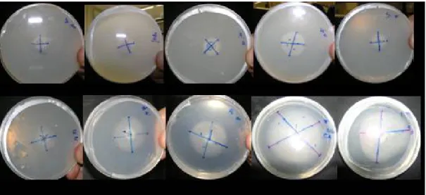
cinerea fungi was studied using PDA agar plates following the modified methodology of Di Piero and Garda (2008). Briefly, a PDA agar disk (0.5cm diameter) of a pure culture of B. cinerea after 4 days of growth was placed onto the center of each PDA plates containing chitosan at 0,5, 1,0, 1,5, 2,0 and 4,0 mg ml-1 (Figure 2). Petri plates were inoculated in five replicates
and incubated at 26C, during 6 days. Radial growth measurements, by measure of the halo diameter (mm), were taken each 24 h until fungi covered the plate. As absolute control (AC) was used only the PDA medium, and added a treatment as relative control (RC) applying acetic acid at 1% in place of chitosan. Growth percentage was determined as the growth in each plate amended with chitosan respect to that of the corresponding control plate.
Figure 2. Evaluation of crustaceous chitosan applied in different concentrations inhibiting the growth of Botrytis cinerea in Petri dishes with BDA medium.
Scanning Electron Microscopy. Mycelia samples collected after 3 d of cultivation at 26oC on the PDB (Potato Dextrose Broth) medium containing low and high molecular weight chitosan at 1, 2 and 4 mg/ml concentrations were washed twice in PBS, pH 7.2, for 10 min. Then they were fixed with 2.5% glutaraldehyde in 0.1 M phosphate buffer, pH 7.4, for 1 h at room temperature. After the stage-setting, all samples were again washed twice with phosphate buffer, for 10 min. This procedure was followed by the post-fixing with osmium tetroxide 1% in phosphate buffer, for 1 h at room temperature, in absence of light. Then the samples were once again washed with 0.1 M phosphate buffer, and submitted to the process of dehydration. The dehydration of the samples was done with ethanol, in concentrations of 50, 70 and 90% (5 min for each exchange) until the proportion of 100% (three times, 10 min each exchange). After this step, the samples were submitted to the drying in hexamethyldisilazane (HMDS), followed by the assembly in support of aluminum and subsequent gold/palladium metallization. Samples were examined and photographed in the Scanning Electronic Microscope, JEOL LV 5600, operating at 22 kV.
RESULTS
Deacetylation degree and Molecular weight. The deacetylation degree is the most used parameter for chitosan
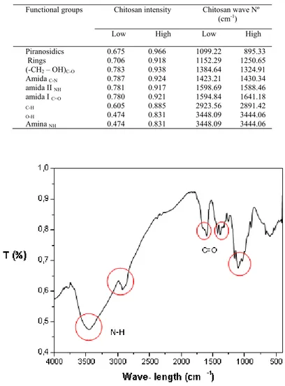
characterization, and is defined as the number of amines group in relation to the number of amides group of the polymeric chain. Infrared spectroscopy (Figure 3) allows the observation and classification of some bands on the vibration characteristics of the functional groups present in the structure of chitosan. In order to determine the exact desacetylation degree the bands in the infrared spectra of ChD and their assignments was determined and reported in Table 1.
Elemental analysis is a tool that can be used to determine the deacetylation degree of chitosan. Although is considered an accurate method, it must be used with great caution due to different moisture contents, which vary depending on storage conditions and pretreatment of the sample. The both analyzed chitosan showed the followed results: C – 390.1 g kg-1; N –
71.2 g kg-1; H – 105.8, g kg-1 and deacetylation degree 80%.
The viscosity of polymeric solutions is the measurement of the hydrodynamic volume of the polymer in solution, due to the large difference in size between the polymer molecules and the solvent. Intrinsic viscosity (η) as function of average molecular weight (M) is represented by Mark-Houwink-Sakurada equations η = KMα where, K and α are constants for a given polymer–solvent–temperature system. Our data was confirmed those supplied by the manufacturer and the found values for the average molecular weight of chitosan ChD are shown in Table 2.
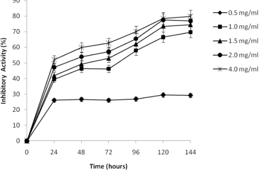
Effect of chitosan on fungal growth. The antifungal effect of chitosan with low and high molecular weight on growth of
Botrytis cinerea at different times and concentrations is shown in Figures 4 and 5. Generally, chitosan with both molecular weights showed satisfactory antifungal activity against B. cinerea and it depends on the concentration and the period of growth. Therefore, antifungal activity of chitosan on growth of B. cinerea was progressively increased during the incubation time.The radial growth of B. cinerea was inhibited by from 69.7% to close 80% after 144 h of incubation when the concentration of chitosan with low molecular weight increased from 1 to 4 mg ml-1 (Figure 4). However, at the lowest
Chitosan inhibiting Botrytis cinerea
Table 1. Infrared band of ChC and ChCD6H.
Functional groups Chitosan intensity Chitosan wave Nº (cm-1)
Low High Low High Piranosidics Rings 0.675 0.706 0.966 0.918 1099.22 1152.29 895.33 1250.65 (-CH2 – OH)C-O 0.783 0.938 1384.64 1324.91 Amida C-N 0.787 0.924 1423.21 1430.34 amida II NH 0.781 0.917 1598.69 1588.46 amida I C=O 0.780 0.921 1594.84 1641.18 C-H 0.605 0.885 2923.56 2891.42 O-H 0.474 0.831 3448.09 3444.06 Amina NH 0.474 0.831 3448.09 3444.06
Figure 3. Infrared spectrum of chitosan.
Table 2. Values of intrinsic viscosity (IV), deacetylation degree (DD), purity and average viscosimetric molecular weight for chitosan studied.
Chitosan IV [] (ml g-1)
Massa Molar
(g mol-1) DD (%) Purity (%)
Low Molecular Weight 3.8278 5.2 x 10³ 80 85
Figure 4. Effect of concentration of low molecular weight chitosan upon micelial growth of Botrytis cinerea during 144 h at 26oC. Growth rates relative to corresponding control plates. Bars represent standard deviations of the means.
For the high molecular weight chitosan, the reduction of fungal growth varied from 60% to close to 80% as the concentration increased from 1 to 4 mg ml-1,after 144 h of incubation (Figure 5). It was also observed that when applied in
small concentration (0.5 mg ml-1) the biopolymer inhibited 39.7% of radial growth within 144 h.
Figure 5. Effects of chitosan concentration with high molecular weight on micelial growth of Botrytis cinerea during 144 h at 26oC. Growth rates relative to corresponding control plates. Bars represent standard deviations of the means.
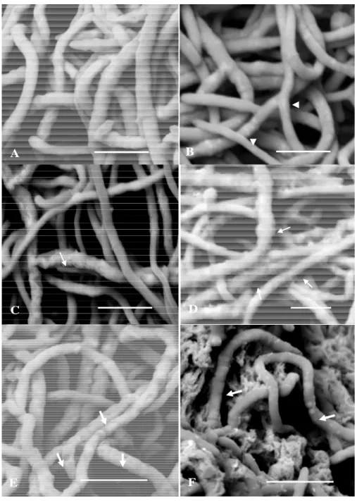
Effect of chitosan on hyphae morphology. Observations of B. cinerea mycelia after exposure to different concentration
of chitosan are presented in Figure 6. In control culture, the presence of regular and homogeneous hyphae, with smooth cell wall, is shown in Figure 6A. Examination of the treated mycelium at concentration 1 mg ml-1 using low molecular weight
Chitosan inhibiting Botrytis cinerea
The increasing of the concentration up to 2 mg ml-1, the hyphae showed a coating with chitosan precipitates. The
precipitate is visualized as lumps and mesh with granular and membranous texture, respectively. The presence of some depressions on the hyphae is also observed (Figure 6F). Additionally, at concentration of 4 mg ml-1 of chitosan no micelial
growth of B. cinerea was observed.
A
B
C
D
E
F
▼ ◄Figure 6. Scanning electron micrographs of B. cinerea mycelia after 72 hours of cultivation on Sabouraud broth with or without low and high molecular weight chitosan at 26oC and orbital shaking of 150 rpm. Control (A); low molecular weight chitosan at 1 mg ml-1 (B); 2 mg ml-1 (C) and 4 mg ml-1 (D). Note thin hyphae (arrowhead) and hyphae showing granular and corrugate surface (arrow). High molecular weight chitosan at 1 mg ml-1 (E) and 2 mg ml-1 (F) showing several damaged hyphae (large arrow). Scale bars = 10µm.
DISCUSSION
Deacetylation degree and Molecular weight. Two methods were used to determine the deacetylation degree of chitosan:
proton nuclear magnetic resonance (NMR 1H) and elemental analysis. NMR 1H is a technique that allows the quantification
of the highest degree of deacetylation. The spectra obtained for ChD showed broad peaks at 3448.09 and 3444.06 cm-1,
the NH amino group (Nunthanid et al. 2001). However, Kasaai (2008) reports that the free primary amine group (-NH2) at
the C2 position of the glucosamine has another peak in the region between 1220 and 1020 cm-1.
The peaks at 2923.56 and 2891.42 cm-1 represent CH aliphatic stretching in the ChD samples, respectively. The fact that
the peaks at 1594.84 and 1641.18 cm-1 correspond to the amine group of the acetylated chitin indicates that the samples are
not fully desacetylated (Rinaudo 2006). However, Kasaai (2008) specifies the peak near the 1655 cm-1 band as one
corresponding the axial deformation of C = O (amide I), and the vibrational mode of angular deformation of the connection NH (amide II) appears as a shoulder at 1607 cm-1.
The peaks at 1384.64 and 1324.91 cm-1, respectively represent the CO stretching of the primary alcoholic group (-CH 2 -
OH) of the ChD samples. The axial deformation of the amide CN appears at 1423.21 and 1430.34 cm-1 for the Aldrich
chitosan desacetylated by the manufacturer. The intense band between 800 and 1200 cm-1 is linked to the pyranosidic rings
(Shigemaza et al. 1996).
Effect of chitosan on fungal growth. Chitosan with low and high molecular weight showed similar antifungal activity.
Furthermore, the results showed that both chitosans at concentration greater than 4 mg ml-1 can significantly inhibit the
growth of B. cinerea. However, since chitosan at the highest concentration within 144 h could not completely inhibit the growth of B. cinerea, denotes that chitosan effect was fungiostaticrather than fungicidal.
Our results corroborate others obtained for other molds, namely those reported by Coqueiro and Di Piero (2011) that evaluated chitosan from Sigma Aldrich with different molecular weights upon the growth of the fungus Alternaria solani, and demonstrated the inhibition effect upon micelial growth and germination of conidia, without significant difference between molecular weights when chitosan was applied in concentrations from 0.2 to 0.5 mg ml-1.
Furthermore, other studies have shown the importance of molecular weight on the antifungal effects of the chitosan. Pacheco et al. (2008) observed the best inhibitory effect of low molecular weight chitosan on the micelial growth and germination of Penicillium digitatum. In addition, Lauzardo et al. (2008) found greater inhibitory effect when applied chitosan with low molecular weight on Rhizopus stolonifer. However, Lauzardo et al. (2011), reported in a review that effect of the molecular weight of chitosan on the growth of different phytopathogens fungi is variable.
On the other hand, Liu et al. (2007) showed the mycelia growth of Botrytis cinerea was completely inhibited by low molecular weight chitosan with 90% deacetylation at 0.5%, whereas in our work did not obtain total growth inhibition of this fungus using low molecular weight chitosan with 75 to 85% deacetylation even at the highest concentration tested. These results demonstrated that deacetylation degree influenced on antifungal activity of chitosan on Botrytis cinerea.
Therefore, we assume that the possible reason for the similar inhibitory effect on growth of B. cinerea may have been the similar deacetylation degree of chitosan used in this study. The results of this study suggest that chitosan may be used as alternative against the fungi pathogenic B. cinerea that cause the gray mold disease in fruits of grape, and constitute a cause of productivity loss in these cultures
Effect of chitosan on hyphae morphology. Comparing the actions of the chitosans under study, the high molecular
weight chitosan affected fungal surface morphology more severely than the low molecular weight chitosan despite the fact similar inhibitory activity for both chitosan. The apparent discrepancy between morphologic and antifungal effects is due to the fact that ultrastructural analysis have been carried out at 72 h of growth and at this moment the high molecular weight chitosan showed higher inhibitory activity than low molecular weight chitosan. Besides that ultrastructural study was performed in liquid medium which allows greater contact of chitosan with the mycelium of the Botrytis cinerea.
Therefore, SEM analysis demonstrated morphological changes for all concentrations and types of chitosan used, but these changes were much more severe with the highest concentrations. Morphological similar changes were reported in Trichoderma harzianum hyphae treated with chitosan, which exhibited hyphae surface intensely corrugated and with an extracellular layer around it (Versentini et al. 2007). Oliveira-Junior.et al. (2012a and 2012b) also observed that chitosan coated the mycelia of the B. cinerea.
All these morphological changes mediated by low and high chitosan observed in this work as distorted and damaged hyphae and a layer on the cell surface of the B. cinerea ultimately can directly or indirectly result in the death of hyphae and therefore lead to growth inhibition of the fungus.
The results showed that the chitosan used in this study is capable of inhibit the growth and damage to the cell structure of B. cinerea, as well as have the ability to form an impervious layer around the cell. Nevertheless, these results are preliminary and based on in vitro testing. Therefore, more studies are required to clarify the antifungic mechanism of chitosan and to determine it is potential as fungicide.
ACKNOWLEDGMENTS
The authors are indebted to the Conselho Nacional de Desenvolvimento Científico e Tecnológico (CNPq), Coordenação de Aperfeiçoamento de Pessoal de Nível Superior (CAPES), Fundação de Amparo à Ciência e Tecnologia do Estado de Pernambuco (FACEPE), Brazil, and Fundação para a Ciência e Tecnologia (FCT) from Portugal for financial support and fellowships.
REFERENCES
Ahmed M (2011). Management of Fusarium wilt of tomato by soil amendment with Trichoderma koningii and a white sterile fungus. Indian
Chitosan inhibiting Botrytis cinerea
Coqueiro DSO, Di Piero RM (2011). Atividade de quitosanas com diferentes pesos moleculares sobre Alternaria solani. Arq Inst Biol, 78, 459-463.
Costa Silva HSR, Santos KSCR, Ferreira EI (2006). Quitosana: Derivados hidrossolúveis, aplicações farmacêuticas e avanços. Química
Nova, 29, 776-785.
Di Piero N, Garda A (2008). Quitosana reduz a severidade da antracnose e aumenta a atividade de glucanase em feijoeiro-comum. Pesq
Agropec Bras, 43, 1121-1128.
Elad Y, Evensen K (1995). Physiological aspects of resistance to Botrytis cinerea. Phytopathology, 85, 637–643.
Fai AEC, Stamford TCM, Stamford TLM (2008). Potencial biotecnológico de quitosana em sistemas de conservação de alimentos. Rev
Iberoamer Polymer, 9, 435-451.
Feng Y, Xia W (2011). Preparation, characterization and antibacterial activity of water-soluble O-fumaryl-chitosan. Carbohyd Polymer, 83, 1169–1173.
Fernandez-Megia E, Novoa-Carballal R, Quiñoa E, Riguera R (2005). Optimal routine conditions for the determination of the degree of acetylation of chitosan by 1H-NMR. Carbohyd Polymer, 61,155–161.
Kasaai MR (2008). A review of several reported procedures to determine the degree of N-acetylation for chitin and chitosan using infrared spectroscopy. Carbohyd Polymer, 71, 497–508.
Kong M, Chen XG, Xing K, Park HJ (2010). Antimicrobial properties of chitosan and mode of action: A state of the art review.
International Journal of Food Microbiology, 144, 51–63.
Lauzardo AHN, Bautista-Baños S, Velázquez-Del Valle MG, Méndez-Montealvo MG, Sánchez-Rivera MM, Bello-Pérez LA (2008). Antifungal effects of chitosan with different molecular weights on in vitro development of Rhizopus stolonifer (Ehrenb.:Fr.) Vuill.
Carbohyd Polymer, 73, 541-547.
Lauzardo AHN, Velázquez-del Valle MG, Guerra-Sánchez MG (2011). Current status of action mode and effect of chitosan against phytopathogenic fungi. African Journal of Microbiology Research, 25, 4243-4247.
Liu J, Tian S, Meng X, Xu Y (2007). Effects of chitosan on control of postharvest diseases and physiological responses of tomato fruits.
Postharvest Biol. Technol, 44, 300-3006.
Nunthanid J, Puttipipatkhachorn S, Yamamoto K, Peck GE (2001). Physical Properties and Molecular Behavior of Chitosan Films. Drug
Development Indust Pharm, 27, 143-157.
Oliveira Junior EN, Melo IS, Franco TT (2012a). Changes in hyphal morphology due to chitosan treatment in some fungal species.
Brazilian Arch. Biol Technol, 55, 637-646.
Oliveira Junior EN, El Gueddari NE, Moerschbacher BM, Franco TT (2012b). Growth rate inhibition of phytopathogenic fungi by characterized chitosans. Brazilian J Microbiol, 800-809.
Pacheco N, Larralde-Corona CP, Sepulveda J, Trombotto S, Domard A, Shirai K (2008). Evaluation of chitosans and Pichia guillermondii as growth inhibitors of Penicillium digitatus. Int J Biol Macromol, 43, 20-26.
Pande S, Galloway J, Gaur PM, Siddique KHM, Tripathi HS, Taylor P, MacLeod MJ, Basandrai AK, Bakr A, Kishore JGK, Isenegger DA, Rao JN, Sharma M (2006). Botrytis grey mould of chickpea: a review of biology, epidemiology, and disease management. Aust. J. Agric.
Res, 57, 1137-1150.
Prashanth KVH, Tharanathan RN (2007). Chitin/chitosan: modifications and their unlimited application potential—an overview. Trends in
Food Science & Technology, 18, 117–131.
Rinaudo M (2006). Chitin and chitosan: Properties and applications. Progr Polym Sci, 31, 603–632.
Shigemaza Y, Matsura H, Sashiva H, Saimoto H (1996). Advances in Chitin Science, In: Domard A, Jeuniau C, Muzzarelli RAA, Roberts G An improved IR spectroscopic determination of degree of deacetylation of chitin. Jacques Andre´ Publishers, Lyon, France. 204-209. Silva MCF, Stamford TCM, Franco LO, Campos-Takaki GM (2006). Effects of salinity and glucose on chitin and chitosan production by
Cunninghamella elegans J Asian Chitin, 2, 29-38.
Staats M, Van Baarlen P, Van Kan JAL (2005). Molecular phylogeny of the plant pathogenic genus Botrytis and the evolution of host specificity. Mol. Biol. Evol, 22, 333–346.
Stamford TCM, Stamford TLM, Stamford NP, Neto BB, Campos-Takaki GM (2007). Growth of Cunninghamella elegans UCP 542 and production of chitin and chitosan using yam bean medium. Electronic Journal of Biotechnology, 10, 61–69.
Stamford TCM, Stamford-Arnaud TM, Cavalcante HMM, Oliveira R, Campos-Takaki GM (2013). Microbiological Chitosan: Potential Application as Anticariogenic Agent. In: Andrade AO, Pereira AA, Naves ELM, Soares AB (Org.). Practical Applications in Biomedical Engineering. 1ed. Rijeka: InTech - Open Access Publisher, 2, 229-244.
Synowiecki J, Al-Khatteb NAA (2003). Production, properties, and some new applications of chitin and its derivatives. Crit Rev Food Sci
Nut, 43, 144-171.
Tolaimate A, Desbrieres J, Rhazi M, Alagui A (2003). Contribution to the preparation of chitins and chitosans with controlled physico-chemical properties. Polymer, 44, 7939–7952.
Vesentini D, Steward D, Singh AP, Ball R, Daniel G, Franich R (2007). Chitosan-mediated changes in cell wall composition, morphology and ultrastructure in two wood-inhabiting fungi. Mycol Res, 874-890.
Yadav AV, Bhise SB (2004). Chitosan: a potential biomaterial effective against typhoid. Curr Sci, 87, 1176 - 2004.
Yang HC, Wang WH, Huang KS, Hon MH (2010). Preparation and application of nano chitosan to finishing treatment with anti-microbial and anti-shrinking properties. Carbohyd Polymer, 79, 176-179.
Yen MT, Yang JH, Mau JL (2009). Physicochemical characterization of chitin and chitosan from crab shells. Carbohyd Polymer, 75, 15–21. Zavami VP, Wagner B, Marcelo ABM (2010). Efeito de casca de camarão, hidrolisada de peixe e quitosana no controle da murcha de