Rev Odontol UNESP. 2012 Sept-Oct; 41(5): 312-317 © 2012 - ISSN 1807-2577
ARTIGO ORIGINAL
Chitosan-based biomaterials used in critical-size bone defects:
radiographic study in rat’s calvaria
Biomateriais a base de quitosana na correção de defeitos ósseos críticos criados em calvaria de ratos:
avaliação radiográica
Rubens SPIN-NETO
a, Felipe Leite COLETTI
b, Rubens Moreno de FREITAS
b, Chaíne PAVONE
b,
Sérgio Paulo CAMPANA-FILHO
c, Rosemary Adriana Chiérici MARCANTONIO
daPhD Student, Oral Radiology, Department of Dentistry, Aarhus University, Aarhus, Denmark
bPhD Student, Department of Periodontology, Araraquara Dental School, UNESP – São Paulo State University,
14801-903 Araraquara - SP, Brazil
cDepartment of Organic Chemistry, São Carlos Chemistry Institute, USP – University of São Paulo,
13560-970 São Carlos - SP, Brazil
dDepartment of Periodontology, Araraquara Dental School, UNESP – São Paulo State University,
14801-903 Araraquara - SP, Brazil
Resumo
Objetivo: Este estudo avaliou através de imagens radiográficas digitais, a ação de biomateriais de quitosana e de cloridrato de quitosana, com baixo e alto peso molecular, utilizados na correção de defeitos ósseos de tamanho crítico (DOTC)em calvária de ratos. Material e método: DOTCs com 8 mm de diâmetro foram criados cirurgicamente na calvária de 50 ratos Holtzman. Em 10 animais o defeito foi preenchido foram preenchidos com coágulo sanguíneo (controle negativo). Os 40 animais restantes foram divididos de acordo com o biomaterial utilizado no preenchimento do defeito (quitosana de baixo peso e de alto peso molecular, e cloridrato de quitosana de baixo e de alto peso molecular), e foram avaliados em dois períodos experimentais (15 e 60 dias), totalizando 5 animais/ biomaterial/período de avaliação. Resultado: A avaliação radiográfica foi feita utilizando duas radiografias digitais do crânio do animal: uma tomada logo após o defeito ósseo ser criado e a outra no momento do sacrifício. Nessas imagens, foi avaliada a densidade óssea radiográfica inicial e a final na área do defeito, que foram comparadas. As análises na densidade óssea radiográfica indicaram aumento da densidade óssea radiográfica dos DOTCs tratados para todos os biomateriais testados, em ambos os períodos. Resultados semelhantes foram encontrados no grupo controle. Conclusão: Conclui-se que os biomateriais de quitosana testados não foram capazes de aumentar a densidade radiográfica em DOTC realizados em calvária de ratos.
Descritores: Materiais biocompatíveis; regeneração óssea; quitosana; quitina; interpretação de imagem radiográfica assistida por computador.
Abstract
Objective: This study evaluated, using digital radiographic images, the action of chitosan and chitosan hydrochloride biomaterials, with both low and high molecular weight, used in the correction of critical-size bone defects (CSBD’s) in rat’s calvaria. Material and method: CSBD’s with 8 mm in diameter were surgically created in the calvaria of 50 Holtzman rats and these were filled with a blood clot (Control), low molecular weight chitosan, high molecular weight chitosan, low molecular weight chitosan hydrochloride and high molecular weight chitosan hydrochloride, for a total of 10 animals, which were divided into two experimental periods (15 and 60 days), for each biomaterial. The radiographic evaluation was made using two digital radiographs of the animal’s skull: one taken right after the bone defect was created and the other at the moment of the sacrifice, providing the initial and the final radiographic bone density in the area of the defect, which were compared. Result: Analysis of radiographic bone density indicated that the increase in the radiographic bone density of the CSBD’s treated with the proposed biomaterials, in either molecular weight, in both observed periods, where similar to those found in control group. Conclusion: Tested chitosan-based biomaterials were not able to enhance the radiographic density in the CSBD’s made in rat’s calvaria.
INTRODUCTION
Diferent techniques have been used aiming to correct critical-size bone defects (CSBD’s) in the cranium-facial region1,
and the autogenous bone grat has become the most foreseeable and best documented method, considered as the gold-standard for correcting this type of defect2. However, this technique is
associated with morbidity and pain, and it is limited with regard to the quantity of available donor material, as well as the necessity of creating an additional surgical site3.
To minimize these problems, new biomaterials have been developed, in an attempt of substituting the bone tissue without the necessity of creating a donor site4. To successfully execute the
expected biological functions, the biomaterial should portray characteristics such as biocompatibility, foreseeability, clinical applicability, biological and chemical stability, good mechanical properties and must be low cost5,6.
Recently, researchers have shown interest in new materials that enhance bone formation, especially natural biopolymers, such as chitosan, which seems to have potential for bone-defect repair7. Chitosan is a hydrophilic biopolymer obtained from
chitin, the second abundant polysaccharide in the nature ater cellulose in annual production quantity8. Its primary natural
source is the crustacean carapace and it presents a great variety of applicability, mainly in the textile, food and cosmetics industry. However, its main application is in the production of chitosan, a biocompatible and biodegradable substance that has a considerable amount of applicability, among them, in agriculture, in the food industry and recently, in the medical ield9-11.
he chitosan-based biomaterials are being tested in the treatment of periodontal bone defects7,12 and a considerable variety
of clinical studies realized to promote their use have not reported any inlammatory or allergic reaction ater its implantation, injection, topical application or ingestion into the human body13. Chitosan’s
chemical structure, similar to that of hyaluronic acid, reinforces this biopolymer’s indication for use as a repairing and healing agent, because chitosan is capable of increasing the inlammatory cell’s function as leucocytes and macrophages, promoting cellular organization and acting in ample wound repair14,15.
Although literature shows studies using chitosan as a biomaterial, research realized so far fails to characterize the chitosan used, not relating factors such as the molecular weight or concentration, harming the reproducibility of the studies and the inluence of these parameters in the results obtained, thus opening more ields for studies regarding its properties. herefore, in vivo studies focusing on all these characteristics are still needed in literature.
he present study radiographically evaluated the action of chitosan and chitosan hydrochloride biomaterials, with both low and high molecular weight, used in the correction of critical-size bone defects (CSBD’s) in rat’s calvaria.
MATERIAL AND METHOD
he study protocol was approved by the Ethics in Animal Research Committee of the Araraquara Dental School (UNESP, Brazil), Process # 24/2006, in compliance with the
applicable ethical guidelines and regulations of the International Guiding Principles for Biomedical Research Involving Animals (Geneva, 1985).
1.
Biomaterials
To attain chitin, shrimp crust obtained from São Paulo State – Brazil – south coast producers were stocked in freezers and washed in running water to remove impurities, crushed in a blender, deproteinized with 1 M sodium hydroxide solution and demineralized with 0.25 M hydrochloric acid, providing the powdered chitin.
For the attainment of chitosan, 5 g of chitin were suspended in 200 mL of a 40% 1 M NaOH solution, at the temperature of 115 °C, during 6 hours and under constant stirring, promoting its deacetylation, with a inal DA of 80%. his reaction was made in duplicity, to produce chitosan with diferent molecular weights, and for that in one of the reaction wells it was introduced Sodium Borohydride (NaBH4) to reduce chain depolymerization, producing a high molecular weight chitosan (4 × 10⁵ kDa). In the reaction well
where this substance was not used, chitosan presented a molecular weight of 9 × 10⁴ kDa. he inal products were re-suspended in
an acetic acid 1% solution for 24 hours, iltered and neutralized by NH4OH. his reaction induced the chitosan precipitation, which was washed in distilled water, iltered, and dried, and so it was available for the biomaterials production.
A water-soluble chitosan derivate, chitosan hydrochloride, was also produced, by diluting both high and low molecular weight chitosan in a 0.1 M acetic acid solution, dialyzing them against a 0.2 M NaCl solution for 72 hours and freezing the samples in liquid nitrogen. Freezed samples were lyophilized, originating chitosan hydrochloride sponges.
For attainment of the gels, chitosan was diluted at a concentration of 20 mg/mL in 0.1 M acetic acid solution. he chitosan hydrochloride sponges were diluted in water, at the same concentration, for the attainment of their gels. All solvents were sterile, as so as the hardware used. It were produced four diferent biomaterials (gels) – High molecular weight chitosan (HMWC), Low molecular weight chitosan (LMWC), High molecular weight chitosan hydrochloride (HMWCH) and Low molecular weight chitosan hydrochloride (LMWCH), with a medium pH of 6.0, and a stable viscosity at 37 °C. All gels were exposed to ultra-violet radiation for a period of 12 hours before in vivo application.
2.
Study Design
In this study, 50 Rattus norvegicus rats were used (Holtzmann,
albinus, male, adults, and weighing around 350 g). he rats were kept at a special facility at São Paulo State University - UNESP, Araraquara Dental School, in a room with a 12 hours light/dark cycle and temperature between 22 and 24 °C. hey were fed regular rodent chow and water ad libitum.
sub-groups for euthanasia at either 15 or 60 days post-operative, in a total amount of 5 animals/biomaterial/period of observation.
Animals were anesthetized by an intramuscular injection of xylazine (6 mg/kg body weight, Agner União S.A., São Paulo, SP, Brazil) and ketamine (70 mg/kg body weight, Laboratórios Calier S.A., Barcelona, Spain). Ater aseptic preparation, a straight incision was made in the scalp in the anterior region of the calvarium allowing relection of a full-thickness lap. An eight millimeter in diameter CSBD was made with a trephine (3i Implant Innovations Inc., Saint-Laurent, Quebec, Canada) used in a low-speed hand piece under continuous sterile saline irrigation. In Group C, the surgical defect was illed with a blood clot only, and in the other groups, the defect was illed with the biomaterial that named the group. All defects were then covered by a collagen membrane (Genius-Baumer, São Paulo, SP, Brazil), cut with round edges and hydrated in sterile saline solution. Sot tissues were then repositioned and sutured to achieve primary closure (Vycril 4.0, Ethicon, São Paulo, SP, Brazil). Each animal received an intramuscular injection of 24,000 IU penicillin G-benzathine (Pentabiótico Veterinário Pequeno Porte, Fort Dodges Saúde Animal Ltd., Campinas, SP, Brazil), followed by a single oral gavage of Paracetamol (600 mg/kg liquid - Tylenol; McNeil-PPC, Ft. Washington, USA) post-surgically.
It is important to emphasize that all bone defects were made by the same trained operator, who also checked the consistency of the defects (i.e. the complete removal of the host bone in the whole area of the critical size bone defect).
3.
Radiographic Bone-density – Image Acquisition
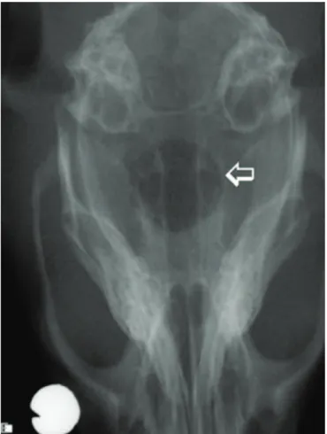
For the radiographic evaluation, two digital radiographs of the CSBD illed with the biomaterial were taken, one radiography taken immediately ater the surgery (initial radiography), and the second taken at the moment the animal was sacriiced ater 15 or 60 days (inal radiography). To make the radiographs, it was used complementary metal-oxide semiconductor (CMOS) equipment (Schick Technologies Inc., Dialom Dental Products, Long Island City, NY), which was positioned parallel to the surface of the created bone defect and this method was standard for all animals using a positioner. he vertical long axis of the implant positioned perpendicularly to the central X-ray beam and parallel to the sensor at 40-cm focus-object distance. he X-ray unit was operated at 70 KVp, 10 mA, and 0.3 seconds (Expectro 70×, Dabi Atlante, Ribeirão Preto, SP, Brazil). Image resolution was 635 ppi (pixels per inch), the size of the image was 900 × 641 dpi and the pixel size was 40 μm. Images (Figure 1) were stored in the TIFF (Tagged Image File Format) without compression (8 bits with 600 dpi resolution).
4.
Image Analysis
he radiographic bone density in the defects was determined by the analysis of gray levels inside the CSBD, in an area containing, in average, 35.000 pixels. his area was selected since it represented the full area of a 8-mm (in diameter) circular area, determining that the whole defect would be assessed, in all cases, standardizing the region that was measured. his analysis was
done by a blinded evaluator using the image-analysis sotware Image Tool 2.03 (UTHSCA, San Antonio, Texas, EUA), which provided the gray scale average and standard deviation in these pre-determined region by means of a histogram graph. he average gray level of the evaluated region was divided by the value of a metallic standard, inserted in all radiographic acquisitions, to compensate minimal diferences among radiographs, since the density of the metallic standard was similar in all specimens16,17.
5.
Statistical Analysis
he Kolgomorov-Smirnov test was used to verify data distribution, and data from radiographic bone density were analyzed statistically by analysis of variance (ANOVA), followed by Tukey post test, or using paired t test when 1 x 1 comparison were made. Signiicance level was set at 5%. Comparisons between the diferent groups and periods were done.
RESULT
he results obtained (Figure 2) indicated that, in the initial images, all defects presented statistically similar radiographic densities. In the inal images, all inal results were statistically equal in the early period of observation (15 days), but in the late period of observation (60 days), both Control and LMWC groups difered from LMWCH group (p < 0.05 – ANOVA, followed by Tukey test).
In the early period of observation (15 days), with the exception of the LMWC and HMWC groups, tested biomaterials presented a statistically signiicant (p < 0.01 – paired t test) increase of the radiographic bone density in the evaluated area, when comparing the initial and inal images, as well as the Control group. At the 60-day period of observation, with the exception of the Control and LMWC groups, no statistically-signiicant increase or decrease could be seen.
DISCUSSION
he biomaterials tested in this study presented considerably poor results, as none of them were capable of signiicantly increase the radiographic bone density in the CSBD area in comparison to the control group.
In bone cavities grating materials normally delay the healing process, since they need to be replaced, incorporated or eliminated, with the aim of promoting bone repair, and this process begins with an inlammatory activity (variable, depending on the biomaterial) which evolves to the resolving of the defect18. Our indings in this study corroborate what we
have already reported before, with no signiicant bone formation following chitosan and chitosan hydrochloride gels application in CSBDs, and defects repaired by connective tissue, with variable degrees of inlammation19.
Literature is controversial in relation to chitosan-based biomaterials attainment and their usage results. hese biomaterials are capable of inluencing all tissue reparation stages in experimental animal models18. In the inlammatory stage,
the chitosan’s astringent properties are independent from the conventional healing cascade. In vivo, this polymer can stimulate the proliferation of ibroblasts and modulate the migration of neutrophils and macrophages, thus modifying the subsequent repair processes such as ibroplasias and tissue neoformation20.
Chitosan polymers have been tested in periodontal defects treatment not demonstrating the stimulation of important allergic or inlammatory reactions ater its implantation, topical injection or ingestion7,12,13. In 2007, Asikainen et al.21 evaluated
the biocompatibility of chitosan ibers and bioactive glass-based biomaterials implanted in the subcutaneous tissue of rats. hey related the formation of a moderate inlammatory iniltration from the initial period, which was maintained even ater the bioactive glass was absorbed, indicating that probably this reaction was caused by the chitosan ibers. Over a long period a moderate chronic inlammatory iniltrate continued to be evident. It is important to say that this inlammation, although necessary in the beginning of the regeneration process, delays and even inhibits the new bone formation18.
Literature has stated that chitosan and its derivates are capable of activate macrophage activity and that they can initiate inlammatory reactions. In 2005, Mori et al.22 evaluated the mechanism by
which chitosan and its derivates could cause macrophages activation. Authors compared pure chitin, high-molecular-weight chitosan and low-molecular-weight chitosan placed in peritoneal macrophage cell cultures, and concluded that, in vivo, all the substances induced the activation of the primary histocompatibility complex I and II, where the low-molecular-weight chitosan was the one with lowest activation property. hey concluded that the cellular activation alterations can, in a certain way, accelerate the tissue repair. However, if the damage is very severe or prolonged, it can cause a chronic inlammatory process, which will impede more complex regenerative processes, and could lead to a miscarriage in bone formation, which would be an explanation for our results, with no advantage for chitosan-based material over the blood clot.
he fact that the an increase in the bone density did not occur in the CSBD’s illed with the evaluated biomaterials, at least from what was radiographically observable, may have occurred due to either the biomaterial’s own characteristics, a reaction of rat’s body to the presence of this biomaterials, or even due to the bone defect type utilized.
Bone defects can be regenerated whenever there are cells and original element tissue that can reposition, or can be repaired by the substitution of the injured tissue with another illing or support tissue. Bone is a tissue that presents a unique potential to restore its original structure, within certain limitations, considering that the reconstruction in the original organizational level occurs in sequence and exactly repeats the bone’s development and growth pattern. In this way, the autogenous grat is the most complete among all other grating materials due to its osteoconductor, osteoinductor and osteogenic properties23.
As demonstrated above, literature has related some articles where the use of chitosan gel was favorable to bone-defect regeneration. However, the parameters of the studied biomaterial were not always delimited, and so information concerning the
concentration, molecular weight and sterilization method were lacking. In this study, despite all the factors relating to the gel’s attainment being well delimited and described in the methodology, the molecular weight of the chitosan and chitosan-hydrochloride gel did not lead to signiicant modiications in the biological properties of the biomaterials tested and none of these biomaterials or their variations contributed to the increase of the radiographic bone density in the CSBD’s created, and, therefore, its indication is not advisable.
CONCLUSION
Based on the limitations of the model that was used and considering the presented results, it is concluded that none of the biomaterials utilized in the study signiicantly increased radiographic bone density in the CSBD’s, when compared to the blood clot alone, and the molecular weight did not seem to interfere in the results in a relevant manner. hese biomaterials need to be submitted to further studies before they can be safely and positively used in tissue engineering.
REFERENCES
1. Yamada Y, Ueda M, Naiki T, Nagasaka T. Tissue-engineered injectable bone regeneration for osseointegrated dental implants. Clin Oral Implants Res. 2004; 15: 589-97. PMid:15355402. http://dx.doi.org/10.1111/j.1600-0501.2004.01038.x
2. Buser D, Dula K, Hess D, Hirt HP, Belser UC. Localized ridge augmentation with autograts and barrier membranes. Periodontol 2000. 1999;19: 151-63. PMid:10321222. http://dx.doi.org/10.1111/j.1600-0757.1999.tb00153.x
3. Summers BN, Eisenstein SM. Donor site pain from the ilium. A complication of lumbar spine fusion. J Bone Joint Surg Br. 1989; 71: 677-80. PMid:2768321.
4. Gross JS. Bone grating materials for dental applications: a practical guide. Compend Contin Educ Dent. 1997; 18: 1013-8, 20-2, 24, passim; quiz.
5. Boss JH, Shajrawi I, Aunullah J, Mendes DG. he relativity of biocompatibility. A critique of the concept of biocompatibility. Isr J Med Sci. 1995; 31: 203-9. PMid:7721555.
6. Service RF. Tissue engineers build new bone. Science. 2000; 289(5484): 1498-500. PMid:10991738. http://dx.doi.org/10.1126/ science.289.5484.1498
7. Park JS, Choi SH, Moon IS, Cho KS, Chai JK, Kim CK. Eight-week histological analysis on the efect of chitosan on surgically created one-wall intrabony defects in beagle dogs. J Clin Periodontol. 2003; 30: 443-53. PMid:12716338. http://dx.doi.org/10.1034/j.1600-051X.2003.10283.x
8. Senel S, McClure SJ. Potential applications of chitosan in veterinary medicine. Adv Drug Deliv Rev. 2004; 56: 1467-80. PMid:15191793. http://dx.doi.org/10.1016/j.addr.2004.02.007
9. Senel S, Ikinci G, Kas S, Yousei-Rad A, Sargon MF, Hincal AA. Chitosan ilms and hydrogels of chlorhexidine gluconate for oral mucosal delivery. Int J Pharm. 2000; 193: 197-203. http://dx.doi.org/10.1016/S0378-5173(99)00334-8
10. Shahidi F, Abuzaytoun R. Chitin, chitosan, and co-products: chemistry, production, applications, and health efects. Adv Food Nutr Res. 2005; 49: 93-135. http://dx.doi.org/10.1016/S1043-4526(05)49003-8
11. Singla AK, Chawla M. Chitosan: some pharmaceutical and biological aspects--an update. J Pharm Pharmacol. 2001; 53: 1047-67. PMid:11518015. http://dx.doi.org/10.1211/0022357011776441
12. Muzzarelli RA, Mattioli-Belmonte M, Tietz C, Biagini R, Ferioli G, Brunelli MA, et al. Stimulatory efect on bone formation exerted by a modiied chitosan. Biomaterials. 1994; 15: 1075-81. http://dx.doi.org/10.1016/0142-9612(94)90093-0
13. Chatelet C, Damour O, Domard A. Inluence of the degree of acetylation on some biological properties of chitosan ilms. Biomaterials. 2001; 22: 261-8. http://dx.doi.org/10.1016/S0142-9612(00)00183-6
14. Ueno H, Murakami M, Okumura M, Kadosawa T, Uede T, Fujinaga T. Chitosan accelerates the production of osteopontin from polymorphonuclear leukocytes. Biomaterials. 2001; 22: 1667-73. http://dx.doi.org/10.1016/S0142-9612(00)00328-8
16. Giro G, Goncalves D, Sakakura CE, Pereira RM, Marcantonio Junior E, Orrico SR. Inluence of estrogen deiciency and its treatment with alendronate and estrogen on bone density around osseointegrated implants: radiographic study in female rats. Oral Surg Oral Med Oral Pathol Oral Radiol Endod. 2008; 105: 162-7. PMid:18230387. http://dx.doi.org/10.1016/j.tripleo.2007.06.010
17. Sakakura CE, Giro G, Goncalves D, Pereira RM, Orrico SR, Marcantonio E, Jr. Radiographic assessment of bone density around integrated titanium implants ater ovariectomy in rats. Clin Oral Implants Res. 2006; 17: 134-8. PMid:16584408. http://dx.doi.org/10.1111/j.1600-0501.2005.01224.x
18. Okamoto Y, Shibazaki K, Minami S, Matsuhashi A, Tanioka S, Shigemasa Y. Evaluation of chitin and chitosan on open would healing in dogs. J Vet Med Sci. 1995; 57: 851-4. PMid:8593291. http://dx.doi.org/10.1292/jvms.57.851
19. Spin-Neto R, de Freitas RM, Pavone C, Cardoso MB, Campana-Filho SP, Marcantonio RA, et al. Histological evaluation of chitosan-based biomaterials used for the correction of critical size defects in rat’s calvaria. J Biomed Mater Res A. 2010; 93: 107-14. PMid:19536827.
20. Kosaka T, Kaneko Y, Nakada Y, Matsuura M, Tanaka S. Efect of chitosan implantation on activation of canine macrophages and polymorphonuclear cells ater surgical stress. J Vet Med Sci. 1996; 58: 963-7. PMid:8915995. http://dx.doi.org/10.1292/jvms.58.10_963
21. Asikainen AJ, Hagstrom J, Sorsa T, Noponen J, Kellomaki M, Juuti H, et al. Sot tissue reactions to bioactive glass 13-93 combined with chitosan. J Biomed Mater Res A. 2007; 83: 530-7. PMid:17508414. http://dx.doi.org/10.1002/jbm.a.31225
22. Mori T, Okumura M, Matsuura M, Ueno K, Tokura S, Okamoto Y, et al. Efects of chitin and its derivatives on the proliferation and cytokine production of ibroblasts in vitro. Biomaterials. 1997; 18: 947-51. http://dx.doi.org/10.1016/S0142-9612(97)00017-3
23. Triplett RG, Schow SR. Surgical advances in implant dentistry. Tex Dent J. 1994; 111 (10):12-3, 5-7, 9.
CONFLICTS OF INTERESTS
he authors declare no conlicts of interest.
CORRESPONDING AUTHOR
Rosemary Adriana Chiérici Marcantonio
Department of Periodontology, UNESP – São Paulo State University, Rua Humaitá, 1680, 14801-903 Araraquara - SP, Brazil e-mail: [email protected]