DEVELOPMENTAL BIOLOGY AND CYTOGENETICS OF
BURSAPHELENCHUS XYLOPHILUS
KOICHI HASEGAWA1, 2, MANUEL MOTA3, KAZUYOSHI FUTAI4 AND JOHJI MIWA1, 4, 5
1
Institute for Biological Function, Chubu University, 1200 Matsumoto, Kasugai 487-8501 Japan 2
Laboratory of Environmental Mycoscience, Graduate School of Agriculture, Kyoto University, Sakyo-ku, Kyoto 606-8502 Japan 3NemaLab-ICAM, Departmento de Biologia, Universidade de Évora, 7002-554 Évora, Portugal 4
Laboratory of Developmental Genetics, Graduate School of Bioscience and Biotechnology, Chubu University, 1200 Matsumoto, Kasugai 487-8501 Japan
ABSTRACT
The pine wood nematode Bursaphelenchus xylophilus reproduces bisexually: a haploid sperm fertilizes a haploid oocyte, and the two pronuclei rearrange, move together, fuse, and begin diploid development. Early embryonic events taking place in the B. xylophilus embryo are similar to those of Caenorhabditis elegans, although the anterior-posterior axis appeares to be determined oppositely to that observed for C. elegans. Thai is, in the B. xylophilus embryo, the male pronucleus emerges at the future anterior end, whereas the female pronucleus appeares laterally. To understand the evolution of nematode developmental systems, we cloned the full length of Bx-tbb-1 (beta tubulin) from B. xylophilus cDNA and attempted to apply reverse genetics analysis to B. xylophilus. Several lengths of double stranded RNA (dsRNA) for the Bx-tbb-1 gene were synthesized by in vitro transcription, and both B. xylophilus and C. elegans were soaked in dsRNA for RNAi. Both nematodes could suck up the dsRNA, and we could detect the abnormal phenotypes caused by Bx-tbb-1 dsRNA in C. elegans, but not in B. xylophilus. We suspect that systemic RNAi might be suppressed in B. xylophilus and are attempting to establish other methods for functionally analyzing B. xylophilus genes.
INTRODUCTION
The pine wood nematode Bursaphelenchus xylophilus is thought to cause a pine wilt disease, which destroys millions of pine trees every year in East Asia (Mamiya, 1983). And it is spreading its devastating malady in Europe (Mota et al., 1999). Although parasitic in nature, B. xylophilus is easily maintained in the laboratory and is experimentally amicable, having a large brood size with short generation time and a simple and transparent body and embryo (Hasegawa et al., 2004; Hasegawa et al., 2006). Here we show some embryological and cytological aspects of B. xylophilus development, as it is compared with those of the model organism Caenorhabditis elegans. And we also attempted to establish methods for the functional analysis of B. xylophilus genes.
NEMATODES
The B. xylophilus isolate S-10 (originally isolated from Shimane Prefecture, Japan) was used. Methods for culturing, handling, and observing B. xylophilus follow the methodology described by Hasegawa et al. (2004). C. elegans var. Bristol, strain N2 was used (Brenner, 1974).
IMMUNOSTAINING FOR MICROTUBULES AND ACTIN
Early B. xylophilus embryos were collected and fixed as described by Hasegawa et al. (2004). Mouse monoclonal anti-α-tubulin antibody (DM1α, Sigma) and rabbit monoclonal anti-actin antibody (Sigma) were used as primary antibodies, and FITC-conjugated goat anti-mouse IgG (Sigma) and Cy3-conjugated sheep anti-rabbit IgG (Sigma) were used as secondary antibodies. Antibody staining was performed as described by Hasegawa et al. (2004). Stained embryos were washed with PBS for several seconds and mounted in Vectashield (Vector Laboratories, Inc.) for viewing with a ZEISS Axiovert 200 microscope equipped with a confocal laser-scanning module (ZEISS LSM510).
DSRNA SYNTHESIS
The plasmid pBxTBB5R-1 was used as a PCR template. This plasmid was constructed by inserting a 5’ fragment of Bx-tbb-1 cDNA (1079 bp) into the pPCR SCRIPT SK (+) MCS (Stratagene) (Hasegawa and Miwa, unpublished data). Three different lengths of DNA templates were prepared from pBxTBB5R-1 with the following primers: for the 1kbp (1,079bp) DNA template, the T7 primer (5’ -GTA ATA CGA CTC ACT ATA GGG C- 3’) and Cmo422 (5’- GCG TAA TAC GAC TCA CTA TAG GGA ACA AAA GCT GGA GCT- 3’) (Mello, personal communication); for the 500 bp (464bp) DNA template, T7BXTBB5R_SP500 (5’ –GTA ATA CGA CTC ACT ATA GGG CCA CTC TTT CCG TC- 3’) and Cmo422; and for the 100bp (117bp) DNA template, T7BXTBB5R_SP100 (5’ –GTA ATA CGA CTC ACT ATA GGG CTG CTT GTG ATC CT- 3’) and Cmo422. Double-stranded RNA (dsRNA) was synthesized by in vitro transcription with T7 RNA Polymerase (Takara Bio) for each length. In addition to
the three dsRNA lengths, we prepared siRNA (about 22bp) from 1 kbp dsRNA fragment, digested with Takara siRNA Cocktail Kit (Takara Bio) for 3.5 hours at 30°C.
SOAKING RNAi
Soaking RNAi of C. elegans L4 animals was performed following the method of Maeda et al. (2001), and B. xylophilus was performed following the method of Urwin et al. (2002), with some modification. For checking the soaking efficiency, the soaking buffer (Maeda et al., 2001) with or without 10 mM octopamine was mixed with FITC dye (1 µg/µL), and nematodes were dipped for 24 hours at 20°C (C. elegans) or 25°C (B. xylophilus) and viewed by fluorescence microscopy (Nikon E600). Double-stranded RNA for each size (1 kb, 500 bp, 100 bp) was dissolved in soaking buffer (0.5- 1 µg/µL), and both B. xylophilus (<50 mixed stage individuals) and C. elegans (6 to 10 L4 individuals) (parental generation) were soaked in dsRNA for RNAi. After 24 hours of incubation, C. elegans were transferred onto a fresh NGM plate seeded with Escherichia coli OP50, and incubated overnight at 20°C for recovery. After that, healthy C. elegans adults were individually transferred onto NGM spot plates for checking the hatching rate of F1 eggs. In case of B. xylophilus, nematodes were incubated in dsRNA for 24 to 48 hours at 25°C. After soaking, B. xylophilus were transferred onto a fresh 1/10 PDA plate seeded with Botrytis cinerea, and incubated 1 day at 25°C for recovery and mating. After that, mating sets of one adult female and 2 to 5 adult males were transferred onto agar plates seeded with B. cinerea for checking F1 hatching rate. Resulting F1 phenotypes were viewed by Nomarski optics (Nikon E600).
RESULTS AND DISCUSSION
FIRST CELL DIVISION AND AP AXIS FORMATION
The B. xylophilus embryo is long and slender, about 25 µm wide and 60 µm long. As in C. elegans, the first cell division was unequal, producing the larger AB cell and the smaller P1 cell (Hasegawa et al., 2004; Hasegawa et al., 2006), which were destined to a certain fate, although still reprogrammable at this stage (Gönczy & Rose, 2005). This division reveals the anterior-posterior (AP) axis of a nematode in that the AB and P1 cells indicate, respectively, the future anterior and the posterior sides (Hasegawa et al., 2004).
Here we analyzed the relations of the male and female pronuclei, cell cytoskeleton formation, and AP axis in B. xylophilus embryos from fertilization to the 2-cell stage.
The cortical membrane of a newly laid egg was rumpled and active, and seemingly random cytoplasmic streaming was visible (Fig. 1A). At this time, the DAPI-stained sperm was seen as a dot positioned at the future anterior side of the embryo (Fig. 1a). Meiosis was propelled by a small seemingly acentriolar meiotic spindle positioned at the lateral mid-point position of the embryo (Fig. 1a). The 1st and 2nd polar bodies were extruded out, while vesicular male and female pronuclei were reconstituted (Fig. 1B, b). Duplicated centrosomes were on the surface of the male pronucleus, interacting with the cortical actin (Fig. 1b). The sperm entry point appeared to become the future anterior end of the B. xylophilus embryo, although the B. xylophilus sperm also brought a centrosome into the oocyte. The rumpled cortical membrane became smooth, and male and female pronuclei moved toward each other and eventually met (Fig. 1C, c). Chromosomes became condensed in the prophase stage, and six chromosomes were visible in each pronucleus (Fig. 1c). They moved to the center where they rotated 90 degrees (Fig. 1D, d), and fused to become one. The pair of male and female pronuclear-derived chromatids was fused and aligned along the metaphase plate (Fig. 1E, e). Two centrosomes were located at the center of the embryo along its longitudinal axis (Fig. 1e). In anaphase, the posteriorly-positioned centrosome moved more posteriorly to divide the chromosomes antero-posteriorly (Fig. 1F, f). Subsequently, the embryo divided unequally (Fig. 1G, g) to form the larger anterior AB cell and the smaller posterior P1 cell (Fig. 1H, h), thereby determining the anterior-posterior axis.
Polarity formation is an important feature for establishing many different cell types through several rounds of asymmetric cell divisions. The initial asymmetric cues such as sperm entry (Goldstein & Hird, 1996) establish the cell polarity followed by cytoskeleton reorganization and polarized localization of several cortical proteins in the C. elegans oocyte. A mature oocyte is fertilized as it passes through the spermatheca by sperm located there (Ward & Carrel, 1979). Compared with sperm, the oocyte is much larger and contains more cellular factors necessary for cell division, cell-fate determination, and morphogenesis. Although almost all factors required for early embryogenesis are supplied maternally (Miwa et al., 1980), paternal factors such as chromosomes, centrosome, and cytoplasm also constitute essential and important contributions (Singson, 2001). Fertilization triggers completion of the final stage of oocyte meiosis and
promotes rearrangement of the cortical actin cytoskeleton, which generates cytoplasmic flows anteriorly in the cortical region and posteriorly inside of the zygote to distribute cell-fate determinants (Munro et al. 2004). At metaphase, two centrosomes are located at the center of the embryo along its longitudinal axis, but in anaphase the posteriorly-positioned centrosome moved posteriorly to divide the chromosomes antero-posteriorly. Asymmetric distribution of cell-fate determinants should eventually lead the first cell division to produce the larger anterior AB cell and the smaller posterior P1 cell, which are programmed to have different fates (Sulston et al., 1983; Miwa, 1986). Although the AP axis determination of the B. xylophilus embryo was entirely opposite to that of the C. elegans embryo, there is a question whether or not the B. xylophilus embryo uses the same types of molecules to generate AP polarity as in the C. elegans embryo? We thus need to compare the functional differences or similarities of the molecules necessary for AP axis determination between B. xylophilus and C. elegans.
ATTEMPT TO INDUCE RNA INTERFERENCE IN BURSAPHELENCHUS XYLOPHILUS Next we attempted to analyze the function of Bx-tbb-1, a homologue of the gene encoding beta tubulin, which is an important molecule for cell division. The Bx-tbb-1 gene has been isolated from B. xylophilus, and its structure and expression patterns were analyzed (Hasegawa & Miwa, personal communication). Its nucleotide sequence shares 78% homology with tbb-1 of C. elegans (Hasegawa & Miwa, personal communication). Transcriptional and functional analyses of B. xylophilus genes were initiated by constructing ESTs (Kikuchi et al., 2004; Kikuchi et al., 2005), and we attempted to introduce the reverse genetics technique in B. xylophilus for analyzing the gene function. Although conventional genetics (forward genetics) provides the most powerful tool to understand most biological phenomena, only a few organisms are endowed with this potential. The recently discovered RNAi (double strand RNA-mediated interference) is now considered by many to be a “wonder magic” for genetic analysis in non-model organisms for which classical genetics is unsuitable, and is sometimes referred to as “reverse genetics,” since we first start with materials like RNA and then find a phenotype: that is , the process is a reversal of classical genetics. The phenomenon of RNAi was discovered first in C. elegans (Fire et al. 1998). The RNAi-based “reverse genetics” in C. elegans offers three different ways for obtaining the interfered phenotype induced by tailor-made double stranded (ds) RNA material: by (1) microinjection of dsRNA into the
nematode body (Fire et al. 1998), (2) feeding E. coli that expresses dsRNA (Timmons et al. 1998), and (3) soaking nematodes in dsRNA (Tabara et al., 1998; Maeda et al., 2001). For feeding and soaking RNAi, dsRNA is absorbed from the gut and transported to other parts of the body. Because B. xylophilus is very slender, it is quite difficult to perform microinjection. In this experiment we tried to perform soaking RNAi with Bx-tbb-1, the gene encoding β-tubulin and necessary for microtubule formation.
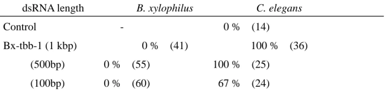
First, to check the soaking efficiency, the dsRNA buffer was mixed with FITC dye (1 µg/µL), and nematodes were dipped for 24 hours at 25°C. In the presence of the neurotransmitter octopamine (10mM), B. xylophilus could suck up the dsRNA more efficiently; FITC green signal was detected clearly in the pharyngeal and intestinal lumen in all nematodes (checked more than 30 individuals) (Fig. 2). Then, to apply reverse genetics to B. xylophilus as a functional analysis of Bx-tbb-1, both B. xylophilus and C. elegans animals were soaked in solutions containing several different lengths of dsRNA (1 kbp, 500bp, 100bp, and siRNA) for RNAi. Because it is known that introduced dsRNA is first processed into siRNA (short interfering RNA) by RNase III and siRNA with other components then forms RISC (RNA-induced silencing complex) to bind and degrade target mRNA (Hammond, 2005), we prepared Bx-tbb-1 siRNA to facilitate the processes. 100% RNAi phenotype was detected in C. elegans by soaking 1 kbp and 500bp Bx-tbb-1 dsRNA, but none was detected in B. xylophilus. After Bx-tbb-1 RNAi treatment, all C. elegans F1 eggs seemed to be arrested at microtubule formation and could not perform pronuclear meeting and chromosome division (Fig. 3). Although male and female pronuclei appeared, they could not move and meet each other. The embryos, nevertheless, started disordered cell division (Fig. 3). The shorter 100bp dsRNA and siRNA were less effective for RNAi in C. elegans; RNAi efficiencies decreased to 67 % and 22 %, respectively (Table 1). We could not detect knock-down phenotypes in B. xylophilus, however, at any of the various dsRNA lengths (Table 1). We also examined soaking RNAi in B. xylophilus with several other genes (Bx-par-1, Bx-mex-3, Bx-tbb-1, Bx-unc-22) in several lengths (from 1.5 kb to siRNA), but could not observe knock-down phenotypes (data not shown). It might be possible that systemic RNAi silencing, whereby mobile dsRNA is taken up by gut cells or transferred among different tissues, was suppressed in B. xylophilus, and thus soaking RNAi was not effective.
Successful results of RNAi in other parasitic nematodes have been reported; dsRNA was delivered by soaking (for example, Urwin et al., 2002; Fanelli et al., 2005), or by
electroporation (Issa et al. 2005). C. elegans and perhaps also these parasitic nematodes have a systemic nature of RNAi; that is, delivered dsRNA moves from one introduced cell to the others, and that has not been reported so far in other organisms except for plants (Grishok, 2005). We need to continue seeking to establish other methods for functionally analyzing B. xylophilus genes to understand its developmental program and the evolution of nematode developmental systems by comparing similarities and differences between the two nematodes B. xylophilus and C. elegans.
ACKNOWLEDGEMENT
We thank Ms. Satsuki Miwa, Viva Informatica, for useful comments and careful reading of this manuscript. This work was supported by a Special Research Fund of Chubu University and a research grant from the Chubu University Institute for Biological Function to JM and a JSPS research fellowship to KH.
REFERENCES
Brenner, S. (1974) The genetics of Caenorhabditis elegans. Genetics 77, 71-94.
Fanelli, E., Di Vito, M., Jones, J. T. and De Giorgi, C. (2005) Analysis of chitin synthase function in a plant parasitic nematode, Meloidogyne artiellia, using RNAi. Gene 349, 87-95.
Fire, A., Xu, S., Montgomery, M. K., Kostas, S. A., Driver, S. E. and Mello, C. C. (1998) Potent and specific interference by double-stranded RNA in Caenorhabditis
elegans. Nature 391, 806-811.
Goldstein, B. and Hird, S. N. (1996) Specification of the anteroposterior axis in
Caenorhabditis elegans. Development 122, 1467-1474.
Grishok, A. (2005) RNAi mechanisms in Caenorhabditis elegans. FEBS Letters 579, 5932-5939.
embryo. In: The C. elegans Research Community (ed) WormBook (October 15, 2005) doi/10.1895/wormbook.1.30.1, http://www.wormbook.org.
Hammond, S. M. (2005) Dicing and silencing. The core machinery of the RNA interference pathway. FEBS letters 579, 5822-5829.
Hasegawa, K., Miwa, S., Futai, K. and Miwa J. (2004) Early embryogenesis of the pinewood nematode Bursaphelenchus xylophilus. Dev. Growth Differ. 46, 153-161.
Hasegawa, K., Mota, M. M., Futai, K. and Miwa, J. (2006) Chromosome structure and behavior in Bursaphelenchus xylophilus (Nematoda: Parasitaphelenchidae) germ cells and early embryo. Nematology 8, 425 - 434.
Issa, Z., Grant, W. N., Stasiuk, S. and Shoemaker, C. B. (2005) Development of methods for RNA interference in the sheep gastrointestinal parasite, Trichostrongylus
colubriformis. Int. J. parasitol. 35, 935-940.
Kikuchi, T., Jones, J. T., Aikawa, T., Kosaka, H. and Ogura, N. (2004) A family of glycosyl hydrolase family 45 cellulases from the pine wood nematode Bursaphelenchus
xylophilus. FEBS Letters 572, 201-205.
Kikuchi, T., Shibuya, H. and Jones, J.T. (2005) Molecular and biochemical characterization of an endo-beta-1,3-glucanase from the pinewood nematode
Bursaphelenchus xylophilus acquired by horizontal gene transfer from bacteria.
Biochemical J. 389, 117-125.
Maeda, I., Kohara, Y., Yamamoto, Y. and Sugimoto, A. (2001) Large scale analysis of gene function in Caenorhabditis elegans by high-throughput RNAi. Current Biology 11, 171-176.
Miwa, J. (1986) Making of the roundworm II. Kagaku 56, 162-170 (in Japanese)
Mota, M. M., Braasch, H., Bravo, M. A., Penas, A. C., Burgermeister, W., Metge, K. and Sousa, E. (1999) First report of Bursaphelenchus xylophilus in Portugal and in
Europe. Nematology 1, 727-734.
Munro, E., Nance, J. and Priess, J. R. (2004) Cortical flow powered by asymmetrical contraction tansport PAR protein to establish and maintain anterior-posterior polarity in the early C. elegans embryo. Dev. Cell 7, 413-424.
Singson, A. (2001) Every sperm is sacred: Fertilization in Caenorhabditis elegans. Dev. Biol. 230, 101-109.
Sulston, J. E., Schierenberg, E., White, J. G. and Thomson, J. N. (1983) The embryonic cell lineage of the nematode Caenorhabditis elegans. Dev. Biol. 100, 64-119.
Tabara, H., Grishok, A. and Mello, C. C. (1998) RNAi in C. elegans: soaking in the genome sequence. Science 282, 430-431.
Timmons, L. and Fire, A. (1998) Specific interference by ingested dsRNA. Nature 395, 854.
Urwin, P. E., Lilley, C. J. and Atkinson, H. J. (2002) Ingestion of double-stranded RNA by preparasitic juvenile cyst nematodes leads to RNA interference. MPMI 15, 747-752.
Ward, S. and Carrel, J. S. (1979) Fertilization and sperm competition in the nematode
Caenorhabditis elegans. Dev. Biol. 73: 304-321
Table 1: Percentage of abnormal RNAi phenotype
dsRNA length B. xylophilus C. elegans
Control - 0 % (14)
Bx-tbb-1 (1 kbp) 0 % (41) 100 % (36)
(500bp) 0 % (55) 100 % (25)
(siRNA) 0 % (29) 22 % (9)
All abnormal phenotypes were embryogenesis arrest. Control was soaking buffer only. Numbers in parentheses indicate the number of examined animals.