In Vivo Efficacy of Latex from
Calotropis procera
in Ameliorating Fever
—
Biochemical
Characteristics and Plausible Mechanism
Vijay L. Kumar1&B. Guruprasad1&
Syed Meraj A. Fatmi1&Priyanka Chaudhary1& Nylane Maria Nunes Alencar2&
José Vitor Moreira Lima-Filho3&Márcio Viana Ramos4
Received: 19 July 2016 / Accepted: 29 December 2016 / Published online: 11 January 2017
#Springer Science+Business Media New York 2017
Abstract Calotropis procera latex fractions possessing anti-inflammatory property were characterized for their biochemical properties, compared for their efficacy in ameliorating fever in rats and their mechanism of action was elucidated. Aqueous fraction and methanol extract (AqDL and MeDL) were derived from the dried latex (DL) and proteins were separated from the fresh latex (LP). Polyacrylamide gel electrophoresis carried out under denaturing conditions showed the presence of proteins with some similarity in LP and AqDL and both of these fractions exhibited proteinase activity by gelatin zymography. A further analysis revealed that only the LP fraction possesses cysteine proteinase activity. Oral administration of both AqDL and MeDL produced a dose-dependent reduction in body temperature in rats where fever was induced by yeast and their effect was comparable to that of standard drug paracet-amol while intravenous administration of LP was not so effective. Both AqDL and MeDL produced a significant reduction in the levels of TNF-α, PGE2, and immunoreactivity of
COX-2 in the hypothalamus as compared to yeast control group. This study shows that both AqDL and MeDL, the orally effective anti-inflammatory fractions of latex, have therapeutic potential in treating various febrile conditions.
DOI 10.1007/s12010-016-2395-y
* Vijay L. Kumar
kumarvl98@hotmail.com; vlkumar@aiims.ac.in
1
Department of Pharmacology, All India Institute of Medical Sciences, Ansari Nagar, New Delhi 110 029, India
2 Departamento de Fisiologia e Farmacologia, Universidade Federal do Ceará, Campus do Porangabu,
Fortaleza, CE CEP 60430-270, Brazil 3
Departamento de Biologia, Universidade Federal Rural de Pernambuco, Campus Dois Irmãos, Recife, PE CEP 52171-900, Brazil
4
Keywords Calotropis procera. Fever . Latex . Proteinases . Cox-2 . Hypothalamus
Introduction
Plant derived preparations have been the mainstay for treatment of various ailments including fever for time immemorial. Calotropis procera is a wild growing plant that occurs in abundance in the wasteland and grows as a weed. In traditional system, the plant has been used for treating diseases of liver and abdomen, piles, ulcers, tumors, and various inflamma-tory and painful conditions [1]. The decoction of its aerial parts has been used for the treatment of joint pain, fever, and muscular spasm, and fresh or warmed leaves have been applied on sores, wounds, and rheumatic joints [2,3]. The aerial parts are rich source of latex that has also been used for curing various infections and painful conditions [2,4]. The latex has been shown to possess anti-inflammatory, analgesic, and antipyretic properties [5–8]. These activities have been demonstrated in the aqueous and methanol extracts of the dried latex (AqDL and MeDL) and proteins isolated from the fresh latex (LP) and all these latex-derived preparations are effective in inhibiting edema formation, cellular influx, mediator release, and oxidative stress at the site of inflammation [9–11]. In various in vivo studies both AqDL and MeDL were found effective following oral administration while the LP fraction being protein in nature was effective when administered parentrally [11,12].
Fever is an autonomic response to acute infection and inflammation where the role of endogenous pyrogens is well established. Cytokines like interferon, interleukins (IL-1, IL-6), and tumor necrosis factor-alpha (TNF-α) have been shown to increase the hypothalamic set
point and induce fever. These endogenous pyrogens exert their effect through cyclooxygenase-2 (COX-cyclooxygenase-2)-mediated prostaglandin E2(PGE2) synthesis and activate central nervous system mechanisms resulting in fever [13]. Yeast induced pyrexia in rats has been widely used for the evaluation of compounds effective in fever [8]. As drugs exhibiting anti-inflammatory and analgesic activity also produce antipyretic effect, the present study was carried out to compare AqDL, MeDL and LP with respect to their biochemical characteristics, antipyretic properties, and to elucidate their mechanism of action.
Materials and Methods
Animals
The study was carried out in Wistar rats of either sex (150–200 g) that had free access to food and water. They were acclimatized for a week during which their rectal temperature was measured daily. The experiments were carried out following due approval of the Institutional Animal Ethics Committee (646/IAEC/11).
Plant Material
identified by the Raw Materials, Herbarium and Museum Division of National Institute of Science Communication, CSIR, New Delhi where a voucher specimen (PID 1739) is pre-served. The latex dried under shade (DL) contains rubber-like isoprene material which separates when it is triturated with water and was discarded. The aqueous suspension devoid of this material was lyophilized to obtain aqueous extract of DL (AqDL). The DL was also soxhlated with petroleum ether to remove rubber-like material and then with methanol. The methanol extract thus obtained was dried (MeDL).
The parentrally effective LP fraction was obtained from the plant growing in Fortaleza, Brazil, identified by a taxonomist and deposited at the Prisco Bezzera Herbarium of the Universidade Federal do Ceará, Brazil (Voucher No. 32663). The latex was collected in distilled water (1:2,v/v) and centrifuged to separate the rubber-rich pellet that was discarded. The supernatant was dialyzed against distilled water using a membrane with a cut off value of 8000 Da. The non-dialyzable material was separated and lyophilized to obtain proteins (LP). The LP fraction comprises of three distinct protein peaks as revealed by ion-exchange chromatography and electrophoresis and these details have been described earlier [15].
Protein Quantitation in Latex Fractions
The latex fractions AqDL (10 mg), MeDL (20 mg), and LP (1 mg) were dissolved in 1 ml of normal saline (NS) and 100-μl aliquot of each was made up to 900μl with NS. After adding
100μl of Bradford’s reagent, the absorbance was read at 595 nm. The protein concentration
(mg/ml) in the sample was determined from the standard curve plotted using different concentrations of bovine serum albumin and protein content (mg) per milligram of each fraction was calculated [16].
Protein Profile and Detection of Proteinases by Zymography
Samples were dissolved in Tris buffer (0.0625 M, pH 6.8) containing 2% sodium dodecyl sulfate (SDS) and electrophoresis was carried out on 12.5% polyacrylamide gel (PAGE). Proteinases were determined in the samples by zymography where 0.1% gelatin was added to the polyacrylamide gel and after electrophoresis the gels were immersed in 2.5% Triton X-100 and shaken for 30 min at 25 °C to wash out SDS. The gels were then immersed in 50 mM acetate buffer (pH 5.0) in the absence or presence of 3 mM dithiothreitol (DTT) for 12 h at 37 °C. All the gels were stained with Coomassie Brilliant Blue (R-350) in solvent comprising of water : acetic acid : methanol (8:1:3.5,v/v/v), and the unbound dye was washed out with the solvent alone. The presence of transparent bands in gelatin containing gel indicates proteinase activity [17].
Assay of Total and Cysteine Proteinase Activity
0.3 ml of 20% trichloroacetic acid and was centrifuged. The supernatant was alkalinized by adding 0.4 ml of 0.2 N NaOH and absorbance was read at 420 nm [18]. For cysteine proteinase activity determination, 50μl aliquots of all the three above mentioned fractions were
pre-incubated with 40μl of a solution containing 3 mM DTT and 2 mM EDTA for 10 min at 37 °C
and 0.2 ml of 1 mM BANA in PBS (pH 5.0, 6.0, and 7.0) was added. The reaction was stopped 30 min later by adding 0.5 ml of 2% HCl in ethanol and 0.5 ml of 0.06% 4-(dimethylamino) cinnamaldehyde, the color was allowed to develop over 40 min, and absorbance was read at 540 nm. All assays were performed in triplicate and one unit of enzymatic activity (UA) was defined as the amount of enzyme that increased the absorbance by 0.01 at the respective wavelengths [19].
Experimental Design for Evaluating Antipyretic Effect
Fever was induced in rats by subcutaneous injection of 20% suspension of freeze-dried Baker’s yeast in 0.9% saline at the dose of 1 g/kg in the nape of the neck, and the rectal temperature was recorded by digital thermometer at 0 h and from 3 to 9 h at an hourly interval [7]. Three consecutive readings were taken at each time interval and their average was taken. A normal control (NC) group was also included for comparison. As the time course study in yeast control group (YC) showed a maximum rise in temperature at 6 h, the latex fractions and the reference drug paracetamol were administered at this time and the rectal temperature was recorded at an hourly interval till 9 h. The LP fraction was administered at 5 and 25 mg/kg doses (LP5 and LP25) through intravenous route and the AqDL (100 and 250 mg/kg; AqDL100 and AqDL250), MeDL (100 and 250 mg/kg; MeDL100 and MeDL250) and paracetamol (150 mg/kg; PCM150) were administered orally in normal saline. The tempera-ture of rats treated with latex fractions and paracetamol was compared with that of saline-treated control rats at 9 h (n= 4–6 per treatment group). At the end of study, the rats were sacrificed and the brain region encompassing the hypothalamus was dissected out and stored at
−80 °C for measuring the levels of TNF-αand PGE2, and in a separate set of experiment, this
region was preserved in buffered formalin for immunohistochemical (IHC) analysis.
Measuring Tissue Levels of TNF-αand PGE2
The tissue was homogenized in cold phosphate buffered saline (PBS, pH 7.4) and centrifuged at 10,000×gat 4 °C for 10 min. The levels of TNF-αand PGE2were measured in supernatant using ELISA kits (Cayman Chemical Company, USA and Gen-Probe, France) and expressed as picograms per milligram tissue.
Immunohistochemical Identification of COX-2
Statistical Analysis
The values are given as mean ± SD with a 95% confidence interval and the groups were compared by one-way ANOVA and post hoc test (LSD) using SPSS version 11.5. The difference between groups was considered statistically significant at p < 0.05, *p < 0.05, and **p< 0.001 vs. YC and##p< 0.001 vs. NC.
Results
Protein Content and Proteinase Activity of Latex Fractions
Three fractions were prepared from the latex collected from the plantC. procera, namely LP, AqDL, and MeDL, and the protein content in these fractions as measured by the Bradford’s method was found to be 1.07 ± 0.16 mg protein/mg of LP and 0.16 ± 0.03 mg protein/mg of AqDL while the MeDL was found to be devoid of any protein. An analysis by SDS-PAGE also revealed the presence of proteins in LP and AqDL with some similarity. All the three fractions were further subjected to zymography using gelatin as a substrate where LP exhibited marked proteinase activity in the presence of DTT, an activator of cysteine proteinase, as revealed by the presence of white smear. Similar but slightly weaker activity was also found in AqDL fraction while MeDL was devoid of any such activity (Fig.1).
The proteinase activity in latex fractions was also determined by colorimetric assay using azocasein as a substrate for total proteinase activity and BANA for cysteine proteinase activity. LP fraction exhibited both total and cysteine proteinase activity in the presence of DTT at pH 6.0, and both AqDL and MeDL were devoid of any proteinase activity (Fig.2).
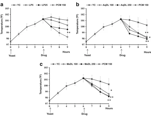
Effect of Latex Fractions on Yeast-Induced Fever
Fever was induced in rats by subcutaneous injection of yeast and a peak effect was obtained at 6 h where the rectal temperature was 101.23 ± 0.63 °F against 97.51 ± 0.64 °F in normal rats. There
was a gradual decline in temperature during subsequent 3 h to 100.22 ± 0.98 °F. Treatment of rats with AqDL and MeDL at 6 h produced a significant reduction in rectal temperature at 9 h as compared to the YC group and the effect of MeDL was comparable to that of paracetamol while that of AqDL was more pronounced than that of paracetamol. Treatment with LP, however, produced only a slight reduction in temperature at 25 mg/kg dose (Fig.3).
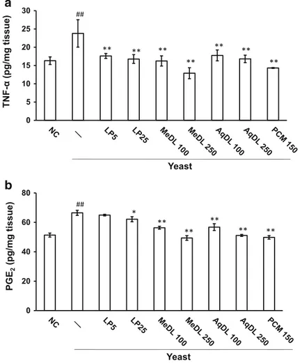
Effect of Latex Fractions on the Level of TNF-αand PGE2
Induction of fever by subcutaneous injection of yeast was associated with a significant increase in the level of TNF-αin the hypothalamic region (23.78 ± 3.75 pg/mg tissue in
0 0.4 0.8 1.2 1.6 2
4 5 6 7 8
LP AqDL MeDL
0 0.2 0.4 0.6
4 5 6 7 8 9 LP AqDL MeDL
0 0.1 0.2 0.3 0.4
4 5 6 7 8
LP AqDL MeDL
U A /µ g Pr o te in U A /µ g Pr o te in U A /µ g Pr o te in pH pH pH c a b
Fig. 2 Proteolytic activity of latex fractions LP, AqDL, and MeDL at different pH using azocasein as substrate in the presence (a) or absence of DTT (b) and BANA as substrate in the presence of DTT (c)
97 98 99 100 101 102 103
0 3 4 5 6 7 8 9 YC LP5 LP25 PCM 150
97 98 99 100 101 102 103
0 3 4 5 6 7 8 9
YC AqDL 100 AqDL 250 PCM 150
Drug Yeast Hours T e m p e ra tu re ( 0F ) ** Hours T e m p e ra tu re ( 0F ) ** ** ** 97 98 99 100 101 102 103
0 3 4 5 6 7 8 9 YC MeDL 100 MeDL 250 PCM 150
Drug Yeast Drug Yeast Hours T e m p e ra tu re ( 0F ) ** ** *
b
a
c
the YC group vs. 16.32 ± 1.04 pg/mg tissue in the NC group). Treatment with all the latex fractions produced a significant reduction in the level of TNF-α and their effect
was comparable to that of paracetamol. The YC group also showed an elevated level of PGE2as compared to the NC group (66.49 ± 1.71 pg/mg tissue vs. 51.36 ± 1.38 pg/mg tissue in the NC group). Treatment with LP25 produced slight though statistically significant reduction in hypothalamic PGE2 levels while treatment with both MeDL and AqDL produced a dose-dependent reduction in PGE2 levels. The PGE2 levels in MeDL250, AqDL250, and PCM150 groups were comparable (Fig.4).
Effect of Latex Fractions on COX-2 Immunoreactivity in Hypothalamus
Immunohistochemical analysis of hypothalamic sections revealed enhanced COX-2 im-munoreactivity following subcutaneous injection of yeast. Treatment of rats with LP25 produced a slight reduction in COX-2 immunoreactivity while AqDL250 and MeDL250
Yeast
T
N
F
-(p
g
/m
g
ti
s
s
u
e
)
a
b
Yeast
PG
E2
(p
g
/m
g
ti
s
s
u
e
)
** ** **
** **
** **
* **
** **
** **
0 20 40 60 80 0 5 10 15 20 25
30 ##
##
markedly reduced COX-2 immunoreactivity and their effect was comparable to that of paracetamol (Fig.5).
Discussion
The plantC. procerais well known for its medicinal properties and its different parts have been used in the traditional medicinal system for the treatment of various disease conditions. The latex produced by this plant is abundantly present in its aerial parts and is a rich source of constituents possessing pharmacological properties. Several studies have been carried out on the fractions prepared from the latex, namely, aqueous extract, methanol extract, and the protein fractions all of which exhibit potent anti-inflammatory and analgesic properties [5,6,8,
9,14]. The aim of the present study was to compare the biochemical characteristics of these fractions, to evaluate their protective effect against febrile response induced in rats, and to elucidate their mechanism of action. As one of the fractions included in this study, LP is protein in nature and has been reported to possess proteinase activity; the presence of proteins and enzymatic activity was also evaluated in the other two fractions that are orally effective, i.e., AqDL and MeDL. Both protein detection assay and SDS-PAGE revealed that the AqDL contains proteins while the MeDL is devoid of any protein constituent of the latex. All the three fractions were also tested for enzymatic activity by gelatin zymography and biochemical analysis. While zymography revealed the presence of proteinase activity in LP and AqDL, the biochemical analysis has shown that only LP fraction possesses cysteine proteinase activity as also reported earlier by Ramos et al., [20]. The results of the present study show that the proteins present in AqDL differ from those present in LP in terms of their enzymatic activity. A recent study on cysteine proteinase profile ofC. proceraaerial parts by transcriptome analysis has highlighted the functional importance of different regions and the role of enzyme purifi-cation method in determining the activity [21].
a b c
d e f
Anti-inflammatory and analgesic drugs are well known to exhibit antipyretic property. The aqueous suspension of the dried latex ofC. procerais one such preparation that has been reported to produce antipyretic effect in rodent model where fever was induced by Baker’s yeast [7]. In the present study, the latex fractions and the standard drug paracetamol were administered in this model at the time of peak hyperthermic response occurring at 6 h and their antipyretic effect was evaluated by recording rectal temperature over a period of 3 h. Of the three fractions tested, the LP fraction produced only a marginal reduction in rectal temperature while the antipyretic effect of AqDL and MeDL was comparable to that of paracetamol. These findings show that latex constituents other than protein produce the central effects and are orally effective while the weak antipyretic effect of LP fraction could have resulted due to peripheral anti-inflammatory effect of this fraction [6]. Although some of the biological activities of the latex have been attributed to the cysteine proteinase activity of its protein constituents, such an activity does not appear to be involved in its antipyretic effect [20]. Besides proteins, the latex of C. proceracontains several other constituents like alkaloids, glycosides, tannins, flavonoids, sterols, and triterpenes [3]. Though the latex fractions AqDL and MeDL have been reported to produce anti-inflammatory and analgesic effect by inhibiting different mediators, the orally effective active principle exhibiting antipyretic effect remains to be identified [12]. These fractions have also been shown to exhibit antioxidant properties and to afford protection in various disease models [4,22]. Further, a study carried out in rats revealed that oral administration of aqueous suspension of dried latex is safe and suggests that it could be used for therapeutic purpose [23].
Fever induced by subcutaneous injection of yeast is associated with the activation of an inflammatory response. The pro-inflammatory mediators released during this process gain access to the central nervous system (CNS) by crossing the blood-brain barrier (BBB) and exhibit their pyrogenic effect by acting on the pre-optic anterior hypothalamic area of the brain [24]. TNF-αis one such mediator that is involved in disease progression,
inflamma-tion, and dysfunction of BBB [25, 26]. It exerts its pyrogenic effect through COX-2 induction and release of central mediator PGE2which increases the temperature set point and produces fever through activation of PGE2 receptor 3 (EPR-3) and initiation of neurotransmitter cascade [13,27,28]. In present study, the hypothalamic levels of PGE2 and TNF-αwere found to be significantly higher in the YC group as compared to the NC
group. Treatment with all the three latex fractions markedly reduced the TNF-αlevel in the
hypothalamus to a significant extent. It is interesting to note that PGE2 levels were significantly reduced only by AqDL and MeDL and not by LP. The effect of the latex fractions in lowering hypothalamic PGE2level was in concurrence with their efficacy in bringing down the body temperature. Our study shows that a small change in PGE2level plays an important role in regulating body temperature than a bigger change in TNF-α. As
release of PGE2through activation of COX-2 plays an important role in induction of fever by pyrogenic cytokines, the present study also evaluated the effect of these fractions on this key step [28]. It was found that COX-2 immunoreactivity was higher in the YC group as compared to the NC group in the hypothalamus and treatment with both AqDL and MeDL markedly reduced it while LP fraction was weak in this regard. Our study shows that by inhibiting TNF-α- and COX-2-mediated PGE2release, both AqDL and MeDL might be
Conclusions
Latex of the plantCalotropis procerais well known to possess potent anti-inflammatory and analgesic activity. It comprises of proteins and non-protein constituents which produce their pharmacological effects following parentral and oral administration. The results of the present study show that the protein fraction possesses proteinase activity and produces weak antipy-retic effect while the orally effective non-protein constituents exhibit potent antipyantipy-retic effect like that of paracetamol. The mechanism for the antipyretic effect of this fraction involves inhibition of PGE2 generation by suppression of COX-2 in the hypothalamus. Further, this study also shows that aqueous and methanol extracts of the dried latex as prepared in this study have a therapeutic potential in ameliorating fever in various inflammatory conditions.
References
1. Kirtikar, K. R., & Basu, B. D. (1935).Indian medicinal plants. Allahabad: Lolit Mohan Basu.
2. Wealth of India. (1992).Raw materials—Vol 3(pp. 78–84). CSIR, New Delhi: Information and Publication
Directorate.
3. Mossa, J. S., Tariq, M., Mohsin, A., Ageel, A. M., Al-Yahya, M. A., Al-Said, M. S., et al. (1991). Pharmacological studies on aerial parts ofCalotropis procera.The American Journal of Chinese Medicine Med., 19, 223–231.
4. Kumar, V. L., & Arya, S. (2006). Medicinal uses and pharmacological properties ofCalotropis procera. In J. N. Govil (Ed.),Recent progress in medicinal plants, Vol. 11(pp. 373–388). Houston: Studium Press. 5. Kumar, V. L., & Basu, N. (1994). Anti-inflammatory activity of the latex ofCalotropis procera.Journal of
Ethnopharmacology, 44, 123–125.
6. Alencar, N. M., Figueiredo, I. S., Vale, M. R., Bitencurt, F. S., Oliveira, J. S., Ribeiro, R. A., et al. (2004). Anti-inflammatory effect of the latex fromCalotropis procera in three different experimental models: peritonitis, paw edema and hemorrhagic cystitis.Planta Medica, 70, 1144–1149.
7. Dewan, S., Kumar, S., & Kumar, V. L. (2000a). Antipyretic effect of latex ofCalotropis procera.Indian Journal of Pharmacology, 32, 252.
8. Dewan, S., Sangraula, H., & Kumar, V. L. (2000b). Preliminary studies on the analgesic activity of latex of
Calotropis procera.Journal of Ethnopharmacology, 73, 307–311.
9. Kumar, V. L., & Roy, S. (2007).Calotropis proceralatex extract affords protection against inflammation and oxidative stress in Freund’s complete adjuvant-induced monoarthritis in rats. Mediators of Inflammation, 2007, 47523.
10. Kumar, V. L., Chaudhary, P., Ramos, M. V., Mohan, M., & Matos, M. P. V. (2011). Protective effect of proteins derived from the latex ofCalotropis proceraagainst inflammatory hyperalgesia in monoarthritic rats.Phytotherapy Research, 25, 1336–1341.
11. Kumar, V. L., Guruprasad, B., Chaudhary, P., Fatmi, S. M. A., Oliveira, R. S. B., & Ramos, M. V. (2015). Protective effect of proteins derived fromCalotropis proceralatex against acute inflammation in rat.
Autonomic & Autacoid Pharmacology, 35, 1–8.
12. Arya, S., & Kumar, V. L. (2005). Anti-inflammatory efficacy of extracts of latex ofCalotropis procera
against different mediators of inflammation.Mediators of Inflammation, 2005, 228–232.
13. Netea, M. G., Kullberg, B. J., & Van der Meer, J. W. M. (2000). Circulating cytokines as mediators of fever.
Clinical Infectious Diseases, 31(Suppl 5), S178–S184.
14. Soares, P. M., Lima, S. R., Matos, S. G., Andrade, M. M., Patrocínio, M. C., de Freitas, C. D., et al. (2005). Antinociceptive activity ofCalotropis proceralatex in mice.Journal of Ethnopharmacology, 99, 125–129. 15. Ramos, M. V., Oliveira, J. S., Figueiredo, J. G., Figueiredo, I. S., Kumar, V. L., Bitencourt, F. S., et al. (2009). Involvement of NO in the inhibitory effect ofCalotropis proceralatex protein fractions on leukocyte rolling, adhesion and infiltration in rat peritonitis model.Journal of Ethnopharmacology, 125, 387–392.
16. Bradford, M. M. (1976). A rapid and sensitive method for the quantitation of microgram quantities of protein utilizing the principle of protein-dye binding.Analytical Biochemistry, 72, 248–254.
17. Macedo, M. L., Freire, M. D., & Parra, J. R. P. (2004). A kunitz-type inhibitor of coleopteran proteases isolated fromAdenanthera pavoninaL. seeds and its effect onCallosobruchus maculatus.Journal of Agricultural and Food Chemistry, 52, 2533–2540.
cowpea (Vigna unguiculata) and the resistance/susceptibility to predation byCallosobruchus maculatus.
Journal of Agricultural and Food Chemistry, 37, 1139–1143.
19. Abe, M., Abe, K., Kuroda, M., & Arai, S. (1992). Corn kernel cysteine proteinase as a novel cystatin superfamily member of plant origin.Molecular cloning and expression studies. European Journal of Biochemistry, 209, 933–937.
20. Ramos, M. V., Araujo, E. S., Juca, T. L., Monteiro-Moreira, A. C., Vasconcelos, I. M., Moreira, R. A., et al. (2013). New insights into the complex mixture of latex cysteine peptidases inCalotropis procera.
International Journal of Biological Macromolecules, 58, 211–219.
21. Kwon, C. W., Park, K. M., Kang, B. C., Kweon, D. H., Kim, M. D., Shin, S. W., et al. (2015). Cysteine protease profiles of the medicinal plantCalotropis procera R.Br. revealed by de novo transcriptome analysis.PloS One, 10, e0119328.
22. Roy, S., Sehgal, R., Padhy, B. M., & Kumar, V. L. (2005). Antioxidant and protective effect of latex of
Calotropis proceraagainst alloxan-induced diabetes in rats.Journal of Ethanopharmacology, 102, 470– 473.
23. Singhal, A., & Kumar, V. L. (2009). Effect of aqueous suspension of dried latex ofCalotropis proceraon hepatorenal functions in rat.Journal of Ethnopharmacology, 122, 172–174.
24. Blatteis, C. M., Sehic, E., & Li, S. (2000). Pyrogen sensing and signalling: old views and new concepts.
Clinical Infectious Diseases, 31(Suppl 5), S168–S177.
25. Montgomery, S. L., & Bowers, W. J. (2012). Tumor necrosis factor alpha and the roles it plays in homeostatic and degenerative processes within the central nervous system.Journal of Neuroimmune Pharmacology, 7, 42–59.
26. Opp, M. R., George, A., Ringgold, K. M., Hansen, K. M., Bullock, K. M., & Banks, W. A. (2015). Sleep fragmentation and sepsis differentially impact blood-brain barrier integrity and transport of tumor necrosis factor-αin aging.Brain, Behavior, and Immunity, 50, 259–265.
27. Candelario-Jalil, E., Taheri, S., Yang, Y., Sood, R., Grossetete, M., Estrada, E. Y., et al. (2007). Cyclooxygenase inhibition limits blood-brain barrier disruption following intracerebral injection of tumor necrosis factor–alpha in the rat.The Journal of Pharmacology and Experimental Therapeutics, 323, 488– 498.
28. Dinarello, C. A. (2004). Infection, fever, and exogenous and endogenous pyrogens: some concepts have changed.Journal of Endotoxin Research, 10, 201–222.
29. Simmons, D. L., Wagner, D., & Westover, K. (2000). Nonsteroidal anti-inflammatory drugs, acetamino-phen, cyclooxygenase 2, and fever.Clinical Infectious Diseases, 31(Suppl 5), S211–S218.
30. Spencer, B. J., & Verma, I. M. (2007). Targeted delivery of proteins across the blood-brain barrier.