Proteolysis of ovine caseins by cardosin A,
an aspartic acid proteinase Jrom Cynara cardunculus L.
Sofia V. Silva, F. Xavier Malcata*
Escola Superior de Biotecnologia, Universidade Católica Portlfguesa, Rua Dr. António Bemardino de Almeida, 4200 Porto, Portugall
(Received 6 October 1997; accepted 3 April1998) c"
Abstract - The breakdown of as -caseins and ~-caseins (in the form of as -caseins, the form of ~-caseins, and the form of a mixture of as- and ~-caseins in Na-caseinate) by cardosin A, one of the major two proteinases present in the flowers of Cynara cardunculus L., was experimentally stu-died via urea polyacrylamide gel electrophoresis. In Na-caseinate, as- and ~-caseins were degraded up to 46 and 76 %, respectively, by 10 h of hydrolysis. In separated form, as-caseins reached a leveI of degradationup to 67 % while ~-caseins were quickly and extensively degraded up to 76 %. Ingeneral, ~-caseins seemed to be more susceptible to proteolysis than as-caseins. @Inra/EIsevier, Paris.
milk protein / enzyme / plant rennet / electrophoresis
Résumé - Protéolyse des caséines de brebis par la cardosine A, une protéase acide aspar-tique de Cynara cardunculus L. La dégradation des caséines as et des caséines
~
(sous la forme de caséines as' de caséines ~, et d'un mélange de caséines as et~d'un caséinate du sodium) par la
car-dosine A, une des protéinasesmajeures présentes dans les fleurs de Cynara cardunculusL., a été étu-diée par électrophorese en gel de polyacrylamide à l'urée. Dans le caséinate de sodium, les caséines as et~
ont été respectivementdégradéesjusqu' à 46 et 76 %, apres 10h d'hydrolyse ; sousforme sépa-rée, les caséines as ont atteint des niveaux de dégradation de 67 % ; pourtant, les caséines~
ont été dégradéesplus rapidementet extensivementjusqu'à 76 %. En général,les caséines~
doivent être plus sensibles à la protéolyse que les caséines as' @Inra/EIsevier, Paris.protéine laitiere / enzyme / présure végétale / électrophorese
1. INTRODUCTION
Milk clotting by proteolytic enzymes is a crucial operation in most forms of cheese-making. Calf rennet was the first (and still considered the best) mixture of milk clot-ting enzymes, mainly chymosin and pep-sino Chymosin accounts for more than 90% of the milk clotting activity in rennets extra-cted from the abomasa of very young ani-mals, but as the animal ages the ratio of chy-mosin to pepsin decreases [8]. The worldwide supply of animal rennet is cur-rently insufficient to satisfy the growing demand by the cheese industry and conse-quently the search for alternative sources of milk clotting enzymes has gained renewed interest. Although rennets from genetically engineered microorganisms have been pro-duced in higher and líigher numbers, seve-ral wild plants can also produce proteoly-tic enzymes which have, nevertheless, not attracted extensive attention. The exception to this rule is a clotting aqueous extract from the flowers of the wild thistle (Cynara car-duncu/us L.), which has been successfully used for centuries in chesemaking in Por-tugal [15] and bordering regions of Spain. The clotting activity of this extract was first attributed to the presence of three proteoly-tic enzymes synthesized by the stigmae of those flowers [3], and such (tentatively ter-med) cynarases 1,2 and 3 were purified and partially characterized [6]. Two new aspar-tic acid proteinases, (tentatively named) car-dosins A and B, were later isolated and are believed to be genetically different from the aforementioned cynarases [17]; in terms of activity and specificity, cardosins A and B are remarkably similar to chymosin and pep-sin, respectively [16].
In addition to this clotting activity, the enzymes present in rennet and entraped in the curd can also bring about protein break-down during cheese ripening [4, 5], and are thus responsible for the release of various peptides with biochemical, rheological and sensory roles in cheese [4]. It has been firmly established that the acid proteinases
present in rennet are highly specific in their
action towards the Phe1O5-Metl06 bond of
K-casein, although they can also attack
caseins other than K-casein at much lower
rates [4]; on the other hand, despite the fact
that the action of cardosins A and B on
bovine asccasein was studied [10],
funda-mental work encompassing the action of
these proteinases on ovine caseins remains
scarce. Hence, the aim of this work was to
study the action of cardosin A on ovine
Na-caseinate, and on families of ovine caseins
isolated therefrom, to eventually better
understand what happens during ripening
of traditionalPortuguese cheeses which use
this enzyme in their manufacture protocol.
4
2. MATERIALS AND METHODS
2.1. Substrate preparation
Ovine sodium-caseinate was prepared from raw mi!k by acidification to pH 4.3 with direct addition of 6 mo!.L-l HC! under stirring, warm-ing up to 45 °C and ho!dwarm-ing at that temperature for 45 minoThe precipitatewas then recoveredby filtration through a clean cloth, washed severa! times with deionized water and resuspended in deionized water, and its pH adjusted to 7.0 with 1 mo!.L-l NaOH. The so!ution was allowed to equilibrate at 4 °C for at !east 2 h and then !yo-philized.
Ovine as-caseinsand j3--caseinswere prepared from !yophilized Na-caseinate via semiprepara-tive separation by chromatography with DEAE-cellu!ose using 0.01 mo!.L-l Tris-HC! buffer (pH 7.0)containing4.5 mo!.L-lurea, 0.01 mo!.L-l imidazo! and 0.1% (w/v) [3-mercaptoethano!as e!uent via a linear gradient of 0-0.4 mo!.L-1of sodium ch!oride within 25 h at the flow rate of 80 rnL.h-l. The various as-casein and [3-casein fractions were independently pooled together, dia!yzed against deionized water and !yophi-lized.
.
2.2. Enzyme preparation
Dried flowers of the thistle (C. carduncu-lus L.) were homogenized at the ratio of 1 g of ground flowers to 12 rnL of citric acid (pH 3.6) and centrifugedat 6 000 g for 20 minoThe
super-Proteolysis of ovine caseins by cardosin A. 515
natant was collected and a 2-mL aliquot was applied to a Highload 26/60 Sephacryl S-200 column (Pharmacia,Uppsala, Sweden) afterpro-per equilibration, and eluted with 20 mmol-L-l Tris-HCI buffer (pH 7.6) at a flow rate of 1.5 mL.min-l. The various fractions correspon-ding to the peak of absorbance eluted at 135 mL were pooled together and applied to a Mono Q HR 5/5 column (Pharmacia), from which they were eluted with the same buffer at the flow rate of 0.75 mL.min-lunder a linear gradientof 0-0.5 mol-L-l sodium chloride within 30 min, and the fraction corresponding to the peaks of absor-bance at the eluted volume of 12.75-16.5 mL was duly collectedas cardosinA; assay for purity was done via polyacrylamidegel electrophoresis with sodium dodecyl sulfate in a Phastsystem (Pharmacia) using the PhastGel gradient 8-25.
2.3. Enzymatic hydrolysis
Whole Na-caseinate, separated as-caseins and separated ~-caseins (as appropriate) were dis-solved in 100 mmol.L-l phosphate buffer (pH 6.5) so as to yield 1% (w/v) final concentration, and sodium azide (0.05% w/v) was added to pre-vent bacterial growth. The preparation of cardo-sin A (I80 ,ug.mL-l) was then added at the ratio of 0.526 mL per mL of substrate solution and the mixture was incubated at 30 oCo Samples were taken after I min, I h, 3 h, 6 h and 10 h of hydrolysis and the reaction was quenched prior to analysis via addition of double concentrated buf-fer at 50% (v/v) [7] in the case of samples for electrophoresis.
2.4. Protein and peptide profiling
by electrophoresis
Urea polyacrylamide gel electrophoresis (urea-PAGE) was performed on samples of casein hydrolyzates following the method of Andrews [I] with slight modifications[12].Elec-trophoresis was carried out in a Protean 11xi cell vertical slab unit (Bio-Rad Laboratories, Wat-ford, UK); the power supply, model 1000/500 (Bio-Rad Laboratories), was set at 280 mV for the stacking gel and then increased to 300 mV for the separating gel. Gels were stained with Coomassie Blue G250 (Bio-Rad Laboratoires) using the method of Blakesley and Boezi [2]. Quantification of as- and ~-caseins was done by densitometry using a model GS-700 imaging densitometer (Bio-Rad Laboratoires, Hercules, CA, USA).
3. RESUL TS AND DISCUSSION
The elec1Tophoretogram of ovine
Na-casei-nate after hydrolysis is shown in figure 1,
whereas those of separated as-caseins and
separated p-caseins after hydrolysis are
shown infigures 2 and 3, respectively. The
ratio of concentrations of intact as - and
p-caseinsto their initial concen1Tations,
both
in ovine caseinate and in separated forms,
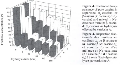
are shown infigure 4.
Sodium caseinate hydrolyzates
contai-ned two major groups of electrophoretic
bands (seefigure 1). Richardson and
Crea-mer [11] pointed out that the group with the
highest electrophoretic mobility is
accoun-ted for by as-caseins, which may be further
subdivided into three variants (asc,
as2-and aS3-casein),whereas the group with the
lowest mobility is accounted
for by
p-caseins, which may be further subdivided
into two variants (Pc and p2-casein), as
represented
as as -casein and p-casein
regions, respectively,infigure 1. AlI caseins
underwent degradation after as little as
1 min of incubation, and p-caseins were
hydrolyzedfasterthan as-caseins (seefigure
4); the percent degradation of the p-casein
family as a whole was 26.6 %, whereas that
of the as-casein family was 17.6 %. After
this time, breakdown of whole ovine
Na-caseinate by cardosin A led to two bands
with higher electrophoretic mobility than
the p-casein region, which disappeared
quickly (see bands A and B of lane 4 in
figure 1). Qne band exists that is located
right after the as-casein region but which
vanishes by 10 h of incubation (see band C
of lane 6 infigure 1), whereas two bands
with highermobilitybecamemore intenseas
hydrolysis time elapsed (see bands D and
E of lanes 2-6 in figure 1). Hydrolysis of
p-caseins occurred to a much greater extent
(up to 75.9 %) than hydrolysis of as-caseins
(up to 46.5 %) (see figure 4), and it also
took place more quickly.
With respect to cleavage of separated
as -caseins by cardosin A, a set of bands
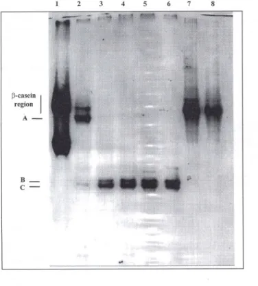
3 4 5 6 7 8 p-ca~ein I reglOn A- B-a,-casein I reglOn
c-
D-
E-2 3 Figure 2. Urea-PAGE e1ectrophoregramof sepa-rated as-caseins after incu-bation for 1rnin, 1 h, 3 h, 6 h and 10 h (lanes 2-6, res-pective1y)with cardosin A; 1ane1 contains plain ovine Na-caseinate, and lanes 7 and 8 contain plain ovine as-caseins, after incubation for 1 min and 10 h, all included as controls.Figure 2.
Diagrammeélec-trophorétique urée-EGPA de caséines as apres incu-bation pendant 1 min, 1 h, 3 h, 6 h et 10h (lignes 2-6, respectivement) avec car-dosine A ; la ligne I con-tient du caséinate de sodium de brebis pur, et les lignes 7 et 8 contiennent des caséines as aprés incu-bation pendant 1 min et 10 h ; utilisés comme contrôles. Ct,-c~sein I regIOn
A-
B-Figure 1. Urea-PAGE electrophoregram of Na-caseinate after incubation for 1 min, 1 h, 3 h, 6 h and 10 h (lanes 2-6, respecti-vely) with cardosin A; lane 1 contains plain ovine Na-caseinate,and lanes7 and 8 contain plain ovine Na-caseinate, after incubation for 1 min and 10 h, all included as controIs.Figure 1.
Diagrammeélec-trophorétique urée-EGPA de caséinate du sodium apres incubation pendant 1 min, 1 h, 3 h, 6 h et 10 h (lignes 2-6, respective-ment) avec cardosine A ; la ligne 1contientdu caséi-nate de sodium de brebis pur, et les lignes 7 et 8 contiennentdu caséinatede sodium apres incubation pendant 1rnin et 10 h ; uti-lisés comme contrôles.
Proteolysis of ovine caseins by cardosin A. 517 2 3 4 5 6 7 8 ~-ca~eiD I reglOD
A-
B-
C-with higher electrophoretic mobility than
the as -casein region can be observed in
figure 2. Two bands were apparentjust after
1 min of hydrolysis but eventually
disap-peared as incubation progressed (see bands
A and B of lane 6 infigure 2), whereas
bands displaying higher mobilities became
thicker and thicker. These results are
com-parable with those obtained by
Ramalho-Santos et aI. [10] pertaining to the action of
cardosin A on bovine asccasein; according
to these authors, Phe23-Phe24 is the most
susceptible peptide bond and leads to
pro-duction of fragments fl-23 and f24-199.
The Trp 164-Tyr166 portion of the latter
fragment is then attacked and four
poly-peptides originate, viz f24-164, f24-165,
fl65-199 and fl66-199; then the
Phe153-Tyr154 bond is broken, thus producing
f24-153,fl54-165 and fl54-166, and finally the
Phe150-Arg151 bond is hydrolyzed, thus
releasingf24-150 and fl51-154. The
appea-rance of the polypeptide Tyr154- Trp 199
Figure 3. Urea-PAGE electrophoregram of sepa-rated ~-caseins after incu-bation for I min, I h, 3 h, 6 h and 10 h (lanes 2-6, respectively) with cardosin A; lane I contains plain ovine Na-caseinate, and lanes 7 and 8 contain plain ovine ~-casein, after incu-bation for I min and 10 h, all inc1udedas contraIs.
Figure 3.
Diagrammeélec-trophorétique urée-EGPA de caséines
~apres
incuba-tion pendant I min, I h, 3 h, 6 h et 10h (lignes 2-6, respectivement) avec car-dosine A ; Ia ligne I con-tientdu caséinatede sodium de brebis pur, et les lignes 7 et 8 contiennent des caséines~apres incubation
pendant I min et 10 h ; uti-lisés comme contrôles.was noticed only due to the slower action
of cardosinA on the region Trp164-Tyr166.
Inspection
of figure
4 indicates
that
as-caseins were degraded up to 67.3 % by
10 h of hydrolysis.
When in separated form, p-caseins were
degraded quickly and extensively and
rea-ched a degradation degree of75.7 % by 10
h of hydrolysis (see figure 4). Hydrolysis
of ovine p-casein (figure 3) led to the
pro-duction of one band with higher
electro-phoretic mobility than that of the p-casein
region (see band A oflane 2 infigure 3) by
1 rnin of incubation,which
disappearedgra-dually after 1 h. Such peptide was
appa-rently equivalent to bovine p-I-casein [13];
in fact, the bonds Leu 192-Tyrl93 and
Ala189-Phe190 of bovine p-casein are the
most susceptible to the catalytic action by
either chymosin or proteinases from C.
car-dunculus [13, 18], and p-I-casein, which
results from c1eavageof those bonds, is thus
a mixture of fl-189 and fl-192; in ovine
~
c .S (::í .... E .., o c o o 100- 90-80, 70 .. c .S ü J: 180 Hydro\ysis time (min)~-casein, Leu 190-Tyr191 and Ala
187-Phe188 are the corresponding bonds
clea-ved [14].Two bands possessinghigher
elec-trophoreticmobility than band A (see bands
B and C oflane 3 infigure 3) could be
noti-ced by 1 h of hydrolysis and became more
intense as time elapsed.
There is a noticeable difference between
the action of this aspartic proteinase upon
ovine caseins in their isolated forms and in
their mixed (or Na-caseinate) counterparts
regarding the rates of proteolysis and
degrees of degradation.
Proteolysis of
~-caseins occurred to similar extents (up to
76 %), but separated ones were hydrolyzed
more quickly than when mixed with
as-caseins in Na-caseinate. With regard to
as-caseins, these proteins were as a whole
more extensivelydegradedin separatedform
than in Na-caseinateformoAlthoughit might
be claimed that the rate of hydrolysis of
either as
-
and ~-caseins is higher insepa-rated form than in Na-caseinate form due
to a dilution factor (recall that as - and
~-caseins exist at the mass ratio of 3:4.7 in
Na-caseinate), it should be noted that the
variation of either protein concentrationwas
in both cases normalized by its initial
concentration;hence, significantdifferences
in proteolysisrates are likely due to the state
of aggregation of the caseins.
Figure 4. Practional disap-pearance of pUfecaseins in separated as -caseins or l3-caseins(a: j3-casein;c: as-casein) and mixed in Na-caseinate form (b: l3-casein; d: as -casein) via hydrolysis effected by cardosin A. Figure 4. Disparition frac-tionnée des caséines en
caséines a ou
13separées
(a : caséine ~ ; c : caséine as) et sous Ia forme d'un mélange en Na-caséinate (b : caséine13 ; d : caséine as) à travers I'hydrolyse
cata-Iysée par cardosine A. ~
d
600
From the results presented above, it is apparent that ~-caseins, either in separated form or mixed with as-caseins in Na-casei-nate, are more susceptible to proteolysis than as -caseins. Enzymes from C. cardun-culus act on bovine as\-' as2- and ~-caseins [9] to a higher extent than the enzymes pre-sent in commercial rennets. Sousa and Malcata [14] reported that crude aqueous extracts of flowers of C. cardunculus attack both ovine as and ~-caseins in cheese, although ~-caseins were reported to be less susceptible to proteolysis than as -caseins.
Although our results were produced in vitro, their extrapolation to cheese ripening can be relatively backed up because 6% of the coagulant used in cheese manufacture is retained in the curd [13]. However, such extrapolation should be cautious because two factors constrain casein proteolysis: the accessibility of the peptide bonds to the enzyme and the actual enzyme specificity. In milk, most caseins exist naturally as micelles; the putative inaccessibility of the cleavage sites to the enzyme when caseins are in micellar form may lead to lower rates of degradation than in separated, soluble form [4]. Furthermore, the complexity of the three dimensional structure of the casein network in cheese will also interfere with accessibility. On the other hand, the
speci-Proteolysis of ovine caseins by cardosin A.
.
ficity of the enzymes in pure form may be
different from that of the enzymes in crude
extracts, although this point requires further
experimentation in order to become dear.
As major condusions, it can be said that
significantly different proteolysis pattems
exist for the actionof cardosinA upon ovine
caseins, and that a.s-caseins, either in
sepa-rated form or mixed with other caseins in
Na-caseinate, are as a whole less
suscep-tible to proteolysis than ~-caseins.
ACKNOWLEDGEMENTS
~
The authors are grateful to Df. José Empis (Instituto Superior Técnico, Lisbon, Portugal) for cosupervision within the scope of the gra-duate program where the author S.V. Silva was enrolled. Financial support for S.V. Silva was provided by an MSc fellowship (BM/8824/96) issued by PRAXIS XXI (JNICT, Portugal). Par-tial financial support for this work was provided by grants through projects PROTOLACTIS: PROdução, por Tecnologias Optimizadas, de LACticínios TradicionaIS (PAMAF, Portugal) and IMPACTO: Investigação dirigida ao Me-lhoramento do queijo serra Por integrAção de abordagensCientíficase TecnOlógicas(pRAXIS XXI, Portugal).REFERENCES
[I] Andrews A.T., Proteinases in normal bovine milk and their action on caseins, 1. Dairy Res. 50 (1983) 45-55.
Blakesley R.W., Boezi I.A., A new staining technique for proteolysis in polyacrylamide gels using Coomassie Brilliant BIue G250, Anal. Biochem. 82 (1977) 580-581.
Cordeiro M., lakob E., Puhan Z., Pais M.S., Brodelius P.E., Milk cIotting and proteolytic acti vities of purified cynarases from Cynara
cardunculus - a comparison to chymosin, MiIchwissenschaft 47 (1992) 681-687. Dalgleish D.E., The enzymatic coagulation of milk, in: Fox P.F. (Ed.), Cheese: Chemistry, Physics and Microbiology, vol. I, Elsevier Applied Science Publishers, London, 1987, pp. 63-96. [2]
.
[3] [4] 519[5] Fox P.E Proteolysis during cheese manufacture and ripening, I. Dairy Sci. 72 (1989) 1379-1400. [6] Heimgartner V., Pietrzak M., Geertsen R., Bro-delius P., Silva Figueiredo A.C., Pais M.S.S., Purification and partial characterization of milk cIotting proteases from fIowers of Cynara
car-dunculus. Phytochemistry 29 (1990) 1405-1410.
[7] McSweeney P.L.H., Olson N.F., Fox P.F., Healy A., Hojrup P., Proteolytic specificity of chymosin on bovine a'l-casein, I. Dairy Res. 60 (1993) 401-412.
[8] Mulvihill D.M., Caseins and caseinates: manu-facture, in: Fox P.F. (Ed.), Developments in Dairy Chemistry, vol. 4, EIsevier Applied Science, London, 1989, pp. 97-130.
[9] Pires E., Faro c., Macedo 1., Esteves c., Mor-gado I., Veríssimo P., Dias-Pereira c., Gomes D., Flor do cardo versus quimosina no fabrico de queijos artesanais, Química 54 (1994) 66-68. [10] Ramalho-Santos M., Veríssimo P., Faro c., Pires
E., Action on bovine asl-casein of cardosins A and B, aspartic proteinases from the fIowers of the cardoon Cynara cardunculus L., Biochim. Biophys. Acta 1297 (1996) 83-89.
[11] Richardson B.C., Creamer L.K., Comparative micelle structure. V. The isolation and charac-terization of the major ovine caseins, N. Z. I. Dairy Technol. 11 (1976) 46-53.
[12] Shalabi S.I., Fox P.E, Electrophoretic analysis of cheese: comparison of methods, Int. I. Food Sci. Technol. 11 (1987) 135-151.
[13] Sousa M.I.C.F., Plant rennet substitute from fIo-wers of Cynara cardunculus, MSc Thesis, Natio-nal Vniversity ofIreland, Cork, Ireland, 1993. [14] Sousa M.J., MaIcata EX., Comparison of plant and animal rennets in terms of microbiological, chemical and proteolysis characteristics of ovine cheese, I. Agric. Food Chem. 45 (1997) 74-81. [15] Vieira de Sã F., Barbosa M., Cheese-making
with a vegetable rennet from cardo (Cynara
car-dunculus), I. Dairy Res. 39 (1972) 335-343.
[16] Veríssimo P., Esteves C., Faro C., Pires E., The vegetable rennet of Cynara cardunculus L. contains two proteinases with chymosin and pepsin-like specificities, Biotechnol. Lett. 17 (1995) 621-626.
[17] Veríssimo P., FaroC., Moir A.J.G., Lin Y., Tang 1., Pires E., Purification, characterization and par-tial amino acid sequencing of two new aspartic proteinases from fresh fIowers of Cynara
car-dunculus L., Eur. I. Biochem. 235 (1996) 762-768.
[18] Visser S., Slangen K.J., On the specificity of chymosin (rennin) in its action on bovine ~-casein, Neth. Milk Dairy I. 31 (1977) 16-30.