KARYOTYPE RESTRUCTURING IN RODENTIA:
FROM EVOLUTION TO CANCER
Universidade de Trás-os-Montes e Alto Douro
Sandra Louzada Gomes Pereira
2013
U
NIVERSIDADE DET
RÁS-
OS-M
ONTES EA
LTOD
OUROK
ARYOTYPE RESTRUCTURING IN
R
ODENTIA
:
FROM EVOLUTION TO CANCER
S
ANDRAL
OUZADAG
OMESP
EREIRAIII
Este trabalho foi expressamente elaborado com
vista à obtenção de grau de Doutor em Genética
Molecular, Comparativa e Tecnológica.
V
Tese de Doutoramento financiada pela Fundação
para a Ciência e Tecnologia, “Programa Operacinal
Potencial Humano – POPH”, co-financiado pelo
Fundo Social Europeu (POPH/FSE) e por fundos
nacionais (POPH-QREN).
VII
Orientadora:
Professora Doutora Raquel Chaves, Centro de Genómica e Biotecnologia, Universidade de Trás-os-Montes e Alto Douro, CGB/IBB/UTADCo-Orientadora:
Doutora Filomena Adega, Centro de Genómica e Biotecnologia, Universidade de Trás-os-Montes e Alto Douro, CGB/IBB/UTADIX
A
GRADECIMENTOSA presente tese de doutoramento não teria sido possível sem a colaboração de muitas pessoas e instituições às quais gostaria de exprimir os meus agradecimentos.
À Universidade de Trás-os-Montes e Alto Douro (UTAD), na pessoa do seu Magnífico Reitor, Professor Doutor Carlos Alberto Sequeira e também ao anterior Reitor, Professor Doutor Armando Mascarenhas Ferreira pela disponibilização dos meios para a realização desta tese. À Escola de Ciências da Vida e do Ambiente da UTAD, na pessoa do seu Presidente Professor Doutor António Fontainhas Fernandes, pela disponibilização dos meios necessários para este trabalho.
Às Comissões Directivas do Curso Doutoral em Genética Molecular Comparativa e Tecnológica, na pessoa do actual Director Professor Doutor Valdemar Carnide, e do antigo Director Professor Doutor Henrique Guedes-Pinto pelas condições que foram fornecidas e que permitiram a realização deste trabalho.
Ao Centro de Genómica e Biotecnologia do Instituto de Bioengenharia e Biotecnologia, na pessoa do Professor Doutor Henrique Guedes-Pinto, pelas condições facilitadas que permitiram a realização deste trabalho.
À Fundação para a Ciência e a Tecnologia, pela confiança depositada na concessão da bolsa de doutoramento (SFRH/BD/25813/2005), sem a qual teria sido impossível a concretização deste trabalho. Quero ainda referir que parte do trabalho se integrou no projecto com a referência POCI/BIA-BCM/58541/2004, financiado pela FCT e COMPETE/QREN/UE.
À Professora Doutora Raquel Chaves, minha orientadora, por me ter aceitado como orientanda e pela confiança que depositou em mim. Agradeço-lhe todos os ensinamentos (quer nas aulas, quer no laboratório), a disponibilidade constante e a preocupação. Foi para mim um privilégio poder trabalhar com uma pessoa que tanto me inspirou pela sua inteligência, sabedoria e capacidade de trabalho e que ao mesmo tempo sempre demonstrou a simplicidade de uma amiga com quem podíamos contar. Obrigada por me guiar durante esta grande aventura.
X
À Doutora Filomena Adega, minha co-orientadora, por me ter acolhido como orientanda, por tudo o que me ensinou quando cheguei ao laboratório, pela motivação constante e pelos conselhos. Agradeço ter sido um valioso exemplo de investigadora e por ter contribuído para o meu sucesso durante esta aventura. Vou para sempre recordar, o carinho, as conversas, as cantorias e sobretudo a amizade que transcendeu a parte profissional e que permanece.
Ao Doutor Jiri Rubes por me ter recebido no seu laboratório e a todos os membros da sua equipe com quem tive o prazer de trabalhar no período de tempo que passei lá, por me terem ajudado, mostrado novas técnicas e pelos bons momentos que passamos juntos.
Aos meus queridos colegas de laboratório, Ana Borges (AnaLu), Ana Vieira da Silva (Netinha), Ana do Paço (Paçito), Jorge Pereira (Jójó), Sara Santos (Sarita), Susana Meles (Susy) e João Coutinho (Jonhy), com os quais formei uma família que me acompanhou ao longo destes últimos anos. Obrigada por tornarem os meus dias tão agradáveis com as conversas, a risota, as “parvoíces”, mas sobretudo pelo apoio, por toda a ajuda, companheirismo e pelo ombro amigo. Todos vocês marcaram a minha vida de diferentes maneiras, e por serem excelentes pessoas (mesmo do melhor que há!), NUNCA vos irei esquecer. Começamos como colegas de doutoramento e hoje a nossa amizade vai para além das bancadas do laboratório. Vou sempre recordar os cafezinhos e as longas conversas com a Ana Lu, as viagens que partilhei com a minha companheira Netinha, os comentários hilariantes e eficiência da Paçito, as piadas do Jorge (mas ele terá um agradecimento especial!), a disponibilidade e apoio constante da Sarita, a boa disposição e as histórias da Susy e o “relax” do Johny. Gostaria de fazer um agradecimento especial ainda á Ana Neta pela rápida e pormenorizada revisão da minha tese.
Aos restantes colegas de curso de Pós-graduação em especial ao Rui Abreu, Cláudia Baptista, Fernando Pimentel e Daniela Sá pela amizade.
À Karine com quem partilhei bancada, pela amizade e apoio.
Aos meus primos, Lídia, Pedro e ao meu querido Tomás, pelos belos e divertidos momentos que passei com vocês nos “intervalos” do trabalho e que muito me ajudaram. Pelo vosso apoio constante e disponibilidade absoluta. Um agradecimento especial ao pequeno Tomás pelas brincadeiras.
XI
Aos meus restantes familiares, aos meus primos (em especial à Marlene que sempre me deu força e incentivo), tios e avós, por todo o apoio e pelos bons momentos. E também á minha “nova” família que me acolheu de braços abertos, em especial aos meus sogros, Maria e Gomes, e à minha cunhadinha Patrícia pela preocupação constante e carinho. Foi na família que encontrei o suporte e o carinho que foram essenciais durante todo este percurso, e devo dizer que me sinto uma sortuda por ter uma família assim.
À minha irmã Joana, umas das pessoas mais importantes da minha vida, por me ter ouvido tantas vezes e pela contenção das minhas angústias, pelos conselhos, por me mostrar que as coisas podem ser vistas de uma outra perspectiva e por acreditar em mim. O teu apoio e amizade foram muito importantes.
Aos meus pais, que eu amo muito, agradeço tudo o que fizeram por mim ao longo da vida, pois é graças a eles que cheguei a esta etapa. Por me terem mostrado o que é importante, por tudo que me ensinaram, pelo apoio incondicional, por terem sempre acreditado no meu valor, e por fazerem tudo para que fosse feliz. Muito muito obrigada.
Ao meu querido esposo Jorge, o meu príncipe e amor da minha vida, por me mostrar o que é a verdadeira felicidade e também por me ter “aturado” nos meus momentos menos bons.
XIII
“I
am among those who think that science has great beauty. A scientist in his
laboratory is not only a technician: he is also a child placed before natural
phenomena which impress him like a fairy tale”.
Marie Curie (1867 - 1934)
XV
A
BSTRACTThe order Rodentia represents the most abundant and diversified order of living mammals. Record high rates of karyotype evolution are found in the rodent’s superfamily Muroidea (the most evolutionary successful mammalian species) making them the perfect organisms for studying chromosome evolution and powerful tools also in the study of chromosome rearrangements and their consequences in cancer. The major goal of the present thesis was the analysis of the karyotype restructuring dynamics, both during species evolution and cancer.
Chromosomal evolutionary events are disclosed by the comparative analysis of different species karyotype using cytogenetic techniques, allowing the fast generation of large scale comparative maps in diverse groups including Rodentia.
The present thesis describes the construction of high resolution chromosome maps of three Rodentia species, namely one Muridae species, Praomys tullbergi, and two Cricetidae Cricetus
cricetus and Peromyscus eremicus. One important outcome presented here is the delineation of the
Ancestral Muroidea Karyotype (AMK), based in the analysis of the Mus musculus syntenic associations outlined by several works and supported by the results obtained. The assembling of these comparative maps permitted the disclosure of genome architecture, as well as the delineation of the chromosome evolutionary history since the common Muroidea ancestor for these species.
Peromyscus eremicus reveled to possess a highly conserved genome sharing most of the identified
syntenic association with the AMK. Cricetus cricetus and Praomys tullbergii, on the other hand, showed to have more derivative genomes accounting a large number of large-scale rearrangements occurred since the AMK, mostly fusion events. The construction of the comparative maps allowed also the identification of the evolutionary breakpoint regions. The presence of repetitive sequences at evolutionary breakpoints has been shown by whole genome alignment studies, and constitutive heterochromatin (CH) has been considered as hotspot for structural chromosome rearrangements. Here we have found a high co-localization of CH with the identified evolutionary breakpoints for the species in analysis (P. tullbergi, C. cricetus and Peromyscus eremicus), clearly indicating its involvement in the structural chromosome rearrangements. Besides, its constituents, such as the satellite DNA, are most likely the responsible for promoting the genomic plasticity and consequently the higher rates of chromosome rearrangements observed.
Satellite DNAs and thus thought to be implicated in karyotype restructuring, both in species evolution and cancer. Satellite DNAs are highly repeated sequences, characterized by a a dynamic behavior and the major constituents of functional centromeres; however being also found in telomeres and interstitial positions. The potential functional importance of satellite DNAs and
XVI
the existence of a whole range of satellite sequences either conserved or divergent, even between closely related species, highlight the importance of studying satellite DNA. In the presented study two repetitive sequences (CCR4/10sat and PMSat) were isolated de novo using laser microdissection, physically mapped and molecularly characterized. Both sequences revealed to be shared by different rodent species enlightening a dynamic behavior and possible implication in karyotype architecture in rodents (Cricetidae). While CCR4/10sat evolution seems to be related with intragenomic movements, the evolutionary pathway of PMSat occurred through copy number variations, culminating in different profiles.
Cancer chromosomes are known to exhibit high levels of complexity and the ability to constantly evolve. Understanding the genetic etiology of the cancer genome is important to comprehend the mechanisms for cancer initiation and progression. The last part of this thesis was dedicated to the genetic/cytogenetic characterization of two DMBA-induced rat mammary tumor cell lines: HH-16 cl.2/1 and HH-16.cl.4. The cytogenetic analysis of both cell lines revealed significant changes in their karyotypes, suggesting the presence of chromosomal instability (CIN) and chromosome structure instability (CSI). It has been demonstrated that CSI can influence tumorigenesis by deregulating expression of specific target genes or by promoting gene fusion. In the present case it was clear the implication of chromosome rearrangements and karyotype restructuring in tumor progression, specifically by causing changes in two oncogenes copy number (Mycn and Erbb2). Both cell lines showed different expression profiles regarding the intensely studied Erbb2. Besides, the expression of Erbb2 in the HH-16.cl.4 rat cell line appears to be affected by global genome demethylation (after 5-Aza-2’-deoxicitidine), suggesting the action of negative regulators of Erbb2 expression. The different outcomes for both tumor cell lines, regarding cytogenetic characterization, gene expression and methylation analysis, suggests different mechanisms involved in tumor progression. This study highlights HH-16 cl.2/1 and HH-16.cl.4 potential as models for studying Erbb2 associated mechanisms and as experimental tools to assist in the generation of new biotherapies.
The present thesis resulted in the elaboration of five articles that were submitted/published in scientific journals.
XVII
S
UMÁRIOA ordem Rodentia representa a mais abundante e diversificada ordem de mamíferos. A análise dos cariótipos/genomas de roedores da superfamília Muroidea tem revelado a ocorrência de elevadas taxas de evolução para estas espécies, o que as torna bons modelos para o estudo da evolução de cromossomas, dos rearranjos cromossómicos e das consequências dos mesmos durante o processo tumoral. O principal objectivo desta tese consistiu na análise da dinâmica envolvida na reestruturação dos cariótipos durante a evolução de algumas espécies de roedores e durante o processo de cancro num modelo celular da espécie Rattus norvegicus.
Os eventos que ocorrem durante a evolução dos cromossomas têm sido desvendados através da análise comparativa dos cariótipos de espécies diferentes usando técnicas de citogenética (“Comparative chromosomics”) que permitem a elaboração de mapas comparativos entre os mais diversos grupos, incluindo os Rodentia. Na presente tese foram construidos mapas cromossómicos de elevada resolução de três espécies de roedores, Praomys tulbergi (PTU, Muridae), Cricetus cricetus (CCR) e Peromyscus eremicus (PER), ambas Cricetidae. Um resultado importante foi o delineamento do cariótipo ancestral putativo dos Muroidea (AMK), baseado nas associações sinténicas do genoma modelo Mus
musculus determinadas neste trabalho e em trabalhos anteriores. A análise destes mapas
comparativos permitiu desvendar a arquitectura, assim como delinear a história evolutiva dos cromossomas desde o ancestral Muroidea até aos cariótipos das espécies em análise. A espécie PER revelou possuir um genoma extremamente conservado, partilhando muitas associações sinténicas com o AMK. As restantes espécies, CCR e PTU possuem genomas mais derivativos, demonstrando a ocorrência de um considerável número de rearranjos desde o AMK. A construção dos referidos mapas permitiu ainda a identificação das regiões de “breakpoint”, regiões estas que apresentam uma grande instabilidade. A presença de sequências repetitivas (constituintes da heterocromatina constitutiva -HC) nesses “breakpoints” evolutivos tem sido demonstrada em vários estudos, tendo sido a HC considerada como uma região propícia (“hotspot”) à ocorrência de rearranjos cromossómicos. Nos genomas analisados (PTU, CCR e PER) foi encontrada uma elevada co-localização da HC com os “breakpoints” evolutivos, indicando o envolvimento da HC na ocorrência dos rearranjos cromossómicos observados, sendo o seu maior constituinte - o DNA satélite - o melhor candidadato a promover esta plasticidade. O DNA satélite é composto por sequências altamente repetidas e dinâmicas e é o principal constituinte dos centrómeros funcionais, tendo também sido descrito em regiões teloméricas e intersticiais.
XVIII
O cariz enigmático das sequências de DNA satélite , conjugado com a existência de uma grande variedade de famílias diferentes, conservadas ou divergentes entre espécies e possível função nos genomas, ilustram a importância do estudo destas sequências. No presente trabalho é descrito o isolamento de novo de duas sequências repetitivas (CCR4/10sat e PMSat) por microdissecção a laser, e a sua caracterização molecular. Ambas as sequências revelaram estar presentes no genoma de diferentes espécies de roedores, apresentando, no entanto diferentes perfis, facto que evidencia a sua importância e dinamismo na reestruturação destes genomas. Enquanto que a evolução do CCR4/10sat parece dever-se a movimentos intragenómicos, as características demonstradas pelo PMSat indicam que o seu percurso evolutivo está relacionado com variações no número de cópias. A constante evolução e o elevado grau de dinamismo são também característicos dos genomas tumorais. A parte final do presente trabalho descreve a caracterização genética/citogenética de duas linhas celulares comerciais de tumor de mama de ratazana: HH-16 cl.2/1 e HH-16.cl.4. A análise citogenética revelou a presença de alterações consideráveis nos seus cariótipos, sugerindo a ocorrência de instabilidade cromossómica (“chromosomal instability”- CIN) e instabilidade estrutural dos cromossomas (“chromosome structure instability”- CSI). Vários estudos revelaram que a CSI pode influenciar a tumorigénese através da desregulação de genes específicos ou mediante fusão de genes. O trabalho permitiu determinar os rearranjos cromossómicos que das linhas celulares em análise, bem como de dois oncogenes - Mycn e Erbb2, afectados por esses mesmos rearranjos. Ambas as linhas celulares revelaram diferentes níveis de expressão do gene Erbb2. Para além disso, na linha HH-16.cl.4 a expressão deste gene parece ser afectada pela desmetilação global do genoma (tratamento com 5-aza-2’-deoxicitidina), sugerindo a acção de reguladores negativos da expressão do Erbb2. Tendo por base os resultados de caracterização citogenética, expressão de genes e análise de metilação, sugeriu-se o envolvimento de mecanismos diferentes na progressão tumoral das duas linhas celulares, evidenciando o potencial das linhas HH-16 cl.2/1 e HH-16.cl.4 como modelo celular para o estudo dos mecanismos epigenéticos associados ao Erbb2, bem como potenciais ferramentas experimentais para o desenvolvimento de novas bioterapias.
A presente tese de doutoramento resultou na elaboração de cinco artigos científicos que foram submetidos/publicados em jornais científicos.
XIX
L
IST OFP
UBLICATIONSThis thesis is based on the collection of the following papers throughout the PhD period:
Paper I: Chaves R, Louzada S, Meles S, Wienberg J, Adega M (2012) Praomys tullbergi (Muridae,
Rodentia) genome architecture decoded by comparative chromosome painting with Mus and Rattus. Chromosome Res. 20(6):673-683.
Paper II: Vieira-da-Silva A*, Louzada S*, Adega F, Chaves R (2012) A high-resolution
comparative chromosome map of Cricetus cricetus and Peromyscus eremicus reveals the involvement of constitutive heterochromatin in breakpoint regions (Submitted)
* Vieira-da-Silva A and Louzada S contributed equally to the present work (2012)
Paper III: Louzada S, Paço A, Kubickova S, Adega F, Guedes-Pinto H, Rubes J, Chaves R (2008)
Different evolutionary trails in the related genomes Cricetus cricetus and Peromyscus eremicus (Rodentia, Cricetidae) uncovered by orthologous satellite DNA repositioning. Micron 39(8): 1149-1155.
Paper IV: Louzada S, Vieira-da-Silva A, Kubickova S, Adega F, Rubes J, Chaves R (2012) An
ancient satellite DNA in Peromyscus genome that evolves by copy number fluctuation: does the sequence matters? (Submitted)
Paper V: Louzada S, Adega F, Chaves R (2012) Defining the Sister Rat Mammary Tumor Cell
Lines HH-16 cl.2/1 and HH-16.cl.4 as an In Vitro Cell Model for Erbb2. PLoS One 7(1):e29923.
From the work described resulted the following communications published in refereed proceedings of conferences:
- Louzada S, Adega F, Chaves R (2010) Molecular cytogenetic characterization of a cell clone from a rat fibrosarcoma cell line. Chromosome Res 18:718.
- Vieira-da-Silva A, Louzada S, Adega F, Guedes-Pinto H, Chaves R (2010) An orthologous satellite DNA family between Muridae and Cricetidae (Rodentia). Chromosome Research 18:7
XX
- Louzada S, Vieira-da-Silva A, Adega F, Guedes-Pinto H, Chaves R (2009) Mouse and rat uncover the chromosome restructuring in Peromyscus eremicus (Cricetidae, Rodentia) – Chromosomes 1 and 5 in focus. Chromosome Research 17(1): S168.
- Vieira-da-Silva A, Louzada S, Adega F, Guedes-Pinto H, Chaves R (2009) Comparative analysis of two Cricetus cricetus chromosomes with Mus musculus and Rattus norvegicus using chromosome painting. Chromosome Research 17(1):S167.
- Adega F, Louzada S, Vieira-da-Silva A, Guedes-Pinto H, Kofler A, Wienberg J, Chaves R (2008) The freewill of evolution in the structuring of Rodentia genomes. Chromosome Research 0: 19.
- Louzada S, Vieira-da-Silva A, Paço A, Kubickova S, Adega F, Guedes-Pinto H, Rubes J and Chaves R (2008). Evolutionary chromosome repositioning of orthologous satellite DNA in the related genomes C. cricetus and P. eremicus (Rodentia,Cricetidae). Chromosome Research16:1046.
- Adega F, Louzada S, Vieira-da-Silva A, Guedes-Pinto H, Kofler A, Wienberg J and Chaves R (2008) The freewill of evolution in the restructuring of Rodentia genomes. Chromosome Research 16: 1046.
- Louzada S, Vieira-da-Silsa A, Kubickova S, Adega F, Guedes-Pinto H, Chaves R, Jíri J (2007)
Cricetus cricetus and Peromyscus eremicus (Rodentia, Cricetidae) share common repetitive DNA
XXI
A
BBREVIATIONSA, C, T, G Adenine, cytosine, thymine, guanine
ACdK Ancestral Cricetidae karyotype
ACnK Ancestral Cricetinae karyotype
AEK Ancestral Ellobius karyotype
AMdK Ancestral Muridae karyotype
AMiK Ancestral Microtus karyotype
AMK Ancestral Muroidea karyotype
AMnK Ancestral Murinae karyotype
ASdK Ancestral Sciuridae karyotype.
BAC Bacterial Artificial Chromosomes
BER Basepair-excision repair
bp Base pair
CCR Cricetus cricetus
CENP-A Centromere protein A
CENP-B Centromere protein B
CH Constitutive heterochromatin
CIN Chromosomal instability
CSI Chromosome structure instability
DMBA 7,12-dimethylbenz[a]anthrazene
DNA Deoxyribonucleic acid
DNMT DNA methyltransferases
DSBs Double stranded breaks
FISH Fluorescent in situ hybridization
HOR Higher-order repeat
HP1 Heterochromatin protein 1
HSR Homogeneously staining region
Kb Kilo bases
LINE1 Long interspersed elements 1
Ma Million years ago
MAR Microtus arvalis
Masat Mouse major satellite
Mb Mega bases
MFISH Multiplex FISH
Misat Minor satellite
MMR Mismatch-repair
MMU Mus musculus
MS3 Mouse satellite 3
MS4 Mouse satellite 4
NER Nucleotide-excision repair
PER Peromyscus eremicus
PRAT Major satellite from Palorus ratzeburgii
PSU Phodopus sungorus
PTU Praomys tullbergi
RAK Ancestral Rodentia karyotype
RNA Ribonucleic acid
RNO Rattus norvegicus
XXII
rRNA Ribosomal RNA
satDNA Satellite DNA
siRNA Small interfering RNAs
XXIII
I
NDEXAGRADECIMENTOS IX
ABSTRACT XV
SUMÁRIO XVII
LIST OF PUBLICATIONS XIX
ABBREVIATIONS XXI
INDEX XXIII
FIGURES AND TABLES INDEX XXV
C
HAPTERI
–
I
NTRODUCTION1
1.Chromosomes and evolution 3
1.1 Genome conservation and karyotype restructuring 3
1.2 Resolving phylogenies in mammalian 5
1.2.1 From homology maps to the ancestral karyotype 5
1.3 Dynamics of chromosome evolution 10
2. Genomic compartments and repetitive sequences 13
2.1 Satellite DNA features and function 15
2.2 Evolutionary dynamics of satellite DNA 16
2.2.1 Concerted evolution 18
2.2.2 Modes of satellite DNA evolution and centromeric function 19
2.2.3 SatDNA dynamics and chromosomal rearrangements 23
3. Cancer is an evolutionary process 27
3.1 Genes that drive cancer 28
3.2 Genomic instability underlying cancer 29
3.2.1 Gene amplification 34
3.2.2 DNA methylation 35
4. Using Rodentia species as model 39
4.1 Rodents evolution and complex phylogeny 39
4.1.1 The superfamily Muroidea 42
4.1.1.1 The Muridae family 42
4.1.1.2 The Cricetidae family 43
4.2 Why study rodents karyotype evolution? 45
4.3 Cellular models for cancer study 46
XXIV
C
HAPTERII
–
R
ESULTS ANDD
ISCUSSION63
1.Comparative chromosomics 63
1.1 Praomys tullbergi (Muridae, Rodentia) genome architecture decoded by comparative
chromosome painting with Mus and Rattus 65
1.2 A high-resolution comparative chromosome map of Cricetus cricetus and Peromyscus eremicus reveals the involvement of constitutive heterochromatin in
breakpoint regions 81
2.Satellite DNA 105
2.1 Different evolutionary trails in the related genomes Cricetus cricetus and Peromyscus eremicus (Rodentia, Cricetidae) uncovered by orthologous satellite
DNA repositioning 107
2.2 An ancient satellite DNA in Peromyscus genome that evolves by copy number
fluctuation: does the sequence matters? 117
3. Cancer chromosomes and cell line modelling 143
3.1 Defining the Sister Rat Mammary Tumor Cell Lines HH-16 cl.2/1 and
HH-16.cl.4 as an In Vitro Cell Model for Erbb2 145
C
HAPTERIII
–
G
ENERALD
ISCUSSION ANDF
UTUREP
ERSPECTIVES165
1.General discussion 165
1.1 Comparative chromosome study in rodentia 165
1.2 Characterization and evolution of satellite DNA in Cricetidae 168
1.3 The role of CH and satDNA in Muroids karyotype restructuring 170
1.4 Cancer chromosomes and cell lines as models 175
1.5 Concluding remarks 178
2. Future Perspectives 180
XXV
F
IGURES ANDT
ABLESI
NDEXC
HAPTERI
–
I
NTRODUCTIONFIGURE 1.1.– Example of the use of Zoo-FISH for the construction of comparative
maps. 4
FIGURE 1.2.–Putative ancestral karyotypes for Rodentia proposed by different authors. 7
FIGURE 1.3.– Putative rodent evolutionary tree. 9
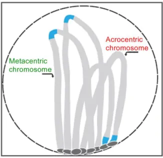
FIGURE 2.1. – The bouquet organization of chromosomes during prophase. 19
FIGURE 2.2.– The Library model. 21
FIGURE 2.3.– Feedback model. 21
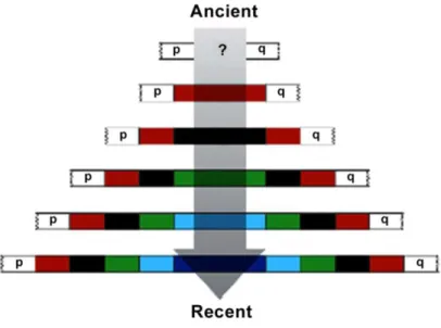
FIGURE 2.4.– Proximal Progressive Expansion mode of evolution of satellite DNA. 22
FIGURE 2.5.– Coevolution of satellite DNA sequences and DNA-binding proteins in the
centromeric region. 23
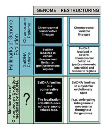
FIGURE 2.6.– The two main outcomes in genome restructuring. 25
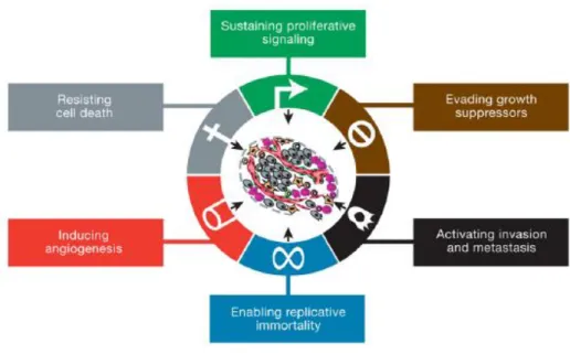
FIGURE 3.1. – The hallmarks of cancer. Distinctive and complementary capabilities that
enable tumor growth and metastatic dissemination. 30
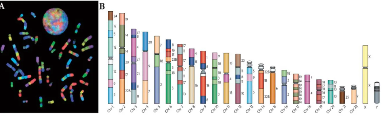
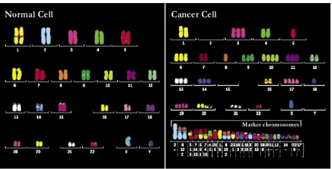
FIGURE 3.2.– Normal and cancer cell karyotypes using multicolor FISH. 31
FIGURE 3.3.– Schematic illustration of mechanisms leading to chromosomal alterations. 32
FIGURE 3.4.– Commonly observed DNA methylation changes in cancer. 37
FIGURE 4.1. – The place of Rodentia in mammal’s evolutionary tree. 40
FIGURE 4.2. – Phylogenetic tree of Rodentia. 41
FIGURE 4.3. – Phylogenetic tree of Muroidea superfamily. 44
C
HAPTERIII
–
G
ENERALD
ISCUSSION ANDF
UTUREP
ERSPECTIVESTABLE 1- Summary of the results obtained with painting experiments and constitutive
heterochromatin analysis. 167
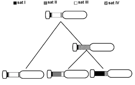
TABLE 2 - Summary table showing the in situ hybridization pattern of CCR4/10sat and
PMSat in Peromyscus eremicus chromosomes. 171
FIGURE 1.1 – Schematic diagram summarizing the different topics studied, the species
C
HAPTER
I|
I
NTRODUCTION
The human genome (entire hereditary information of an organism that is encoded in their DNA) provides the underlying code for human biology (ENCODE Project Consortium 2012). Cells within an organism contain a complete copy of these instructions, written in the four-letter language of DNA (A, C, T and G). These nucleic acids are arranged into units called genes, which are organized in the chromosomes (Reece 2004). This organization ensures not only proper gene function but also an accurate distribution of genes to daughter cells during cell division. Chromosomes are thus the ultimate determinants of the organization of all living organisms (Sumner 2003). The entire chromosome set of a species is known as a karyotype, which can be thought of as a global map of the nuclear genome.
Genomes have the ability to evolve throughout local changes in nucleotide sequences or by changes in the karyotypes, by means of chromosome rearrangements that can result in dramatic phenotypic consequences and are assumed to play an important role in the evolution of species and in cancer. Reproductive isolation and tumorigenic karyotypic transformation can be initiated through the same structural rearrangements, therefore karyotype restructuring can drive both speciation and carcinogenesis (Ye et al. 2009).
The major goal of the present thesis was the analysis of the karyotype restructuring dynamics, both during evolution and cancer, in Rodentia species. Specifically, three objectives may be outlined: i) the comparative analysis of the genome of various Rodentia species by means of comparative chromosome painting in an evolutionary perspective; ii) the molecular and cytogenetic characterization of satellite DNA families, and the analysis of their dynamic behavior in the light of the species’ karyotype evolution; iii) cytogenetic and molecular characterization of two rat tumor cell lines, highlighting the chromosome rearrangements effect in gene expression, and validating the use of these cell lines as cellular models for breast cancer research, namely in the elucidation of the epigenetic events involved in the regulation of Erbb2 expression.
This thesis is divided in three major parts: chromosomal evolution (Section 1; Papers I and II), satellite DNA dynamics and evolution (Section 2; Papers III and IV) and
INTRODUCTION|
- 2 -
performed; the Results chapter will be presented as individual papers being some of them already published, and others submitted. A general discussion is made at the end in order to integrate and correlate all the data achieved.
INTRODUCTION| CHROMOSOMAL EVOLUTION
- 3 -
1.
C
HROMOSOMES ANDE
VOLUTIONIn the 1900s a series of experiments by Theodor Boveri gave the definitive demonstration that chromosomes are the vectors of heredity. Eukaryotic chromosomes present differences in morphology (shape and size) within the karyotype and karyotypes vary in terms of number and organization even between closely related species (Sumner 2003).
Going back to 1859, Darwin introduced the biologists to the concept that allied species are descended from a common ancestor and that species change gradually over long periods of time. The idea of evolution as the principle for the origin of biodiversity can be applied to chromosomes, being the chromosomal diversity found the result of the action of different mechanisms during the process of chromosome evolution, elucidating the high plasticity of the genomes at the chromosomal level. Moreover, the wide diversity of karyotypes found, combined with evidence that chromosomal rearrangements might reduce the fertility of heterozygous hybrids (King 1993), has led some researchers to argue for a causative role of chromosomal change in speciation.
1.1GENOME CONSERVATION AND KARYOTYPE RESTRUCTURING
The first approach in the study of chromosomal evolutionary events was the comparative analysis of several species karyotype. Initial attempts to identify chromosome homologies were based on chromosome banding patterns (Dutrillaux et al. 1980, Nash and O’Brien 1982). Comparative studies were facilitated when molecular techniques were incorporated into cytogenetics, allowing DNA level comparison even between phylogenetically distant or highly rearranged species. With the advent of advanced molecular approaches, a new term emerged - Comparative Chromosomics - used to define the field of cytogenetics using methodologies which allow further resolution of comparative maps (Graphodatsky 2007). Comparative chromosome painting and Zoo-FISH revealed to be a powerful tool in comparative chromosome studies, allowing the construction of large-scale comparative maps mostly in mammalian groups. This technique is based in cross-species fluorescent in situ hybridization (FISH) using chromosome-specific DNA sequences as painting probes allowing the definition of chromosomal homologue segments between species (Figure 1.1) (Wienberg et
INTRODUCTION| CHROMOSOMAL EVOLUTION
- 4 -
Comparative chromosome painting permitted the disclosure of syntenic segments, defined as large blocks of DNA often extending to whole chromosomes or chromosome arms, which are shared by different species. Contiguous syntenic segments which are homologous to regions belonging to different chromosomes in another species are designated as syntenic
associations (exemplified in Figure 1.1B) (Froenicke 2005). Comparative studies showed
remarkable interspecies chromosome segments conservation, but also demonstrated that between species the syntenic blocks are assembled in different combinations, resulting in distinct chromosome number and chromosome morphology, reflected in the karyotype variability (Ferguson-Smith and Trifonov 2007). The reassembling of those segments and consequent karyotype restructuring is promoted by chromosome rearrangements, being the most common the translocations, inversions (paracenric and pericentric), fusions and fissions (Pevzner and Tesler 2003a). Duplications, deletions and heterochromatin additions/eliminations were also responsible for changes in chromosomes during evolution (Bailey et al. 2004, Adega et al. 2009). Conserved segments that are fused together in one species can be separated on different chromosomes in another. Chromosome numbers can increase or decrease by fission or fusion events, and segments within blocks can be inverted and centromeres repositioned. The analysis of the most parsimonious scenarios is the dominant approach in genome rearrangement study uncovering the evolutionary history (Ferguson-Smith and Trifonov 2007).
Comparative chromosome painting allowed the fast generation of large-scale comparative maps in Eutheria (placental mammals), however, its resolution presents some limitations. This methodology fails to determine the orientation of each conserved block within a chromosome, it does not allow the identification of intrachromosomal rearrangements such as inverted segments, and it is not efficient in revealing syntenies between distantly relates species
Figure 1.1| Example of the use of Zoo-FISH for the construction of comparative maps. A) Human metaphase and interphase nucleus after hybridization with a chromosome-specific paint probe set derived from gibbon chromosomes. B) The analysis of the painting experiments enabled the construction of a homology map of gibbon chromosomal segments on human chromosomes where syntenic associations can be observed, e.g. syntenic association 7/9 in chromosome 4 (adapted from Ferguson-Smith and Trifonov 2007).
INTRODUCTION| CHROMOSOMAL EVOLUTION
- 5 -
(Murphy et al. 2005). Cytogenetic analysis can be in some cases complemented with the use of BAC (Bacterial Artificial Chromosomes with cloned DNA fragments), allowing the detection of more detailed homologies (Goureau et al. 2001). With the advent of large-scale genome sequencing of eukaryotic genomes and the use of powerful algorithms to promote their alignment and comparative analysis, an exquisite molecular resolution at the level of single-base pair differences, as well as identification of gene order and changes in synteny was accomplished (Froenicke et al. 2006). Complete sequencing of genomes has confirmed the extensive levels of conserved synteny originally found by cytogenetic comparative mapping, but the high density of markers afforded by complete sequence also results in a more complex view of chromosomal evolution, with remarkable levels of intrachromosomal rearrangement (Eichler and Sankoff 2003). Nevertheless, the drawbacks pointed to comparative chromosome painting do not invalidate this methodology, in fact, several species were analysed using this technique and many more will be, as this is clearly easier and faster than sequencing a species genome. In this way, for the species whose genome has already been sequenced, the use of painting probes derived from different species combined with comparative sequencing projects is definitely the more efficient approach in comparative studies.
1.2 RESOLVING PHYLOGENIES IN MAMMALIA
Traditionally, the analysis of mammalian phylogenies was restricted to fossil records and morphological characters. In the past years, data from a range of research disciplines, such as molecular systematics, genome sequencing and comparative cytogenetics, have disclosed the evolutionary relationships between humans and their mammalian relatives. Mammalian phylogeny and evolution is now the driving force behind comparative genomic analysis, investigating the details of mammalian genomes and how they evolved (e.g. Engelbrecht et al. 2006, Robinson and Ruiz-Herrera 2008). Together, these tools are now converging on a well-established phylogeny and timescale of mammalian species.
1.2.1 FROM HOMOLOGY MAPS TO THE ANCESTRAL KARYOTYPE
The search for the ancestral mammalian karyotype has a long tradition in cytogenetics. The first comparative chromosome maps drawn outlined the segments with conserved homology between human and other species. Chromosome homology maps of higher resolution were also prepared from chromosome-specific paints from other animals, such as the domestic dog (Graphodatsky et al. 2000), gibbon (Müller et al. 2003), the house mouse (Romanenko et al. 2006), several rodent species (reviewed in Romanenko et al. 2012), among others.
INTRODUCTION| CHROMOSOMAL EVOLUTION
- 6 -
ANCESTRAL KARYOTYPE DELINEATION| The cladistic analysis of Zoo-FISH data was used in the construction of ancestral karyotypes. This method relies in the identification of primitive/ancestral chromosome traits (sympleisiomorphies) and shared derived chromosome traits (synapomorphies), assisted by parsimony analyses of the chromosome evolutionary rearrangements direction (Chowdhary et al. 1998, Wienberg et al. 2000). In order to define the conserved and derived syntenic associations, it is important the comparison with an outgroup, a distantly related taxon known to be phylogenetically outside the group of species under study (Wienberg 2004). If the syntenic association is present in the outgroup, then, according with the parsimony principle, it is considered as ancestral, while others are classed as common derived characters.
The first reconstruction of the ancestral eutherian karyotype was based in cladistic analysis of Zoo-FISH data of seven non-primate species, representing three orders, and was performed by Chowdhary and colleagues (1998). Several further analysis of the ancestral eutherian karyotype have been made since then, each providing additional insights into the organization of this ancestral karyotype (e.g. Wienberg 2004, Froenicke 2005, Ferguson-Smith and Trifonov 2007). Using a similar strategy it was possible to reconstruct ancestral karyotypes of different mammalian groups, such as primates, carnivores and rodents (reviewed in Graphodatsky et al. 2011). Regarding rodents, the delineation of the ancestral Muroidea karyotype (AMK) has been proposed by several authors (e.g. Stanyon et al. 2004, Engelbrecht et
al. 2006, Romanenko et al. 2007). Recently, data from all comparative studies in Muroid rodents
was compiled and besides the suggestion of the AMK, were also presented putative ancestral karyotypes for Cricetidae (ACdK) and Muridae (AMdK) (Figure 1.2), all based in the analysis of the syntenic associations defined using mouse (Mus musculus) paints (reviewed in Romanenko et
al. 2012). Also Chaves et al. (2012) proposed an high precision Muroidea ancestral karyotype
(Muridae/Cricetidae and Murine) based in a broad species analysis combining previous reported comparative maps together with newly presented data.
INTRODUCTION| CHROMOSOMAL EVOLUTION
- 7 -
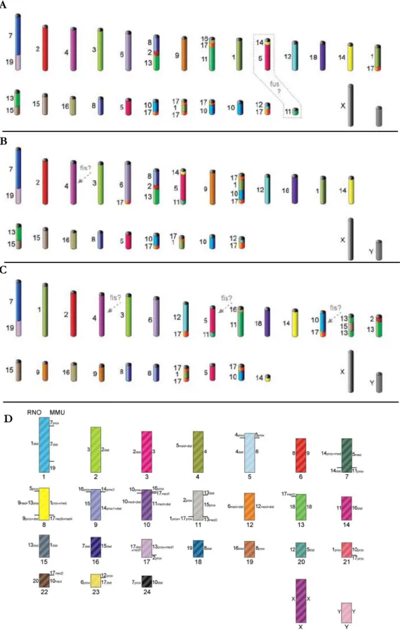
Figure 1.2|Putative ancestral karyotypes for Rodentia proposed by different authors. A) Ancestral Muroidea karyotype (AMK). B) Ancestral Cricetidae karyotype (ACdK). C) Ancestral Muridae Karyotype (AMdK). Different colors correspond to different mouse chromosomes. Some elements state is still ambiguously determined in the ancestral karyotypes, being represented by dashed grey frame and arrows (from Romanenko et al. 2012). D) Putative AMK with Mus musculus (MMU) and Rattus norvegicus (RNO) homologies (from Chaves et al. 2012).
D A
B
INTRODUCTION| CHROMOSOMAL EVOLUTION
- 8 -
CONSTRUCTING COMPARATIVE MAPS IN RODENTIA| Within Rodentia, comparative chromosome studies have been particularly productive in the analysis of non-muroid families, such as the Sciuridae (squirrels), whose karyotypes are highly conserved and retain many ancestral conditions (Stanyon et al. 2003, Li et al. 2006). On the contrary, the superfamily of muroid rodents, including the important laboratory animals, have highly rearranged karyotypes in comparison with humans, and for that reason cross-species chromosome painting with human probes was sometimes difficult to interpret (Ferguson-Smith et al. 1998). This problem was overcome by the use of painting probes from different rodents, such as Mus musculus, in cross-species experiments, being afterwards the human homologies inferred from human-mouse comparative maps based in the genome sequencing data (e.g. Romanenko et al. 2006). Mus
musculus paint probes have been the most commonly used in rodent comparative studies
(Cricetidae and Muridae families), but recent works describe the use of chromosome paints from rodent species belonging to other subfamilies, namely Cricetinae, Arvicolinae and Sigmodontinae (reviewed in Romanenko et al. 2012).
PHYLOGENETIC TREES| The number of syntenic segments per haploid set provides a measure of the relationship between species. When compared with the human genome, most eutherians have 30 to 40 separated segments of homology (O’Brien et al. 1999). Some species are exceptional, such as dogs and gibbons, and have about twice as many conserved segments (Wienberg et al. 1990, Yang et al. 1999), the rat shares about 100 segments with human (O’Brien
et al. 1999), while the mouse is unique in having almost 200 blocks (Nilsson et al. 2001). By
comparing different species chromosomes, a phylogenic tree can be constructed based on the minimum number of rearrangements occurred since the ancestral or the maximum number of shared syntenic segments. Molecular cytogenetic data and the increasing availability of partially or fully sequenced genomes from a variety of vertebrate species have fueled advances in phylogenomics, the phylogenetic reconstructions using genomic data(Robinson and Yang 2012). Because chromosomal rearrangements are such unique events, the probability that they occurred twice in different lineages (convergence) is low (Wienberg 2004). Therefore, the chromosome rearrangements identified by comparative studies revealed to be reliable evolutionary signatures or landmarks and have been used in the construction of phylogenetic trees elucidating phylogenetic relationships between species (e.g. Rokas and Holland 2000, Romanenko et al. 2007, Nie et al. 2012). During these last years, several comparative studies performed attempted to resolve some of the Muroids complex phylogenies, and phylogenetic trees were constructed for different muroid families, as it is exemplified in figure 1.3 (reviewed in Romanenko et al. 2012).
INTRODUCTION| CHROMOSOMAL EVOLUTION
- 9 -
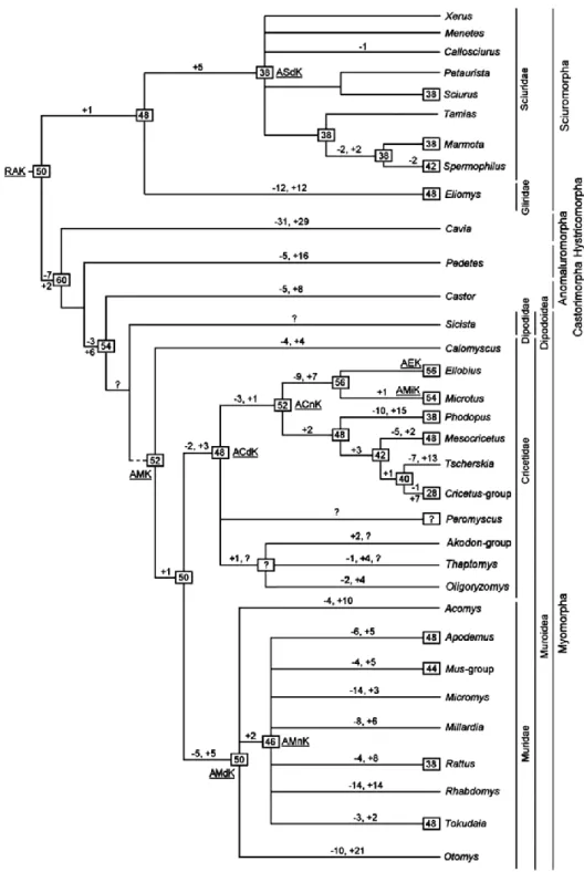
Figure 1.3| Putative rodent evolutionary tree. This tree is based in comparative data from several Muroid rodents, showing chromosome evolution to the genus level. RAK—ancestral Rodentia karyotype; ACdK—ancestral Cricetidae karyotype; ACnK—ancestral Cricetinae karyotype; AEK—ancestral Ellobius karyotype; AMdK—ancestral Muridae karyotype; AMK—ancestral Muroidea karyotype; AMiK—ancestral Microtus karyotype; AMnK—ancestral Murinae karyotype; ASdK— ancestral Sciuridae karyotype. Presumable ancestral diploid number characters for node are shown in black frames. Minus sign indicates chromosome fissions, plus sign indicates chromosome fusions, and question mark indicates unresolved positions (from Romanenko et al. 2012).
INTRODUCTION| CHROMOSOMAL EVOLUTION
- 10 -
RATES OF CHROMOSOMAL REARRANGEMENT| The chromosome painting data now available for many species belonging to different eutherian orders, as well as data from alignment of genome sequencing and radiation hybrid maps, helped to estimate the average rate of evolutionary rearrangements (Murphy et al. 2005). The rates of chromosomal rearrangement vary radically not only among different lineages but also between sex chromosomes and autosomes (Eichler and Sankoff 2003). In most eutherian orders, there are species presenting a slow rate of chromosome evolution considered as the ”default” frequency (Wienberg 2004). The default rate of eutherian chromosome evolution was calculated as approximately one rearrangement within 10 million years. Detailed investigation within groups has suggested that at different times, the rate of evolution, as well as the prevailing type of rearrangement, can change greatly (Murphy et
al. 2005). For example, in the lineage that extends from the eutherian ancestor to the primate
ancestor, during 50 million years, only three rearrangements took place (Froenicke et al. 2006). In contrast, a sudden karyotype diversification occurred in the gibbon lineage, with 24 rearrangements leading to the common gibbon ancestor and then multiple rearrangements subsequently leading to the karyotypes of the extant species (Müller et al. 2003, Wienberg 2005,). During the same period, karyotype evolution within the great apes group was extremely slow.
Record high rates of karyotype evolution are found in muroid rodents (Romanenko et al. 2006, 2007, Sitnikova et al. 2007), canids (Yang et al. 1999, Graphodatsky et al. 2000) and gibbons (Müller et al. 2003). The evolutionary rate between mouse and rat appears to be ten times greater than that found between humans and cat, or between humans and chimpanzees (Stanyon et al. 1999). Nevertheless, each of these mammalian orders contains groups with slower rates of chromosomal rearrangement, namely Sciuridae family among rodents (Stanyon et al. 2003, Li et
al. 2004, 2006), felidae among carnivore (Perelman et al. 2005) and apes among primates
(Froenicke 2005). The variation in the rates of mammalian karyotype evolution remains unexplained. Environmental effects, overall mutation rates, population size and the activation of mobile elements and retroviruses are among the possible contributory factors (Ferguson-Smith and Trifonov 2007).
1.3 DYNAMICS OF CHROMOSOME EVOLUTION
Along this thesis section it was mentioned that every genome rearrangement study involves identification of the syntenic chromosomal segments between species, and solving a combinatorial puzzle to find a plausible series of genome rearrangements to transform one genome into another. Such studies allow also the localization of breakpoint regions which are given by the two boundaries of the syntenic segments (Froenicke 2005), corresponding to
INTRODUCTION| CHROMOSOMAL EVOLUTION
- 11 -
regions where genome synteny has been disrupted by chromosomal rearrangements. A breakpoint or breakpoint region is not a tangible physical entity in a genome; it is an analytical construct arising from the comparison of two genomes (Sankoff 2009). An early work by Ohno (1973) proposed the random breakage model of genomic evolution, postulating that the distribution of chromosome rearrangements breakpoints was uniformly random. The work by Nadeau and Taylor (1984), as well as comparative mapping and sequencing studies among vertebrate species, provided convincing arguments in favor of this model, and the random breakage model become the widely accepted theory of chromosome evolution. When data from the genome sequencing projects was comparatively analyzed by algorithms, it revealed remarkable levels of intrachromosomal rearrangements. The prevalence of short inversions restricted to specific chromosome segments represented a departure from the random breakage model in evidencing several breakpoints in one same region. Besides this, after comparing human and mouse genomes (Pevzner and Tesler 2003a), 281 syntenic blocks were found compared with the 180 known from comparative gene mapping (Nilsson et al. 2001). The explanation found was that breakpoint regions between the synteny blocks would have been disrupted an average of 1.9 times each, showing high density of breakpoints over these regions. This suggested an alternate model for chromosomal evolution, termed fragile breakage model (Pevzner and Tesler 2003b, which considers that there are regions (designated hotspots) throughout the mammalian genome prone to breakage and reorganization (Zhao et al. 2004, Peng et al. 2006). In support of this theory, chromosome breakpoint analyses have identified shared evolutionary breakpoint regions between different species (Murphy et al. 2005). Besides, a recent study reveals a high level of reuse of evolutionary breakpoint regions among muroid rodents, further supporting the fragile breakage model of chromosome evolution (Mlynarski et al. 2010). An interesting finding was that evolutionary breakpoint regions tend to colocalize with the more commonly occurring human cancer-associated breakpoints (Robinson et al. 2006). Furthermore, whole genome alignment studies have shown that evolutionary breakpoints regions are rich in repetitive elements (Murphy et al. 2005 Ruiz-Herrera et al. 2006), such as segmental duplicated regions, centromeric and telomeric regions. The presence of repetitive sequences at evolutionary breakpoint regions is thought to be related to the role that tandem repeats play as a substrate for non-homologous recombination, thereby promoting chromosomal rearrangements (Froenicke and Lyons 2008).
A segmental duplication involves the duplication of a small portion of chromosomal material (with 90% of similarity) either in tandem or transposed to new locations within the genome. Initial analyses of the human genome sequence have identified a large amount of
INTRODUCTION| CHROMOSOMAL EVOLUTION
- 12 -
tandem as well as interspersed segmental duplications (Bailey et al. 2001). These observations raise the possibility that segmental duplications may have played a significant role in gene and genome evolution. Once formed, segmental duplications promote further rearrangement through misalignment and subsequent non-allelic homologous recombination (Stankiewicz and Lupski 2002). Studies in human corroborate this assumption showing that 25-53% of the recurrent breakpoint regions colocalize with human segmental duplications (Armengol et al. 2003, Bailey et al. 2004). Besides, in primates a strong association of segmental duplications with recurrent chromosomal structural rearrangements and also with disease was also demonstrated (Carbone et al. 2006, Marques-Bonet et al. 2009).
Centromeres and telomeres have long been recognized as peculiar dynamic regions of chromosomal evolution. The repetitive nature of these regions extends beyond the classically defined boundaries of centromeric and telomeric sequences; such transition regions, termed pericentromeric and subtelomeric DNA, are hotspots for the insertion or retention of repeat sequences. Among primates, there is now overwhelming evidence that blocks of recently duplicated sequence populate subtelomeric and pericentromeric regions (Eichler and Sankoff 2003). Chromosomal fissions probably require the complex regeneration of centromeres and telomeres, once chromatids have to be capped by telomeres and a new centromere have to be created. Gene map alignments indicate a high rate of de novo centromere formation (Murphy et al. 2005). Because the evolutionary reoccurring breakpoint regions form only a small proportion of a eutherian genome (3%), the colocalization of half of the neo-centromere hotspots with these regions allows us to speculate that an association exists, although only a few neo-centromeres have been observed (Robinson et al. 2006). This colocalization might indicate that neo-centromere generation is linked to the eutherian chromosomal plasticity, and that the potential for neo-centromere generation might also be evolutionarily conserved.
Overall, genomes can be considered a mosaic comprising regions of fragility that are prone to reorganization and regions that do not exhibit the same levels of evolutionary plasticity that have been conserved in different lineages during the evolutionary process.
INTRODUCTION| SATELLITE DNA DYNAMICS AND EVOLUTION
- 13 -
2. G
ENOMICC
OMPARTMENTS ANDR
EPETITIVES
EQUENCESChromatin is found in two forms in eukaryotic genomes: euchromatin and heterochromatin. This classification was based on the observation that euchromatic chromosome regions changed their degree of condensation during the cell division cycle, whereas heterochromatic chromosome regions remained highly condensed throughout the majority of the cell cycle (Heitz 1928). In addition to differences in the timing of chromosome condensation, numerous other dissimilarities have been identified between these two genomic compartments. Euchromatin is enriched with unique coding sequences (genes), which are typically transcribed. Heterochromatin, on the other hand, is considered to be gene poor, being primarily composed of arrays of highly repetitive sequences (Hughes and Hawley 2009). Heterochromatin may be either facultative or constitutive. Facultative heterochromatin is found at developmentally regulated loci, where the chromatin state can change in response to cellular signals and gene activity, while constitutive heterochromatin (CH) occurs as large blocks in regions harboring repetitive sequences such as the pericentromeric regions, interstitial chromosome regions and telomeres (Dimitri et al. 2005, Adega et al. 2007, Paço et al. 2009). Constitutive heterochromatin is a basic component of eukaryotic genomes forming about 5% of the genome in Arabidopsis thaliana, 30% in Drosophila and humans, 60% in rodents and up to 70– 90% in certain nematodes and plants (Sherwood and Patton 1982, Arabidopsis genome initiative 2000, Dimitri et al. 2005). Constitutive heterochromatin can be revealed by preferential “loss” of DNA from non-heterochromatic regions, achieved by conventional C-banding technique (Pathak and Arrighi 1973). Restriction endonuclease digestion followed by C-banding has shown its ability in demonstrate CH heterogeneity by revealing additional heterochromatic bands, cryptic C-bands (Chaves et al. 2004, Adega et al. 2005).
Studies primarily conducted in Drosophila melanogaster have shown that CH plays different roles in important cellular functions, such as chromosome organization, besides containing essential genes for viability and fertility (Dimitri et al. 2009). Pairing of heterochromatic regions is required for the proper segregation of chromosomes that fail to undergo recombination during female meiosis (reviewed in Grewal and Jia 2007).
But how does heterochromatin perform its diverse functions? The formation of heterochromatin requires methylation of histone H3 at lysine 9 and the subsequent recruitment of chromodomain proteins such as heterochromatin protein HP1. Evidence from studies in diverse model systems indicates that heterochromatin serves as a self-assembling framework of histone modifications to recruit effector proteins, which in turn regulate various chromosomal processes (Shimada and Murakami 2010). Given the critical functions of heterochromatic
INTRODUCTION| SATELLITE DNA DYNAMICS AND EVOLUTION
- 14 -
sequences in both meiosis and mitosis and its rapid change in sequence, it has been hypothesized that differences in either heterochromatic sequences or the proteins that maintain them might indeed play a role in species isolation and thus speciation (Hughes and Hawley 2009).
DIFFERENT TYPES OF REPETITIVE SEQUENCES IN THE GENOME| A significant portion of the eukaryotic genome is comprised by repetitive sequences that can be located in both euchromatin and heterochromatin. Repetitive sequences can be categorized into two main classes considering their primary organization in the genome: interspersed DNA and tandemly repeated DNA (reviewed by Slamovits and Rossi 2002). The first class refers to sequences scattered in the genome, generally known as transposable elements due to their ability of “jumping” to different genomic locations (transposition). These elements are divided in different classes according to their mechanism of transposition (reviewed by Wicker et al. 2007): retrotransposons or class I, transpose via an RNA intermediate and as examples are the well-known Alu sequences in humans and the L1 elements in mammals (Capy et al. 1997, Furano 2000); the class II are the DNA transposons, and these elements transpose by excision from their location and integration in other genomic sites without an RNA intermediate (Finnegan 1989, Capy et al. 1997). The second class of repetitive sequences, the tandemly repeated DNA, includes three distinct groups: microsatellites, minisatellites and satellite DNA. Micro and minisatellites are characterized by short repeat units, ranging from up to 100 bp for microsatellites and 1-5 bp for minisatellites (Charlesworth et al. 1994). Array size for both micro and minisatellites varies from 10 to 100 repeat units, and they can be found distributed throughout the genome (Li 1997). Microsatellites appear to be primarily located in euchromatic regions of chromosomes or in the vicinity of genes, such as the human CGG trinucleotide repeats (Riggins et al. 1992), but microsatellite arrays can be also often detected in heterochromatin (Gindullis et al. 2001). Minisatellites can be found irregularly dispersed in euchromatin and largely clustered in subtelomeric chromosomal regions (Royle et al. 1988).
Satellite DNA (satDNA), that is the repetitive sequence in focus in this thesis, is along with
transposable elements, the mainly constituent of constitutive heterochromatin (John 1988, Chaves et al. 2004). SatDNA is characterized by long tandem arrays and it is usually present in the genomes in several million copies (Charlesworth et al. 1994). Early experiments historically coined the term “satellite DNA”, referring to tandemly arranged sequences forming satellite bands which differentiated from the rest of the genomic DNA by density gradient separation (John 1988). Once no protein coding function could be primarily associated with satellite DNAs, it has been early considered as useless genomic elements accumulated as junk (Ohno 1972), or as
INTRODUCTION| SATELLITE DNA DYNAMICS AND EVOLUTION
- 15 -
sequences representing genomic parasites proliferating independently (Orgel and Crick 1980). Nevertheless, evidences emerged that demonstrated the functional significance of satellite DNA sequences, ranging from chromosome organization and pairing, to cell metabolism and speciation. Studies support these functionalist assumptions concerning the association of satellite DNAs with complex features of eukaryotic chromosomes (e.g. Csink and Henikoff 1998, Henikoff et al. 2001, Sullivan et al. 2001), which will be highlighted in the next paragraphs.
2.1 SATELLITE DNA FEATURES AND FUNCTION
Satellite DNAs are highly repeated DNA sequences, typically organized as long arrays of head-to-tail linked repeats (Charlesworth et al. 1994). The amount of satellite DNA content can sometimes exceed 50% of a species total DNA (Mravinac and Plohl 2010), being this genome fraction responsible for the variation of genome size in some eukaryotes (Gregory et al. 2007). The length of the repeating unit (monomer) can range from only few base pairs up to more than 1 kb, thus forming arrays that may reach 100 Mb long (reviewed Plohl et al. 2008). These lengthy arrays of satDNA form conspicuous blocks of differentially condensed chromatin in the chromosomes, mostly in centromeres but also in telomeres and interstitial positions (Chaves et al. 2000, Meles et al. 2008). A satDNA family is defined by a specific sequence and length. Different satDNA families can vary greatly in base composition, with some being rich in AT and others in GC. In the mouse genome, for instance, four distinct satDNAs have been characterized, the major satellite (MaSat) and the minor satellite (MiSat) which are AT-rich, and mouse Satellite 3 (MS3) and mouse Satellite 4 (MS4) which are CG-rich (Kuznetsova et al. 2005). Despite satDNA variation in nucleotide sequences across species, they share some common features such as monomer length, which is generally between 150–180 bp and 300–360 bp, in both plants and animals (reviewed in Plohl et al. 2008). As example, the human alpha-satellite presents a monomer size of 171bp (Manuelidis 1978), the maize CentC and CentO satellites have 156 bp (Birchler et al. 2011), while the pig Mc1 satellite presents a monomer of 340 bp (Adega et al. 2008). This can be explained by the organization of the satDNA around the nucleossomes. The mentioned sizes constitute the required DNA length to be wrapped around one or two nucleosomes (Henikoff et al. 2001). Another feature shared by different satellite DNA families is the presence of a short motif with 17 bp, known as CENP-B box, found in several centromeric satellites, such as human alphoid satellites (Masumoto et al. 1989), the mouse minor satellite (Wong and Rattner 1988) and satellites from other species. CENP-B box represents the binding site for centromere protein B (CENP-B) (Kipling and Warburton 1997), being one of the most well characterized satDNA sequence binding protein (Earnshaw et al. 1989, Sugimoto et al. 1998). Given the conservation of the CENP-B box between diverse mammalian species, a functional
INTRODUCTION| SATELLITE DNA DYNAMICS AND EVOLUTION
- 16 -
constraint in centromere activity was attributed to CENP-B, being considered essential in human assembly of centromeric-specific chromatin (reviewed by Ugarković 2005). It has been demonstrated that the CENP-B box is required for de novo centromere chromatin assembly on human alphoid DNA (Ohzeki et al. 2002, Okada et al. 2007). A recent study demonstrates that the accumulation of CENP-B-containing satDNA in a neocentromeric region, leads to the increase binding of another centromeric protein, CENP-A (centromeric protein A), eventually leading to a mature centromere that binds more CENP-A (Marshall and Choo 2011). This new proposed model explains, among others, the evolutionary conservation of CENP-B.
Given their primary localization in transcriptionally suppressive heterochromatin, transcriptional activity was not expected for satellite DNAs. Although transcripts of satellite DNAs have been reported in several organisms including vertebrates, invertebrates and plants (e.g. Lee et al. 2006, Pathak et al. 2006, Wong et al. 2007). It has been shown that satellite DNAs are temporally transcribed at particular developmental stages or are differentially expressed in some cell types, tissues or organs in most of the species analyzed (reviewed by Ugarkovic 2005). For instance, mouse gamma satellite DNA is differentially expressed during development of the central nervous system, as well as in the adult liver and testis (Rudert et al. 1995). Besides, some satDNA transcripts have shown to be important for epigenetic chromatin modifications, being involved in the initiation of histone H3 methylation, a necessary prerequisite for heterochromatin formation and maintenance (Martienssen 2003). Transcripts of satellite DNAs in the form of small interfering RNAs (siRNA) participate in the epigenetic process of chromatin remodeling and heterochromatin formation (Grewal and Elgin 2007). Some satellite DNA transcripts, particularly from some insects, nematodes and amphibians (Epstein and Gall 1987, Ferbeyre et al. 1998, Rojas et al. 2000), function as ribozymes with self-cleavage activity, whereas human satellite III transcripts are involved in the recruitment of splicing factors during stress (Chiodi et al. 2004). The presented examples suggest an active role for satellite transcripts in several regulatory layers from chromatin modulation to transcription and RNA maturation translation.
2.2 EVOLUTIONARY DYNAMICS OF SATELLITE DNA
Satellite DNA sequences probably arise as the result of large-scale duplication of sequences that are integrated into the genome at a favorable site (Britten and Khone 1968). Once established in the genome how do these sequences spread and amplify? Previously presented features show that satellite DNA seems to be a very distinctive component of the eukaryotic genomes constituting highly dynamic sequences. The variability of satDNAs even among closely