Chromosomal abnormalities in recurrent miscarriages by conventional
karyotyping analysis
Alessandra Bernadete Trovó de Marqui 1
1Departamento de Patologia, Genética e Evolução. Instituto de Ciências Biológicas e Naturais. Universidade Federal do Triângulo Mineiro. Campus I. Praça Manoel Terra, nº 330. Uberaba, MG, Brasil. CEP: 38015-050. E-mail: [email protected]
Abstract
Objectives: to describe the prevalence and types of chromosomal abnormalities in couples with recurrent miscarriage and products of conception.
Methods: electronic searches were performed in the PubMed/Medline database and in the Portal Regional da Biblioteca Virtual em Saúde/BVS (Regional Website of the Virtual Library in Health/BVS) using the descriptors “chromosomal abnormalities and abortions and prevalence”. After applying the inclusion and exclusion criterias, 17 studies were selected.
Results: 11 studies were conducted in couples with recurrent miscarriage and six in pro-ducts of conception. The main results of the couples with recurrent miscarriage were: the frequency of chromosomal abnormalities which varied from 1.23% to 12% and there was a predominance alteration of the chromosomal structures (reciprocal translocations, followed by Robertsonian). In products of conception, the results observed were: the frequency of chro-mosomal abnormality was above 50% in approximately 70% of the studies; there was a predominance alteration of the numerical chromosomal (trisomy - chromosomes 16, 18, 21 and 22, followed by polyploidy and monosomy X).
Conclusions: in summary, cytogenetic alterations represent an importante cause of preg-nancy loss and its detection can help couples with genetic counseling. Therefore, the value of knowledge on the prevalence of cytogenetic abnormalities in miscarriage samples is unques-tionable, once it is permitted a proper genetic counseling for the couple.
Introduction
Recurrent miscarriage (RM), also referred to as recurrent pregnancy loss (RPL) or habitual abortion (HA), is classically defined by Royal College of Obstetricians and Gynaecologists (RCOG) as the occurrence of three or more consecutive abortions prior to 20 weeks of gestation.1 However, the
American Society of Reproductive Medicine (ASRM) has recently redefined recurrent pregnancy loss as two or more abortions.2,3
The RM etiology may be multifactorial and about 40–60% of these patients are non-identifiable causes, in this case the condition is classified as idio-pathic or unexplained RM.4,5
The main etiological factors related to RM are: 1) genetic abnormalities (parental chromosomal rearrangements and abnormal embryonic karyo-types); 2) endocrine abnormalities; 3) anatomical factors; 4) immune factors; 5) inherited throm-bophilic disorders; 6) infective agents; 7) miscella-neous factors (lifestyle and environmental factors); and 8) new risk factors.6-8
A recent study described women with RM epidemiologic and obstetric characteristics and some risk factors were identified such as advanced age, consumption of alcoholic beverages and higher body mass index.9In this sense, lifestyle modifications
should also be implemented to improve reproductive prognosis.7
Genetic factors, mainly chromosomal abnorma-lities, are the most common cause of early miscar-riage (50–60%). The chromosomal abnormalities can be divided in two basic groups: numerical and structural abnormalities. These abnormalities can involve one or more autosomal chromosomes, sexual chromosomes and both simultaneously and are iden-tified by using the conventional cytogenetic methods based on light microscopy.4,5
Conventional karyotyping is traditionally performed to elucidate the possible causes of fetal loss, indicanting if any chromosomal abnormality was responsible for the miscarriage. The use of the classic cytogenetic to assess the fetal karyotype of the miscarriage material is complicated because the sample may be contaminated by the maternal tissue and the associated risk of false negative results.10In
addition, products of conception are characterized by a low sample quality that often leads to a cell culture failure.11,12 In case of culture failure or
maternal contamination, molecular techniques may contribute to detect additional chromosome abnor-malities in these miscarriage samples in addition to standard karyotyping.13
A recent review also summarized a current knowledge on the genetic causes (karyotype abnor-malities, recessive diseases carrier status, dominant
diseases and thrombophilia) of the RM.10Genetic
reasons may involve changes in the genetic embry-onic/fetal or parental material. Therefore, genetic tests may be performed in both parents as well as in the miscarriage material (fetus or afterbirth).10
The presence of karyotype abnormalities in one of the parents is one of the most common known causes of RM. They are most commonly found as balanced rearrangements, i.e. abnormalities cause no clinical symptoms in carriers but possibly induce the production of abnormal reproductive cells containing abnormal amounts of genetic material.10
In couples with RM, one partner – frequently is the woman - will have a genetically balanced structural chromosome rearranged being the most common balanced translocation (reciprocal followed by Robertsonian). The inversions are much rarer but are also associated to an increased risk of RM.6
In products of conception, at least 50% of all miscarriages are associated to numerical chromo-some abnormalities - trisomy, polyploidy and mono-somy X.6Therefore, genetic counselling is
impor-tant when a genetic factor is identified.6
Others genetic factors, such as genetic
polymor-phisms may contribute for RM.4,5A recent
syste-matic review and meta-analysis showed significant associations among RM and 53 genetic polymor-phisms of 37 genes. The genetic variants of HLA-G, IFNG, TNF, IL-6, IL-10, FII, FV, FXIII, ITGB3, MTR, MTHFR, PAI-1, NOS3, KDR, TP53, VEGFA, CYP17, CYP1A1, CYP2D6, ANXA5, and XCI may serve as RM biological markers.14 These genetic
variants were associated to the immune response, thrombophilia, placental function and hormonal and detoxification system and may contribute to the RM pathogenesis.15 Although significant associations
have been found among many genetic variants and RM, further functional research is needed to esta-blish its role as biomarkers and introduce it into a clinical practice routine.15
RM is an important reproductive health issue. Despite various etiologies have been identified, almost half of the cases remain unexplained. Regardless of the cause, a thorough follow-up with an important psychological support can help most couples achieve a successful birth.7
Figure 1
Flow chart illustrating the steps on the electronic search.
Publications in the database identified by electronic search
PubMed / Medline(n=102) BVS (n=137)
Methods
Electronic searches were performed in
PubMed/Medline database (available at
http://www.ncbi.nlm.nih.gov/pubmed) and in the
Portal Regional da Biblioteca Virtual em Saúde/BVS
(Regional Website of the Virtual Library in Health/BVS) (available at bvsalud.org), in June, 2017. What the descriptors used in both searches
were: “chromosomal abnormalities and abortions
and prevalence”. The steps on the electronic search are presented in Figure 1.
The initial screening of the publications was based on the analyses of the titles and/or abstracts. The inclusion criteria were: research articles closely related to the objectives of this research, which used conventional cytogenetic techniques; those published in the last 10 years (that is, between 2007 and 2016), in English, Portuguese and Spanish, which were full text, entirely available and free of charge. The articles that did not fulfill the previous established criteria were excluded.
Conventional karyotyping is defined as the morphological characterization of an individual’s
IDENTIFICATION
TRACKING
ELIGIBILITY
EXCLUSION
INCLUSION
Publications excluded by titles and/or abstracts analysis PubMed / Medline(n=90)
BVS (n=118)
Selected Articles
PubMed / Medline(n=12) BVS (n=19)
Excluded studies:
- absence of full text unavailable PubMed / Medline(n=2) BVS (n=7)
- language
PubMed / Medline(n=1)
- not directly related to the objective of the research and / or classical cytogenetic tech-nique inapplicable
PubMed / Medline(n=1) BVS (n=6)
Included studies: PubMed / Medline (n=8) BVS (n=6)
complement chromosomal, including number, form and size of the chromosomes. It can detect abnor-malities in the entire genome and therefore is used as a standard to detect chromosome abnormalities in miscarriages samples.13
From the 102 and 137 articles identified in PubMed / Medline and BVS, eight and six were included, respectively. A further search was performed from the references of the articles identi-fied in the investigated database and three arti-cles11,12,16 were included, a total of 17 scientific
arti-cles.
Results
From the 17 articles included in this literature
review, 11 were carried out in couples with RM (Table 1) and six in products of conception (Table 2). The frequencies and types of chromosomal abnormalities in couples with RM and miscarriage material are shown in Tables 1 and 2, respectively.
Some studies have shown higher prevalence of chromosomal abnormalities in couples with larger number of miscarriages.16,18 According to Ghazaey
et al.16 the highest percentage of chromosomal
abnormalities was observed in couples with five or more RMs (4.7% - 1 RM, 11% - 2 RMs, 15% - 3
RMs, 15% - 4 RMs and 21.2% - 5 RMs). Another
study showed that chromosome abnormalities were found in 5% of the couples with a history of two miscarriages, in 10.3% with three miscarriages and in 14.3% with four or more miscarriages.18
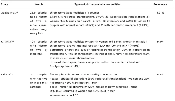
Table 1
Types and prevalence of chromosomal abnormalities in couples with RM.
continue RMs: recurrent miscarriages.
Study Sample Types of chromosomal abnormalities Prevalence
Ozawa et al.17
Kiss et al.18
Palet al.19
2324 couples had a history of two or more conse-cutive preg-nancy loss
108 couples with history of two or more RMs
56 couples who had two or more mis-carriages
chromosome abnormalities: 114 couples
3.18% (74) reciprocal translocations, 0.99% (23) Robertsonian translocations (17 women, 0.73% and 6 men 0.26%), 0.43% (10) inversions and 0.39% (9) others 14 couples with normal variants (0.6%) and 81 with pericentric inversion 9 (3.49%)
chromosome abnormalities: 10 cases (5 women and 5 men) woman-man ratio 1:1 chromosomal analysis (normal results): 46,XX (n=100) and 46,XY (n=103) 5 structural alterations (30% of reciprocal translocation, 20% of Robertsonian translocation, 10% of chromosome inversion) and 5 numerical alterations (50% of mosaicism – sexual chromosomes)
in one of the couples, the woman presented two concomitant alterations 3 polymorphisms (1.4%)
five couples - chromosomal abnormality in one partner
4 cases - structural alterations (60% reciprocal translocations - women and 20% Robertsonian D/D translocations - men)
1 case - numerical abnormality (20% mosaic of Down syndrome - men) 60% (n=3) occurred in women and 40% (n=2) in men
woman-man ratio 1.5:1
4.91%
9.3%
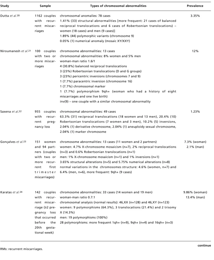
Table 1 continued
Types and prevalence of chromosomal abnormalities in couples with RM.
continue RMs: recurrent miscarriages.
Study Sample Types of chromosomal abnormalities Prevalence
Dutta et al.20
Niroumanesh et al.21
Saxena et al.22
Gonçalveset al.23
Karatas et al.24
1162 couples with recur-rent miscar-riages
100 couples with two or more miscar-riages
955 couples with recur-rent preg-nancy loss
151 women and 94 part-ners (couples with two or more recur-rent first t r i m e s t e r miscarriages)
142 couples with recur-rent miscar-riage (≥2 pre-gnancy loss that occurred before the 20th gesta-tional week)
chromosomal anomalies: 78 cases
1.41% (33) structural abnormalities [more frequent: 21 cases of balanced reciprocal translocations and 6 cases of Robertsonian translocations) – women (18 cases) and men (9 cases)]
1.89% (44) polymorphic variants (chromosome 9) 0.05% (1) numerical anomaly (mosaic XY/XXY)
chromosome abnormalities: 13 cases
chromosomal abnormalities: 8% women and 5% men woman-man ratio 1.6/1
4 (30.8%) balanced reciprocal translocations
3 (23%) Robertsonian translocations (D and G groups) 3 (23%) pericentric inversions (chromosomes 7 and 9) 1 (7.7%) paracentric inversion (chromosome 16) 1 (7.7%) chromosomal marker
1 (7.7%) polymorphism 9qh+ (woman who had a history of eight miscarriages and one live birth)
inv(9) – one couple with a similar chromosomal abnormality
chromosomal abnormalities: 49 cases
63.3% (31) reciprocal translocations (18 women and 13 men), 20.4% (10) Robertsonian translocations (7 women and 3 men), 10.2% (5) inversions, 2.04% (1) derivative chromosome, 2.04% (1) aneuploidy sexual chromosome, 2.04% (1) marker chromosome
chromosome abnormalities: 13 cases (11 women and 2 partners)
women: 4.7% X-chromosome mosaicism (n=7), 2% reciprocal translocations (n=3) and 0.6% Robertsonian translocations (n=1)
men: 1% X-chromosome mosaicism (n=1) and 1% inversions (n=1) 3.65% structural alterations (n=5) and 5.75% numerical alterations (n=8) normal variations in the chromosomes structure: 4.6% (women, n=7) and 6.4% (men, n=6), more frequent: 9qh+ (9 cases)
chromosome abnormalities: 33 cases (14 women and 19 men) woman-man ratio 0.7:1
chromosomal analysis (normal results): 46,XX (n=128) and 46,XY (n=123) women: 9 polymorphisms (64.3%), 3 translocations (21.4%) and 2 trisomy X (14.3%)
men: 19 polymorphisms (100%)
28 polymorphisms: more frequent 1qh+ (n=9), 9qh+ (n=4) and 16qh+ (n=3)
3.35%
12%
1.23%
7.3% (woman) 2.1% (man)
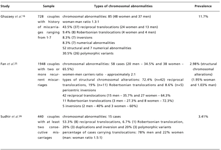
Table 1 concluded
Types and prevalence of chromosomal abnormalities in couples with RM.
RMs: recurrent miscarriages.
Study Sample Types of chromosomal abnormalities Prevalence
Ghazaey et al.16
Fan et al.25
Sudhir et al.26
728 couples with history of miscarria-ges ranging from 1-7
1948 couples with two or more recur-rent miscar-riages
440 couples with at least two conse-cutive mis-carriages
chromosomal abnormalities: 85 (48 women and 37 men) woman-man ratio 1.3:1
43.5% (37) reciprocal translocations (24 women and 13 men) 9.4% (8) Robertsonian translocations (4 women and 4 men) 8.3% (7) inversions
8.3% (7) numerical abnormalities
52 structural and 7 numerical abnormalities 30.5% (26) polymorphic variants
chromosomal abnormalities: 58 cases (20 men – 34.5% and 38 women – 65.5%)
women-men carriers ratio - approximately 2:1
types of structural chromosomal alterations: 72.4% (n=42) reciprocal translocations, 19% (n=11) Robertsonian translocations and 8.6% (n=5) pericentric inversions
42 reciprocal translocations (15 men – 35.7% and 27 women – 64.3% 11 Robertsonian translocations (3 men – 27.3% and 8 women – 72.3%) 5 inversions (2 men – 40% and 3 women – 60%)
chromosomal abnormalities: 15 cases
53.3% (8) reciprocal translocations, 6.7% (1) Robertsonian translocation, 20% (3) duplications and inversion and 20% (3) polymorphic variants percentage of cases carrying translocations: 78% men and 22% women (man: woman ratio 1.5:1)
11.7%
2.98% (structural chromosomal
alterations) (1.95% woman and 1.03% man)
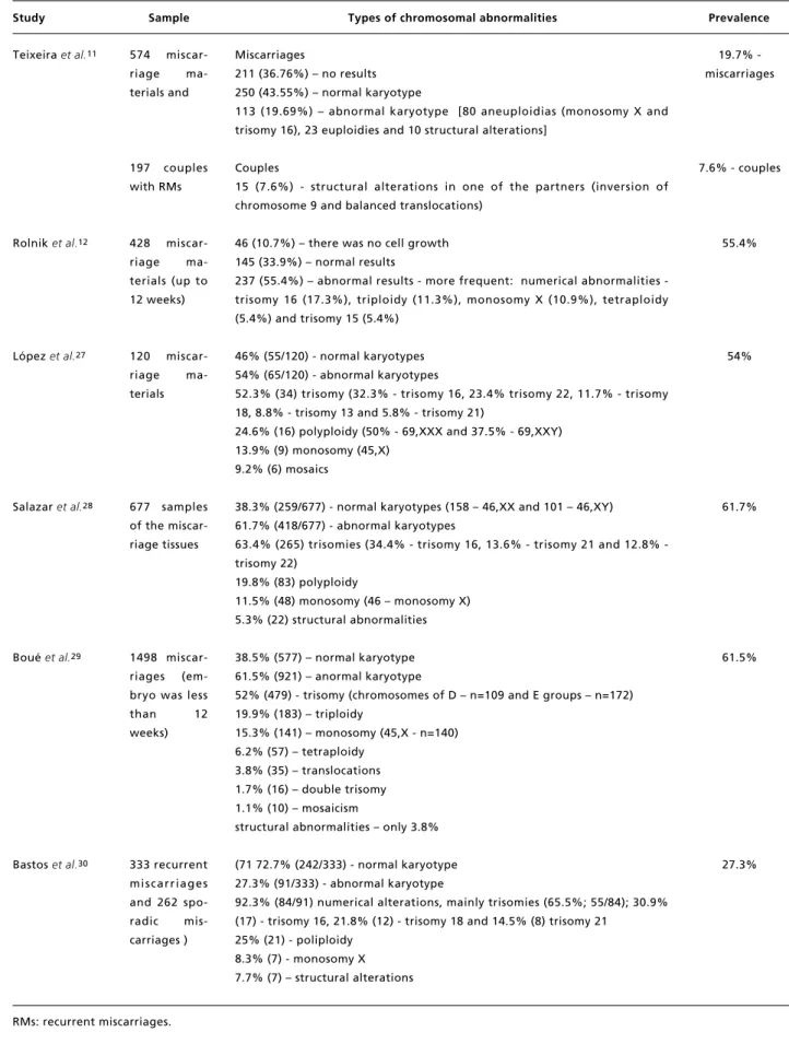
Table 2
Types and prevalence of chromosomal abnormalities in products of conception.
Study Sample Types of chromosomal abnormalities Prevalence
Teixeira et al.11
Rolnik et al.12
López et al.27
Salazar et al.28
Boué et al.29
Bastos et al.30
574 miscar-riage ma-terials and
197 couples with RMs
428 miscar-riage ma-terials (up to 12 weeks)
120 miscar-riage ma-terials
677 samples of the miscar-riage tissues
1498 miscar-riages (em-bryo was less than 12 weeks)
333 recurrent miscarriages and 262 spo-radic mis-carriages )
Miscarriages
211 (36.76%) – no results 250 (43.55%) – normal karyotype
113 (19.69%) – abnormal karyotype [80 aneuploidias (monosomy X and trisomy 16), 23 euploidies and 10 structural alterations]
Couples
15 (7.6%) - structural alterations in one of the partners (inversion of chromosome 9 and balanced translocations)
46 (10.7%) – there was no cell growth 145 (33.9%) – normal results
237 (55.4%) – abnormal results more frequent: numerical abnormalities -trisomy 16 (17.3%), triploidy (11.3%), monosomy X (10.9%), tetraploidy (5.4%) and trisomy 15 (5.4%)
46% (55/120) - normal karyotypes 54% (65/120) - abnormal karyotypes
52.3% (34) trisomy (32.3% - trisomy 16, 23.4% trisomy 22, 11.7% - trisomy 18, 8.8% - trisomy 13 and 5.8% - trisomy 21)
24.6% (16) polyploidy (50% - 69,XXX and 37.5% - 69,XXY) 13.9% (9) monosomy (45,X)
9.2% (6) mosaics
38.3% (259/677) - normal karyotypes (158 – 46,XX and 101 – 46,XY) 61.7% (418/677) - abnormal karyotypes
63.4% (265) trisomies (34.4% trisomy 16, 13.6% trisomy 21 and 12.8% -trisomy 22)
19.8% (83) polyploidy
11.5% (48) monosomy (46 – monosomy X) 5.3% (22) structural abnormalities
38.5% (577) – normal karyotype 61.5% (921) – anormal karyotype
52% (479) - trisomy (chromosomes of D – n=109 and E groups – n=172) 19.9% (183) – triploidy
15.3% (141) – monosomy (45,X - n=140) 6.2% (57) – tetraploidy
3.8% (35) – translocations 1.7% (16) – double trisomy 1.1% (10) – mosaicism
structural abnormalities – only 3.8%
(71 72.7% (242/333) - normal karyotype 27.3% (91/333) - abnormal karyotype
92.3% (84/91) numerical alterations, mainly trisomies (65.5%; 55/84); 30.9% (17) - trisomy 16, 21.8% (12) - trisomy 18 and 14.5% (8) trisomy 21
25% (21) - poliploidy 8.3% (7) - monosomy X 7.7% (7) – structural alterations
19.7% -miscarriages
7.6% - couples
55.4%
54%
61.7%
61.5%
Discussion
RM continues to be a challenging reproductive problem for the patient and clinician. It is a trau-matic event for couples and has psychological impli-cations, primarily depression and anxiety, and inter-feres in the couples’ relationship.31-33Identifying a
cytogenetic cause for a miscarriage can be psycho-logically important to overcome grief and loss, as well as to decide whether or not to try again.34
All the studies included in this review employed the karyotype test, which is the most common tech-nique of conventional cytogenetics. It is laborious technique and requires cell culture and the results can take 10 to 15 days. However, it can detect different types of chromosomal abnormalities. In couples with recurrent miscarriage, a lymphocyte culture was carried out from the peripheral blood, with analyses of approximately 20 to 30 metaphases. In case of miscarriage material, the tissue culture (chorionic villus) is used.
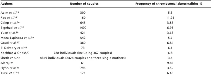
The frequency of chromosomal abnormalities among couples with RMs varied from 1.23% to 12% (Table 1). The results in this present study are similar to those conducted previously (Table 3).
There was a predominance of structural chromo-somal abnormalities in couples with recurrent miscarriage.16,17,19-22,24-26These findings were in
accordance with the literature.34,35,40-44Only in two
studies had higher frequency of numerical chromo-somal alterations23or the same percentage of
nume-rical and structural alterations.18
Regarding to the type of the structural alteration, the most frequent ones were the reciprocal transloca-tions, followed by the Robertsonians16-23,25,26 as
reported in the literature (Azim et al.35– 1.6%
reci-procal translocations versus 0.6% Robertsonian translocations; Kochhar & Ghosh42 – 5.9%
reci-procal translocations versus 0.7% Robertsonian translocations; Sheth et al.43 – 24.7% reciprocal
translocations versus 17.64% Robertsonian translo-cations). In the reciprocal translocation there is an exchange of two terminal segments from different chromosomes. Robertsonian translocation involves two acrocentric chromosomes with the loss of short arms and their fusion by or near the centromere. Both reciprocal and Robertsonian translocations are balanced rearrangements, that is, individuals with these translocations do not present phenotypic alte-rations resulting from them. The existing risks are restricted to the offspring, because, depending on the segregation occurred during the gametogenesis there may be chromosomally unbalanced fetuses forma-tion, consequently non-viable.18The translocations
were more common in women compared to men.16,17,19,20,22-25The incidence of translocation is
more in women than in men according to the litera-ture.42,43Only one study showed that the
percen-tagem of men (78%) carrying translocations was higher than in women (22%).26 Therefore, the
genetic counseling for couples with structural chro-mosomal abnormalities should consider the gender of the carriers.25According to some authors, as men
translocations carriers demonstrate reduced fertility.47,48A possible explanation for this
diffe-rence is that the chromosomal abnormalities such as in men carriers of autosomal reciprocal transloca-tions may cause severe meiotic disorders and stop-page of spermatogenic, but the oogenesis usually is conserved and results in production of gametes with a high risk of presenting unbalanced chromosomal abnormalities.47,48
It is worth mentioning that most of the studies in Table 1 included the frequency of chromosomal abnormalities of those alterations considered vari-ants of normality (polymorphisms).16-18,20,21,23,24,26
The frequency of polymorphisms ranged from
0.6%17 to 100%24 (Table 1). However, some
research has shown a possible association between polymorphic variants and infertility.49-51 A recent
study showed an increase in the frequency of poly-morphic variants among infertile patients (19.4% in the study group vs. 13.4% in the control group; p<
0.01).51
Of the 17 studies included in this review, only six have assessed miscarriage material (Table 2). Two of them referred not having reached the results and the cell culture failure in the cytogenetic analysis (CA).11,12 The CA of products of
concep-tion presents at least two main challenges, cell culture failure and excess of normal woman kary-otypes related to maternal cell contamination. Although the CA of abortive material is highly recommended, alternative complementary tech-niques for CA such as Fluorescence in situ Hybridization / FISH,52,53 Multiplex
Ligation-dependent Probe Amplification / MLPA,54
Quantitative fluorescent polymerase chain reaction /
QF-PCR55,56 and array Comparative Genomic
Hybridization / CGH57have been used for genetic
When the cytogenetic studies are successful, the newer techniques may have limited additional clin-ical use. However, when the tissue culture fails, the molecular techniques are very useful, although it is important to understand the limitations of each tool. In this manner, a combined approach using conven-tional and molecular methods will elucidate the cause of the miscarriage on almost all the samples. In a clinical setting this would be optimum.58
Chromosomal abnormalities in miscarriage material was found above 50% in approximately 70% of the studies.12,27-29A frequency of 61% of
chromosome abnormalities in products of concep-tion was detected by CA.55Other studies using
cyto-genetics found lower frequencies (33.24% and 48%) of chromosomal abnormalities.56,59
Two studies published in 2014 and 2017 employed CA and QF-PCR.55,56 The first applied CA
on 534 miscarriages, 73% (390/534) of them was successful. One hundred and forty-four miscarriages (27%, 144/534) did not grow in culture. A total of 27 cases were analysed by QF-PCR for chromosomes 13, 18, 21, X and Y and 30% (8 of 27 cases) showed a numerical chromosome abnormality by QF-PCR. Two hundred and thirty-seven cases (61%, 237/390) presented chromosomally alterated by CA.55 The
other was conducted in 884 products of conception, 204 of which were analyzed by cytogenetics and 680
by molecular biology based on QF-PCR.56Despite
using different techniques, the abnormal results were similar (40% by QF-PCR and 48% by cytogenetics).56A recent study, using only
conven-tional CA, with 457 products of conception showed that 382 cases were sucessfully karyotyped while 75 cases of cell culture failed (culture failure rate: 16.42%). Cytogenetic abnormalities were detected in 127 of the 382 cases (33.24%).59
Unlike the findings presented in Table 1, there was a predominance of numerical chromosomal alterations in the studies about miscarriage mate-rial.The frequency of numerical chromosomal alte-rations was higher than 92%,28-30reaching 100%27
in four of the six studies presented in Table 2. On the other hand, the frequency of structural alterations was lower and ranged from 3.8% to 7.7%.28-30In all
the studies in Table 2, the structural chromosomal abnormalities were little frequent in products of conception according to the literature.55,59When a
structural chromosomal alteration is found in the miscarriage material, the karyotype of both parents should be done, in order to assess the inherited nature or the abnormality found in the pregnancy loss.30
In general, the trisomy was the most common chromosome abnormality detected in the miscarriage material, followed by polyploidy and monosomy X.12,27-30 The most frequent trisomy was the 16 11,12,27,28,30and others trisomies, especially those
involving chromosomes 18, 21 and 22 are also implicated in the miscarriage.27,28,30 Autosomal
trisomies were the predominant chromosomal abnor-malities with a frequency of 48.8% (trisomy 16 – 12.6%; trisomy 22 – 7.9%; trisomy 21 – 5.5%; trisomy 13 – 3.1%; trisomy 10 – 3.1%), followed by
Table 3
Frequencies of chromosomal abnormalities in previous studies.
Authors Number of couples Frequency of chromosomal abnormalities %
Azim et al.35
Rao et al.36
Celep et al.34
Elgehzal et al.37
Yuce et al.38
Meza-Espinoza et al.39
Goud et al.40
El Dahtory et al.41
Kochhar & Ghosh42
Sheth et al.43
Alaraji44
Flynn et al.45
Turki et al.46
300 160 645 1400
421 542 380 73
788 individuals (including 367 couples) 4859 individuals (2428 couples and three single mothers)
61 795 171
5.3 11.25
3.86 6.93 3.68 5.7 6.84
References
1. Royal College of Obstetricians and Gynaecologists (RCOG) (April 2011). “The investigation and treatment of couples with recurrent first-trimester and second-trimester miscarriage”. Green-top Guideline No. 17. Royal College of Obstetricians and Gynaecologists (RCOG).
2. Practice Committee of American Society for Reproductive Medicine. Definitions of infertility and recurrent pregnancy loss: a committee opinion. Fertil Steril. 2013; 99 (1): 63. 3. Practice Committee of the American Society for
Reproductive Medicine. Evaluation and treatment of recur-rent pregnancy loss: a committee opinion. Fertil Steril. 2012; 98 (5): 1103-11.
4. Agenor A, Bhattacharya S. Infertility and miscarriage: common pathways in manifestation and management. Womens Health (Lond). 2015; 11 (4): 527-41.
5. Diejomaoh MF. Recurrent spontaneous miscarriage is still a challenging diagnostic and therapeutic quagmire. Med Princ Pract. 2015; 24 (Suppl. 1): 38-55.
6. Garrido-Gimenez C, Alijotas-Reig J. Recurrent miscar-riage: causes, evaluation and management. Postgrad Med J. 2015; 91 (1073): 151-62.
7. El Hachem H, Crepaux V, May-Panloup P, Descamps P, Legendre G, Bouet PE. Recurrent pregnancy loss: current perspectives. Int J Womens Health. 2017; 9: 331-45.
poliploidy (18.9% - triploidy and tetraploidy) and 45, X (16.5%). Structural chromossomal abnormali-ties were rare (9.5%).59Trisomy was also the most
common chromosome abnormality detected in the miscarriage material, accounting for 63% (232 of 368), followed by polyploidy (18.8%; 69 of 368) and
monosomy X (16.6%; 61 of 368).56 The most
frequent trisomy was the 16 (17.4%), followed by trisomy 22 (17.1%).56Another study55showed that:
(1) trisomy was the most common chromosome abnormality and accounted for 53% (125/237) of the abnormal karyotypes; (2) chromosomes 16, 22, 15 and 21 were most frequently involved in the aneu-ploidies; (3) fifty-four cases (23%, 54/237) with a polyploidy and 7% (16 cases) of monosomy X were also found and (4) individual unbalanced structural chromosome abnormality represented 4% (10/237) of the abnormal karyotypes.55Russo et al.53applied
FISH in the interphase of 855 formalin-fixed paraffin embedded miscarriage materials and the aneuploidy rate was detected at 50.3%. The most frequent chromosomal abnormalities were: auto-somal trisomies (60%), polyploidies (23.2%), and monosomy X (14%). Among the autosomal trisomies, chromosome 22 was the most frequently involved (33.7%) followed by trisomy of chromo-somes 16 (23.3%), 21 (19.4%), 15 (13.3%), 18 (5.34%), and 13 (5.0%).53
Two studies16,18showed a higher prevalence of
chromosomal abnormalities in couples with higher numbers of miscarriages. These findings corrobate previously published data,41that was observed in
(2/27) = 7.4% of the couples with a history of two miscarriages, in (3/23)=13% with three miscarriages and in (4/23)=17.39% with four or more miscar-riages.41 On the other hand, other studies showed
that the prevalence of chromosomal abnormalities
does not appear to be related to the number of miscarriages.37,42
The present findings also confirm that the chro-mosomal analysis in couples with RM are an impor-tant and necessary part of the etiological investiga-tion in fetal loss. For this sense, it is essential that gynecologists/obstetricians refer to CA couples who had 2 or more recurrent miscarriages in order to confirm or exclude the contribution of chromosomal abnormalities. When a chromosomal abnormality is found in one of the partners and is precisely identi-fied, a more exact prognosis for future pregnancies can be given. The genetic counselling with an option of prenatal diagnosis should be offered to couples with chromosomal abnormalities.22
Conclusions
According to the data presented, it can conclude that: (1) chromosomal abnormalities, primarily balanced rearrangements are common in couples with RM; (2) the most common parental abnormalities are the balanced translocations; (3) the most frequent auto-somal abnormality observed in products of concep-tion is trisomy 16, followed by other autossomal aneuploidies.
8. Shahine L, Lathi R. Recurrent pregnancy loss: evaluation and treatment. Obstet Gynecol Clin North Am. 2015; 42 (1): 117-34.
9. Costa OL, Santos EM, Netto EM. Epidemiological and obstetrics aspects in women with recurrent pregnancy losses at a public maternity in the Brazilian Northeast. Rev Bras Ginecol Obstet. 2014; 36 (11): 514-8.
10. Kacprzak M, Chrzanowska M, Skoczylas B, Moczulska H, Borowiec M, Sieroszewski P. Genetic causes of recurrent miscarriages. Ginekol Pol. 2016; 87 (10): 722-6. 11. Teixeira ACZ, Oliveira ARCP de, Pereira TM, Jesus NA de,
Rodrigues MG, Salvador R, Agostinho MA de B, Rodini ESO. Estudo citogenético de abortos espontâneos. Arq Ciênc Saúde. 2009; 16 (2): 59-61.
12. Rolnik DL, Carvalho MHB, Catelani ALPM, Pinto APAR, Lira JBG, Kusagari NK, Belline P, Chauffaille M de L. Análise citogenética em material de abortamento espon-tâneo. Rev Assoc Med Bras. 2010; 56 (6): 681-3. 13. van den Berg MM, van Maarle MC, van Wely M, Goddijn
M. Genetics of early miscarriage. Biochim Biophys Acta. 2012; 1822 (12): 1951-9.
14. Shi X, Xie X, Jia Y, Li S. Maternal genetic polymorphisms and unexplained recurrent miscarriage: a systematic review and meta-analysis. Clin Genet. 2017; 91 (2): 265-84. 15. Tur-Torres MH, Garrido-Gimenez C, Alijotas-Reig J.
Genetics of recurrent miscarriage and fetal loss. Best Pract Res Clin Obstet Gynaecol. 2017; 42: 11-25.
16. Ghazaey S, Keify F, Mirzaei F, Maleki M, Tootian S, Ahadian M, Abbaszadegan MR. Chromosomal analysis of couples with repeated spontaneous abortions in north-eastern iran. Int J Fertil Steril. 2015; 9 (1): 47-54. 17. Ozawa N, Maruyama T, Nagashima T, Ono M, Arase T,
Ishimoto H, Yoshimura Y. Pregnancy outcomes of reci-procal translocation carriers who have a history of repeated pregnancy loss. Fertil Steril. 2008; 90 (4): 1301-4. 18. Kiss A, Rosa RF, Dibi RP, Zen PR, Pfeil JN, Graziadio C,
Paskulin GA. Chromosomal abnormalities in couples with history of recurrent abortion. Rev Bras Ginecol Obstet. 2009; 31 (2): 68-74.
19. Pal S, Ma SO, Norhasimah M, Suhaida MA, Siti Mariam I, Ankathil R, Zilfalil BA. Chromosomal abnormalities and reproductive outcome in Malaysian couples with miscar-riages. Singapore Med J. 2009; 50 (10): 1008-12. 20. Dutta UR, Rajitha P, Pidugu VK, Dalal AB. Cytogenetic
abnormalities in 1162 couples with recurrent miscarriages in southern region of India: report and review. J Assist Reprod Genet. 2011; 28 (2): 145-9.
21. Niroumanesh S, Mehdipour P, Farajpour A, Darvish S. A cytogenetic study of couples with repeated spontaneous abortions. Ann Saudi Med. 2011; 31 (1): 77-9.
22. Gada Saxena S, Desai K, Shewale L, Ranjan P, Saranath D. Chromosomal aberrations in 2000 couples of Indian ethnicity with reproductive failure. Reprod Biomed Online. 2012; 25 (2): 209-18.
23. Gonçalves RO, Santos WV, Sarno M, Cerqueira BA, Gonçalves MS, Costa OL. Chromosomal abnormalities in couples with recurrent first trimester abortions. Rev Bras Ginecol Obstet. 2014; 36 (3): 113-7.
24. Karatas A, Eroz R, Albayrak M, Ozlu T, Cakmak B, Keskin F. Evaluation of chromosomal abnormalities and common trombophilic mutations in cases with recurrent miscarriage. Afr Health Sci. 2014; 14 (1): 216-22.
25. Fan HT, Zhang M, Zhan P, Yang X, Tian WJ, Li RW. Structural chromosomal abnormalities in couples in cases of recurrent spontaneous abortions in Jilin Province, China. Genet Mol Res. 2016; 15(1).
26. Sudhir N, Kaur T, Beri A, Kaur A. Cytogenetic analysis in couples with recurrent miscarriages: a retrospective study from Punjab, north India. J Genet. 2016; 95 (4): 887-94. 27. López AGA, Huerta SB, Gálvan RH, Posadas RA, del
Ángel AG, González PG. Diagnóstico citogenético en aborto espontáneo del primer trimestre. Ginecol Obstet Mex. 2011; 79 (12): 779-84.
28. Salazar A, Álamos C, Arriagada M, Selman E. Estudio Citogenético en 677 casos de aborto espontáneo. Revista Anacem. 2011; 5 (2): 74-7.
29. Boué J, Boué A, Lazar P. Retrospective and prospective epidemiological studies of 1500 karyotyped spontaneous human abortions. 1975. Birth Defects Res A Clin Mol Teratol. 2013; 97 (7): 471-86.
30. Bastos R, Ramalho C, Dória S. [Prevalence of chromo-somal abnormalities in spontaneous abortions or fetal deaths]. Acta Med Port. 2014; 27 (1): 42-8.
31. Serrano F, Lima ML. Recurrent miscarriage: psychological and relational consequences for couples. Psychol Psychother. 2006; 79 (Pt 4): 585-94.
32. Mevorach-Zussman N, Bolotin A, Shalev H, Bilenko N, Mazor M, Bashiri A. Anxiety and deterioration of quality of life factors associated with recurrent miscarriage in an observational study. J Perinat Med. 2012; 40 (5): 495-501. 33. Legendre G, Gicquel M, Lejeune V, Iraola E, Deffieux X,
Séjourné N, Bydlowski S, Gillard P, Sentilhes L, Descamps P. [Psychology and pregnancy loss]. J Gynecol Obstet Biol Reprod (Paris). 2014; 43 (10): 908-17.
34. Celep F, Karagüzel A, Ozeren M, Bozkaya H. The frequency of chromosomal abnormalities in patients with reproductive failure. Eur J Obstet Gynecol Reprod Biol. 2006; 127 (1): 106-9.
35. Azim M, Khan AH, Khilji ZL, Pal JA, Khurshid M. Chromosomal abnormalities as a cause of recurrent abor-tions: a hospital experience. J Pak Med Assoc. 2003; 53 (3): 117-9.
36. Rao L, Murthy K, Babu A, Venkata P, Deenadayal M, Singh L. Chromosome inversions and a novel chromosome inser-tion associated with recurrent miscarriages in South India. Arch Gynecol Obstet. 2005; 272 (4): 273-7.
37. Elghezal H, Hidar S, Mougou S, Khairi H, Saâd A. Prevalence of chromosomal abnormalities in couples with recurrent miscarriage. Fertil Steril. 2007; 88 (3): 721-3. 38. Yuce H, Tekedereli I, Elyas H. Cytogenetic results of
recur-rent spontaneous abortions in Turkey. Med Sci Monit. 2007; 13 (6): CR286-89.
40. Goud TM, Mohammed Al Harassi S, Khalfan Al Salmani K, Mohammed Al Busaidy S, Rajab A. Cytogenetic studies in couples with recurrent miscarriage in the Sultanate of Oman. Reprod Biomed Online. 2009; 18 (3): 424-9. 41. El-Dahtory FA. Chromosomal abnormalities as a cause of
recurrent abortions in Egypt. Indian J Hum Genet. 2011; 17 (2): 82-4.
42. Kochhar PK, Ghosh P. Reproductive outcome of couples with recurrent miscarriage and balanced chromosomal abnormalities. J Obstet Gynaecol Res. 2013; 39 (1): 113-20.
43. Sheth FJ, Liehr T, Kumari P, Akinde R, Sheth HJ, Sheth JJ. Chromosomal abnormalities in couples with repeated fetal loss: An Indian retrospective study. Indian J Hum Genet. 2013; 19 (4): 415-22.
44. Alaraji SMH. Chromosomal abnormalities associated with recurrent spontaneous abortions in Iraqi women. Med J Babylon. 2014; 7: 2.
45. Flynn H, Yan J, Saravelos SH, Li TC. Comparison of repro-ductive outcome, including the pattern of loss, between couples with chromosomal abnormalities and those with unexplained repeated miscarriages. J Obstet Gynaecol Res. 2014; 40 (1): 109-16.
46. Turki RF, Banni HA, Assidim M, Al-Qahtani MH, Abduljabbar HS, Jamel HS, Rouzi AA, Abuzenadah AM. Analysis of chromosomal and genetic disorders in patients with recurrent miscarriages in Saudi Arabia. BMC Genomics. 2014; 15 (Suppl. 2): P73.
47. Dong Y, Du RC, Jiang YT, Wu J, Li LL, Liu RZ. Impact of chromosomal translocations on male infertility, semen quality, testicular volume and reproductive hormone levels. J Int Med Res. 2012; 40 (6): 2274-83.
48. Flannigan R, Schlegel PN. Genetic diagnostics of male infertility in clinical practice. Best Pract Res Clin Obstet Gynaecol. 2017; 44: 26-37.
49. Minocherhomji S, Athalye AS, Madon PF, Kulkarni D, Uttamchandani SA, Parikh FR. A case-control study identi-fying chromosomal polymorphic variations as forms of epigenetic alterations associated with the infertility
pheno-type. Fertil Steril. 2009; 92 (1): 88-95.
50. Mierla D, Stoian V. Chromosomal polymorphisms involved in reproductive failure in the romanian population. Balkan J Med Genet. 2012; 15 (2): 23-8.
51. Morales R, Lledó B, Ortiz JA, Ten J, Llácer J, Bernabeu R. Chromosomal polymorphic variants increase aneuploidies in male gametes and embryos. Syst Biol Reprod Med. 2016; 62 (5): 317-24.
52. Jia CW, Wang L, Lan YL, Song R, Zhou LY, Yu L, Yang Y, Liang Y, Li Y, Ma YM, Wang SY. Aneuploidy in Early Miscarriage and its Related Factors. Chin Med J (Engl). 2015; 128 (20): 2772-6.
53. Russo R, Sessa AM, Fumo R, Gaeta S. Chromosomal anomalies in early spontaneous abortions: interphase FISH analysis on 855 FFPE first trimester abortions. Prenat Diagn. 2016; 36 (2): 186-91.
54. Kim JW, Lyu SW, Sung SR, Park JE, Cha DH, Yoon TK, Ko JJ, Shim SH. Molecular analysis of miscarriage pro-ducts using multiplex ligation-dependent probe amplifica-tion (MLPA): alternative to convenamplifica-tional karyotype analysis. Arch Gynecol Obstet. 2015; 291 (2): 347-54. 55. Jenderny J. Chromosome aberrations in a large series of
spontaneous miscarriages in the German population and review of the literature. Mol Cytogenet. 2014; 7: 38. 56. Teles TM, Paula CM, Ramos MG, Costa HB, Andrade CR,
Coxir SA, Penna ML. Frequency of Chromosomal Abnormalities in Products of Conception. Rev Bras Ginecol Obstet. 2017; 39 (3): 110-4.
57. Shen J, Wu W, Gao C, Ochin H, Qu D, Xie J, Gao L, Zhou Y, Cui Y, Liu J. Chromosomal copy number analysis on chorionic villus samples from early spontaneous miscar-riages by high throughput genetic technology. Mol Cytogenet. 2016; 9: 7.
58. Hardy K, Hardy PJ. 1(st) trimester miscarriage: four decades of study. Transl Pediatr. 2015; 4 (2): 189-200. 59. Yakut S, Toru HS, Çetin Z, Özel D, Şimşek M,
Mendilcioğlu İ, Lüleci G. Chromosome abnormalities iden-tified in 457 spontaneous abortions and their histopatholo-gical findings. Turk Patoloji Derg. 2015; 31 (2): 111-8.
______________
Received on October 15, 2017