DICOM-RT standard in
Radiotherapy Information Systems
A National Study
Celeste Oliveira
DICOM-RT standard in
Radiotherapy Information Systems
A National Study
Celeste Oliveira
Oct|2011
To Marcos for being an unconditional source of patience and tenderness.
Acknowledgements
It is a pleasure to thank those who made this thesis possible, who contributed for it in some way. I would like to acknowledge all the institutions that collaborated in the study, as well as professionals for providing all the information used in this research through the questionnaire that supported this work.
My special thanks to my supervisor Pedro Pereira Rodrigues for all his support, guidance and optimism given along this thesis.
Also, I would like to acknowledge the contribution provided by the radiotherapy experts Luís Cunha, António Gonçalves and Ana Barros, and the help afforded by Jorge Gomes in the MedQuest utilization.
I am grateful to all colleagues of radiotherapy team of “Instituto Português de Oncologia do Porto” by incentives, to Joana Lencart by provision of books of radiotherapy, imaging and medical physics and to the institution itself as it presents a real school of life.
I would like to recognize my deep appreciation to Ana Filomena for unconditional support shown. Last, but not the least, I would like to express my sincere gratitude to my husband, who even proposed to marry this year, and to my parents and brothers for being who they are.
Context
Being graduated in radiotherapy at the University of Aveiro and working in radiotherapy (RT) department of the Portuguese Oncology Institute [Instituto Português de Oncologia] – Porto, the author started in 2009 a master‟s degree in Medical Informatics at the Faculty of Medicine and Faculty of Sciences, University of Porto. This academic degree increased the author‟s interest in information systems (IS) and interoperability.
At the discipline of “Medicine and Health Systems” from the master‟s degree, the author had to work in partnership with a professional of informatics, who presented the health issues with guidelines and the author presented the informatics domain specifying the IS involved. In this work, entitled "Therapeutic approach for breast cancer," the author realized that the standard used by IS in RT is the RT extension of the standard DICOM, namely DICOM-RT, extension not well known by the author. Knowing different workflows of various RT departments, the author found it interesting to analyze the RT departments at the level of their IS and interoperability, particularly in the DICOM-RT utilization.
This thesis challenged the author‟s knowledge in the informatics domain obtained in this degree together with the knowledge obtained in the radiotherapy domain obtained in the “licentiate” degree, in the curricular traineeship performed in four institutions, and in the exercise of his profession in two institutions.
Abstract
Currently, in radiotherapy (RT) departments, there are different proprietary and stand-alone Information Systems (IS) for single-purpose applications, having most of the data distributed through multiple systems with limited interface between them. The IS are focused on the system instead of being patient-centered, limiting the data availability due to incompatibles formats between the equipment workstations and the IS. The need to integrate all the scattered information from different IS of RT department is steadily recognizable because of its imaging intensive nature and ever increasing demands for better treatment equipment and complete information. Besides transfer of RT data between IS there is the problem of clinical data exchange, between departments and hospitals impeding the research collaboration between institutions. This lack of interoperability in IS, causes discontinuity in health care leading to redundant clinical evaluations and clinical decisions based on incomplete information limiting clinical trials and scientific investigations.
The digital imaging and communications in medicine - radiation therapy (DICOM-RT) extensions consists of various DICOM-RT objects that provide a standardized mean of transferring much of the information circulating in the RT workflow. DICOM-RT plays an important task in enabling application interoperability (“plug and play”), however its implementation has some problems, the “communication” part works but the “interoperation” part is difficult at the RT workflow.
This thesis aims to investigate the relevance of DICOM-RT in the RT workflow, integration profiles of integrating the healthcare enterprise – radiation oncology (IHE-RO) task force, and in the data model of DICOM-RT based electronic patient record (ePR). To accomplish this aim, the work was divided in different phases: a comprehensive review of the state of the art focusing this issues, a bivalent study with: a survey of Portuguese RT departments characterizing facilities in terms of treatment equipments, imaging modalities and IS, and its compliance standards; and a study about DICOM-RT expert‟s opinion about interoperability in RT context and the relevance of DICOM-RT.
This study is cross-sectional representing the reality found in RT departments and the opinion given by the DICOM-RT experts from the participating departments, and results presented in this master thesis are relative to the period May-September 2011. It was created a questionnaire online that was sent by email for one expert of each department.
The response rate of the target population (all RT departments from Portugal) was 70% (n=14) and results show that RT departments have IS and equipments from different manufacturers, but there are few departments with multiple-treatment units from different vendors. This fact reveals that departments have the tendency of purchasing treatment machines from the same manufacturer to not have to resolve integration problems between them and other equipments and IS. Regarding expert‟s opinion about interoperability and DICOM-RT, results shows that they trust in the benefits of integration between the IS and equipments provided by DICOM-RT but with lack of specific information about this pertinent issue.
According to the results, a list of recommendations was established to advise RT professionals regarding interoperability issues. Recommendations for a good policy in RT department are listed for definition of IS specifications, new equipments/IS purchasing, problems occurrence, etc. Compliance with DICOM-RT is recommended, when buying new RT equipments preferably with all DICOM-RT
objects available, and compliance with DICOM v3.0 is suggested, when acquiring new imaging equipments. It is important to adapt the existing RT workflows to IHE-RO integration profiles for optimization of the RT interoperability in Portuguese departments.
Resumo
Atualmente, nos serviços de radioterapia existem diferentes Sistemas de Informação (SI) “proprietários” que funcionam de forma isolada com aplicações muito específicas, sendo que grande parte da informação disseminada em vários sistemas com fraca interface entre eles. Estes SI são muito orientados para o próprio sistema em vez de serem centrados no paciente, o que condiciona a disponibilidade da informação devido a incompatibilidades de formatos entre as estações de trabalho dos equipamentos e dos SI. A necessidade de integrar toda a informação dispersa dos múltiplos SI do serviço de radioterapia é amplamente reconhecida devido à sua natureza imagiológica e a demanda pela completude da informação clínica e melhoria tecnológica a nível de equipamentos. Para além do problema existente na transmissão de informação específica de radioterapia entre os SI do serviço, existe a limitação de partilha desta informação com outros serviços de radioterapia e instituições condicionando a investigação nesta área. Esta visível falta de interoperabilidade nestes SI causa descontinuidade nos cuidados de saúde ao doente oncológico, condicionando os ensaios clínicos, sujeitando-o a avaliações clínicas repetidas e decisões clínicas baseadas em informação incompleta.
A norma “imagem digital e comunicações em medicina” [digital imaging and communications in medicine – DICOM] possui uma extensão dedicada à radioterapia (DICOM-RT), que consiste numa série de objetos DICOM-RT que providenciam uma normalização da transmissão de grande parte da informação que circula no fluxo de trabalho da radioterapia. A norma DICOM-RT para além de permitir a conetividade entre os sistemas potencia a interoperabilidade entre as aplicações contudo, a sua implementação tem alguns problemas associados.
A presente tese visa investigar a relevância das extensões da norma DICOM-RT no fluxo de trabalho da radioterapia, nos perfis de integração da “iniciativa de integração da saúde para a radioterapia” [integrating the healthcare enterprise: radiation oncology- IHE-RO] e no modelo de informação do registo clínico eletrónico baseado em DICOM-RT. Para se atingir este objetivo, o trabalho foi dividido em diferentes fases. A primeira fase aborda uma revisão bibliográfica compreensiva do estado da arte da norma DICOM-RT. A segunda fase estuda a realidade dos serviços de radioterapia através de um levantamento nacional relativamente aos equipamentos de tratamento implementados, modalidades de imagem utilizadas, SI existentes e respetiva conformidade à norma DICOM. A terceira fase analisa a opinião dos peritos dos departamentos participantes acerca da interoperabilidade no contexto da radioterapia e importância da norma DICOM-RT.
O desenho do estudo é transversal e visa a representação da realidade encontrada nos serviços de radioterapia e da opinião dada pelos peritos das instituições participantes. Reporta resultados relativos ao período de maio a setembro de 2011. Um questionário foi criado e disponibilizado online para ser enviado via correio electrónico para um perito de cada instituição.
A taxa de respostas da população alvo (todos os departamento portugueses) foi de 70% e os resultados evidenciam que os serviços de radioterapia possuem SI e equipamentos de diferentes fornecedores, mas que existem poucos serviços com aceleradores lineares de diferentes fornecedores. Este facto revela que os serviços de radioterapia tendem a adquirir equipamentos do mesmo fornecedor para não terem que resolver os problemas de integração. Em relação às opiniões dos peritos acerca da interoperabilidade e da norma DICOM-RT, os resultados demonstram que eles acreditam nos benefícios decorrentes da
integração entre os SI e equipamentos com a norma DICOM-RT, contudo revelam pouco conhecimento acerca da interoperabilidade.
De acordo com os resultados, uma lista de recomendações foi criada para aconselhar os profissionais envolvidos nestas questões de interoperabilidade na área da radioterapia. Recomendações para uma boa política na gestão de um serviço de radioterapia são enumeradas para diversas situações, tais como: na definição das especificações dos SI, na compra de novos equipamentos ou SI, na ocorrência de problemas de integração, etc. A conformidade com a extensão DICOM-RT e disponibilidade de todos os objectos DICOM-RT para a radioterapia externa é uma característica altamente recomendável para a aquisição de novos equipamentos de radioterapia, ao passo que a conformidade com a norma DICOM v.3.0 é essencial para os equipamentos imagiológicos. Um aspecto que também se considera importante na radioterapia é a adaptação dos fluxos de trabalho e de informação existentes aos perfis de integração do IHE-RO para a optimização da interoperabilidade nos departamentos de radioterapia.
After all, science is essentially international, and it is only through lack of the historical sense that national qualities have been attributed to it.
Contents
Acknowledgements ... v Context ... vi Abstract ... vii Resumo... ix Contents ...xiiiList of Abbreviations and Acronyms ... xv
List of Figures ... xvii
List of Tables ... xviii
Thesis Outline ... xix
Scientific Results and Financing... xx
1. Introduction ... 1
1.1 Computers in Radiotherapy ... 1
1.2 Problem Setting ... 2
1.3 Aim ... 2
2. State of the Art ... 7
2.1 Radiotherapy ... 7 2.1.1 History ... 7 2.1.2 Workflow ... 8 2.1.3 Equipments ... 11 2.1.4 Information Systems ... 12 2.2 DICOM-RT ... 16 2.2.1 History ... 16
2.2.2 The DICOM Standard ... 17
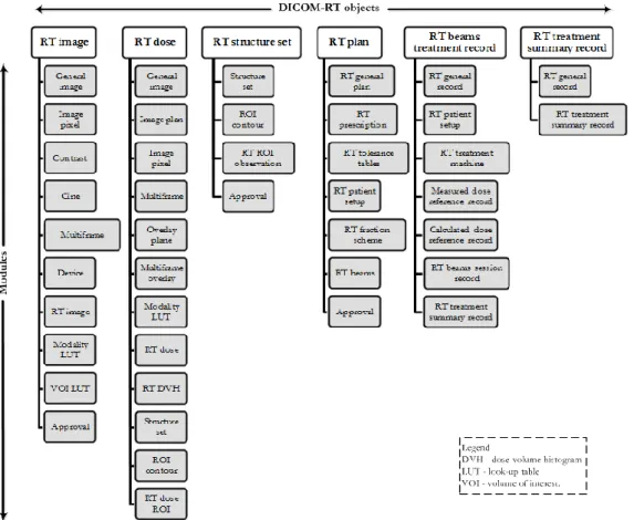
2.2.3 RT Objects ... 20 2.2.4 RT Modules ... 23 2.2.5 Benefits ... 24 2.2.6 Problems... 25 2.3 IHE-RO ... 26 2.3.1 Mission... 26 2.3.2 Structure ... 27 2.3.3 Integration Profiles ... 27 2.3.4 Benefits ... 29
2.4 DICOM-based ePR in Radiotherapy ... 29
2.4.2 Database Schema ... 31
2.4.3 Benefits ... 32
3. Bivalent Study on Radiotherapy Information Systems and DICOM-RT ... 37
3.1 Introduction ... 37 3.2 Global Methodology ... 39 3.2.1 Participants ... 39 3.2.2 Study Design ... 41 3.2.3 Questionnaire ... 41 3.2.4 Data Collection... 42 3.2.5 Statistical Analysis ... 42 4. Results ... 45 4.1 RT National Survey ... 45 4.1.1 Descriptive Results ... 46 4.1.2 Associative Results... 47
4.2 DICOM-RT Expert‟s Opinion ... 49
4.2.1 Descriptive Results ... 50
4.2.2 Associative Results... 53
4.2.3 Question Correlation Results ... 56
5. Discussion ... 63 5.1 Main Contributions ... 65 5.2 Research Limitations ... 65 5.3 Recommendations ... 66 5.4 Future Work ... 68 References ... 73 Appendix ... 81
A. Questionnaire “DICOM-RT in the radiotherapy information systems” ... 83
B. Email sent to the National Coordination for Oncological Diseases ... 97
C. Examples of documents requested to conduct the survey... 99
1. Example of an email to request authorization for the chairman of the board of directors ... 99
2. Example of a specific request authorization ... 100
3. Example of a request for authorization approved ... 101
D. Example of an email sent for the experts ... 105
List of Abbreviations and Acronyms
1-D 2-D 3-D AAPM one-dimensional two-dimensional three-dimensionalAmerican Association of Physicists in Medicine
ACR American College of Radiology
AE ANSI ASTRO
application entity
American National Standards Institute American Society for Radiation Oncology
BEV beam‟s eye view
CEN/TC CT
European Committee of Standardization/Technical Committee [Comité Européen de Normalisation]
computerized tomography
DICOM digital imaging and communications in medicine
DICOM-RT DICOM radiation therapy extensions
DRR digitally reconstructed radiograph
DVH EPID
dose-volume histogram
electronic portal imaging device
ePR ESTRO
electronic patient record
European Society for Therapeutic Radiology and Oncology
GUI graphic user interface
HIMSS HIS HL7 HTTP
Healthcare Information and Management Systems Society hospital information system
health level 7
hypertext transfer protocol
ICRU International Commission on Radiation Units and Measurements
IEC IEEE IGRT IHE
international electrotechnical commission institute of electrical and electronics engineers image guided radiotherapy
integrating the healthcare enterprise
IHE-RO integrating the healthcare enterprise in radiation oncology
IMRT intensity modulated radiotherapy
IP IPEM IS ISO IT JPEG internet protocol
Institute of Physics and Engineering in Medicine information system
International Standards Organization information technology
joint photographic experts group
MLC MRI
multi-leaf collimator magnetic resonance imaging
MU NEMA OSI PACS
monitor unit
National Electrical Manufacturers Association open system interconnection
picture archiving and communication system
PC PET
planning committee
positron emission tomography
R&V REV RIS
record and verify room‟s eye view
radiology information systems
RO RSNA RTOG RT-PACS SCP SCU SOP SPECT SRO TC TCP TDS TMS TPS URL VS WG WS XML x,y,z cm kV mm MeV MV α γ-rays radiation oncology
Radiological Society of North America Radiation Therapy Oncology Group radiotherapy-dedicated PACS service class provider
service class user service-object pair
single-photon emission computed tomography spatial registration object
technical committee
transmission control protocol treatment delivery system treatment management system treatment planning system uniform resource locator virtual simulation working group workstation
extensible markup language point coordinates
centimeters kilovolt millimeters mega electron volt megavolt
significance level gamma-radiation
List of Figures
Figure 1. Chain of RT adapted from (Schlegel et al., 2006) ... 9 Figure 2. Chart illustrating RT workflow with DICOM-RT objects adapted from (Law and Liu, 2009) ....10 Figure 3. Diagram of DICOM data at RT workflow adapted from (Mayles et al., 2007) ...21 Figure 4. DICOM-RT data model of external beam RT adapted from (Dicom Standards Committee, 2009) ...22 Figure 5. Chart illustrating the six DICOM-RT objects of external beam RT and their associated modules adapted from (Law and Liu, 2009) ...23 Figure 6. Data cycle of RT instruments (Tee, 2010) ...24 Figure 7. Diagram of the IHE-RO integration profile “basic RT treatment planning” adapted from (I.H.E., 2011b) ...28 Figure 8. DICOM-RT based ePR system components adapted from (Law et al., 2009) ...30 Figure 9. Arquitecture of the web application server of the DICOM-RT-based ePR system adapted from (Law et al., 2009) ...31 Figure 10. RT workflow based on GUI adapted from (Law, 2005) ...32 Figure 11. Geographical distribution of Portuguese RT departments ...40
List of Tables
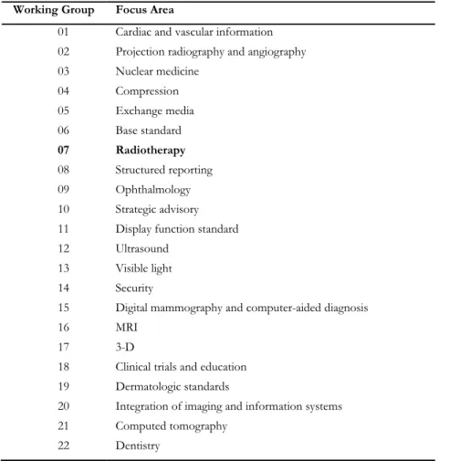
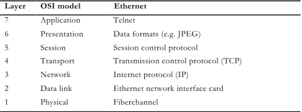
Table 1. RT techniques which require dedicated TPS (Podgoršak, 2005) ...13 Table 2. Working groups of DICOM 3.0 (Dicom Standards Committee, 2009) ...17 Table 3. An OSI model realization with Ethernet adapted from (Schlegel et al., 2006) ...19 Table 4. Structure of the DICOM 3.0 (Dicom Standards Committee, 2009) ...19 Table 5. The four object levels of the DICOM model (Huang, 2010) ...20 Table 6. Portuguese institutions with RT departments listed by geographic distribution ...40 Table 7. Structure of the questionnaire “DICOM-RT in the radiotherapy information systems” ...41 Table 8. Generic identification of the equipment/IS manufacturers of RT departments ...47 Table 9. Quantification of personnel and technologies by health system ...48 Table 10. Techniques, equipments and imaging modalities by health system ...48 Table 11. PACS utilization, type of treatment units, and type of TPS workstations by health system ...49 Table 12. Results from questions 4.2 and 7 (group IV) of the questionnaire ...51 Table 13. Results from questions 1.1 and 1.2 (group V) of the questionnaire ...51 Table 14. Results from questions 2 and 3 (group V) of the questionnaire ...52 Table 15. Results from questions 4 and 5 (group V) of the questionnaire ...53 Table 16. DICOM-RT utilization at RT workflow and IS behavior by linac‟s context of RT department .54 Table 17. Preponderance of factors in interoperability approach and services involved in the IS integration by years of professional experience of experts ...54 Table 18. Importance attributed to possible scenarios of treatment units for a good policy in RT departments by linac‟s context ...55 Table 19. Importance attributed to procedures to achieve interoperability with DICOM-RT by academic qualification of the experts...55 Table 20. Importance attributed to factors for the immaturity of the DICOM-RT implementation by professional experience of the experts ...56 Table 21. Importance of DICOM-RT benefits by years of professional experience of the experts ...56 Table 22. Correlation between questions about the IS behavior in RT department where the expert works and factors preponderant in interoperability approach when purchasing new IS for RT department ...57 Table 23. Correlation between questions about the professionals that defines the IS integration when implementing new IS in RT department ...57 Table 24. Correlation between various scenarios of treatment unit‟s context that contribute for a good policy in RT department ...58 Table 25. Correlation between procedures to achieve interoperability with DICOM-RT ...58 Table 26. Correlation between factors for the immaturity of the DICOM-RT implementation ...58 Table 27. Correlation between levels of importance attributed to benefits of DICOM-RT ...59
Thesis Outline
Chapter 1 Introduces a brief history of computers in radiotherapy, shows the problem setting and explains the aim of the thesis.
Chapter 2 Contains the state of the art on radiotherapy focusing the information systems used in
the workflow, on DICOM-RT describing the contents for understanding the standard, on IHE-RO initiative presenting the integration profiles, and on DICOM-RT based ePR showing a potential model.
Chapter 3 Covers the bivalent study on radiotherapy information systems survey and DICOM-RT
relevance in interoperability, presenting an introduction of the two issues and a global methodology, containing the participant‟s characterization, definition of study design, questionnaire description, data collection details and statistical analysis.
Chapter 4 Presents the results from the RT national survey, showing the Portuguese radiotherapy
departments in terms of facilities characterization, including treatment equipments, imaging modalities and information systems. Also presents DICOM-RT expert‟s opinions about interoperability and DICOM-RT relevance.
Chapter 5 Discusses the results from the bivalent study according to the state of the art, describes
the main contributions and the research limitations of the work, proposes a list of recommendations for radiotherapy professionals, and gives some directions for future work.
Scientific Results and Financing
The present work was submitted for publication in the “HealthInf 2012 - International Conference on Health Informatics”, with preliminary results of the study titled “The relevance of DICOM-RT in radiotherapy information systems - preliminary results from a national survey” (see Appendix E).
Preliminary results of the thesis were presented in “4th Medical Informatics Symposium”, Faculty of Sciences, University of Porto, October 8, 2011.
The work of this thesis did not receive funding from any organization, but the author as a student of the master‟s degree in Medical Informatics received financial support from the master programme, for the following poster presentations in “Healtinf 2011” and subsequent publications:
Celeste Oliveira and Pedro Pereira Rodrigues. “Automatic organ delineation of computed tomography images for radiotherapy planning in prostate cancer: an overview”. In Proceedings of
the International Conference on Health Informatics - HEALTHINF 2011, 482-485, ISBN
978-989-8425-34-8, INSTICC Press. Rome, Italy. January 2011.
Cátia Pereira, Celeste Oliveira, Cláudia Vilaça and Ana Ferreira. “Protection of clinical data: comparison of European with American legislation and respective technological applicability”.
In Proceedings of the International Conference on Health Informatics - HEALTHINF 2011, 567-570,
ISBN 978-989-8425-34-8, INSTICC Press. Rome, Italy. January 2011.
The article “Automatic organ delineation of computed tomography images for radiotherapy planning in prostate cancer: an overview” was presented in “3rd Medical Informatics Symposium”, Faculty of Sciences, University of Porto, October 29, 2010 and in “Journeys of Oncology”, IPO Porto, Solverde Espinho hotel, July 8,
I shall particularly insist on the following fact, which appears to me very important and quite outside the range of phenomena one might expect to observe.
1. Introduction
This introductory chapter describes a brief history of computers in radiotherapy, explains the problem setting and the aim of the present thesis.
1.1 Computers in Radiotherapy
Radiotherapy (RT), also referred as radiation therapy, radiation oncology (RO) or therapeutic radiology, is one of the three principal modalities used in the treatment of cancer, the other two being surgery and chemotherapy (Podgoršak, 2005). The word radiotherapy comes from the Greek radius, a ray and therapeia, cure (Expert Working Group on Radiation Oncology Services, 2003). For more than a century, ionizing radiation has been used against diseases that usually are malignant. RT is, after surgery, the most successfully treatment modality used for cancer (Schlegel et al., 2006). The power of radiotherapy is that the radiation can be precisely adjusted in space and time, unlike chemotherapy, where drugs are administrated hoping that will be selectively absorbed by the malignant cells (Kalet, 2008). The goal of RT has always been to maximize the probability of controlling the tumor and minimizing normal tissue complications. The achievement of this goal is the key component driving the technological developments (Dyk, 2005). In contrast to other medical specialties that are based mainly on clinical knowledge and experience, RT relies greatly on modern technology and multidisciplinary team approach (Podgoršak, 2005).
In the 1960s RT was considered an empirical discipline with low probability of success, but from its early commercial introduction, linear accelerators (linacs) providing megavoltage X-rays and electron beams has undergone notable expansion. In the 1970‟s the introduction of the computerized tomography (CT) and the help of computers for the radiation calculations it produced universal atlases of isodose distributions. The practice of RT defined itself and developed into a distinct discipline was in the 1980‟s. In the middle 1980‟s, the first treatment management systems (TMS) with low-functionality were custom systems developed by academic departments or equipment manufacturers (Dyk, 1999, Schlegel et al., 2006, Wu, 2008) .
RT is an early example of computer programming application to help the clinical treatment decision. The computer modeling focused on the physics of radiation absorption in human tissue relative to the geometry of radiation beams and on the design of interactive systems that could be used for manual computer aided design (Kalet, 2008). Advances in RT have always resulted from successful combinations of technological progress combined with improved biological understanding. The computer revolution characterized by the development of powerful computers had a major impact on planning and delivery of the radiation treatment (Bentzen et al., 2008). In to sum up, RT is based on physics, radiation biology, mathematics, computer science, electrical and mechanical engineering, making it an interdisciplinary field (Schlegel et al., 2006).
Currently, RT can be applied so accurately that the tumor control and the probability of cure, has significantly increased for many cancers due to physics and technology innovations. The development and implementation of modern informatics have intensely changed the management and practice of RT (Schlegel et al., 2006).
1.2 Problem Setting
Nowadays, in RT departments, the treatments are based on technological advancements in diagnostic imaging, image processing and high computerization technology, such as the treatment planning system (TPS), leading to an increase of the complexity of storage and availability of RT data (Law et al., 2009).
Often there are different proprietary and stand-alone information systems (IS) for single-purpose applications. Each system has its own “storage area” and oftentimes has limited interface with other systems. These IS, focused on the system instead of being patient-centered, acquire the necessary information during the RT treatment course, being most of the data distributed through each IS. This data isn‟t immediately available due to incompatibles formats between the equipment workstations (WS) and the IS, leading to utilization of multiple IS in the search of clinical information of a particular patient. For example, radiation oncologists need to go to dedicated WS to delineate volumes, to approve treatment plans or portal images, because there is no “home base” for all images of the patient. The need to integrate all the scattered information from different IS of RT department is steadily recognizable because of its imaging intensive nature and ever increasing demands for better treatment equipments and complete information. RT involves radiobiologic factors which demand the record of all treatment parameters for patient future reference. One problem in RT concerns about the exchange of specific information between IS, departments and hospitals. This has impeded the research collaboration between institutions. This lack of interoperability between the IS causes discontinuity in health care, leading to redundant clinical evaluations and clinical decisions based on incomplete information limiting clinical trials and scientific investigations(Law, 2005, Liu et al., 2007, Law et al., 2009, Shakeshaft, 2010).
“Quality standards” of the RT is the set of recognized criteria against which the quality of the RT workflow can be evaluated, being part of the quality assurance program that supports the IS maintenance too. Various organizations, such as the American Association of Physicists in Medicine (AAPM) and the European Society for Therapeutic Radiology and Oncology (ESTRO) have issued recommendations for standards in RT, whereas other organizations, such as the International Electrotechnical Commission (IEC) and the Institute of Physics and Engineering in Medicine (IPEM), have addressed recommendations for certain parts of the RT workflow. RT departments where industry standards are not available, local standards need to be developed, based on a local assessment of requirements (Podgoršak, 2005).
The adoption of the digital imaging and communications in medicine – radiation therapy (DICOM-RT) extensions, integration profiles from Integrating the Healthcare Enterprise – Radiation Oncology (IHE-RO), and of Radiotherapy Picture Archiving and Communication Systems (RT-PACS) allows a system integration infrastructure. All RT information and images from various sources can be converted to the DICOM-RT standard and integrated into a DICOM-based database. Benefits of this integration are the reduction of time and effort in searching for information related to the treatment (Liu et al., 2007, Law and Liu, 2009).
1.3 Aim
The aim of this thesis is to study the relevance of the standard DICOM-RT in the interoperability between the IS used in RT, through the revision of existing reality in the Portuguese RT departments and the analysis of expert‟s opinion about the issue. We intend to clarify the state of art, finding the main problems and recommend some ways to optimize the DICOM-RT utilization in order to achieve interoperability.
To accomplish this aim, the present work was divided in different phases: a comprehensive review of the state of the art focusing radiotherapy, DICOM-RT, IHE-RO and DICOM-based electronic patient record (ePR) in RT, present in chapter 2; a bivalent study with national survey characterizing RT facilities
in terms of equipments and IS, and an expert‟s opinion about the DICOM-RT relevance and interoperability in RT context, present in chapter 3 and 4; and the discussion with the main contributions and suggestion of some recommendations for RT professionals, present in chapter 5.
To the electron: may it never be of any use!
2. State of the Art
This chapter presents the radiotherapy, its workflow, equipment and types of IS dedicated. Also reviews the state of the art of DICOM-RT with a description of DICOM-RT objects, their use in integration profiles from IHE-RO, and in the model creation of a DICOM-RT based ePR.
2.1 Radiotherapy
In modern medicine, one of the most technologically advanced fields is RT, that is a comprehensive and dynamic discipline which plays a major role in cancer care (Schlegel et al., 2006). About 60% of patients with cancer will require RT during the course of their illness and those who are cured, 80-90% of patients underwent RT (Perez and Brady, 1998).
The aim of RT is to deliver the prescribed radiation dose to the tumor, while sparing as much as possible the surrounding normal tissues. This therapy includes two treatment modalities: external beam radiotherapy, also called teletherapy that uses radiation produced by a machine, mainly photons or electrons, where the tumors are treated at a long distance from the radiation source; and internal radiation, designated brachytherapy that administers radiation with small radioactive sources placed on or in the tissue to be irradiated. The brachytherapy can be intracavitary (sources that are loaded within body cavities), interstitial (sources that are implanted into tissues), intraluminal (trains of sources within the lumen of organs) and superficial brachytherapy (sources supported in a mould over a tumor). Whereas teletherapy is designed to produce a homogenous dose distribution in most cases, brachytherapy uses the inhomogeneous dose distributions found around sources to create a high dose in tumors and producing a low dose in normal tissue. External beam RT represents over 90% of the workload in RT departments and it is an image and computer graphic intensive process (Mayles et al., 2007, Cleto et al., 2008, Law and Liu, 2009, Law et al., 2009).
2.1.1 History
The discovery of X-rays in 1895 by Wilhelm Roentgen, in Germany, revolutionized the field of medicine. That same year Henri Becquerel announced his discovery of a new type of invisible radiation emitted from uranium salts. He called this radiation as “Becquerel rays”, later termed “radioactivity” by Marie Curie who studied this type of radiation and set the stage for the development of new tools for diagnosis and therapy. A year later Joseph Thomson, in Cambridge, announced the discovery of the electron (Senior, 1998, Dyk, 1999).
In 1899 was reported the first patient cured by RT and in 1910 was used for the first time the brachytherapy with the insertion of radium needles and tubes (Perez and Brady, 1998). During the 1930‟s and 1940‟s brachytherapy systems of calculation evolved with the Manchester system being one of the predominant systems. In 1951 the first patient was treated with cobalt-60 γ-rays, in London. Between 1910 and 1950, kilovoltage X-ray units played an important role in the early development of RT and later in the treatment of superficial lesions. Teletherapy cobalt-60, machines with penetrating γ-ray beams emitted from a radioactive source was first used for patient treatment in 1951, in Canada. Cobalt units
became the mainstay of external beam therapy for the next 30 years. The linacs, machines with megavoltage X-ray beams and electron beams, which achieved routine clinic status in the 1960‟s, were used with a manual treatment planning through the manipulation of isodose charts into patient body contours that were generated by direct tracing and relied on the careful choice of beam weight and wedging. In the late 1960‟s, the simulator was introduced as an additional device to assist the simulation of external RT treatment. In 1972 Godfrey Hounsfield introduced the CT in the medical community. In the end of the 1970‟s the development of CT along with the advent of accessible computing power led to the development of CT based computerized treatment planning providing the ability to view dose distributions directly superimposed upon a patient‟s axial anatomy. In this decade, diagnostic radiology and RT began to separate into distinct medical specialties. The CT-based treatment planning was later supplemented with magnetic resonance imaging (MRI) in order to understand the tumor morphology more precisely, and thus achieve improved definition of target volumes through image registration. In the early 1980‟s, the introduction of stereotactic into RT allowed the high accuracy execution of the computer plans to the patient. Stereotactic treatment techniques were first developed for single-dose irradiations, called radiosurgery; then for fractionated treatments in the brain and the “head and neck region”, designated stereotactic RT; later for extra-cranial tumor locations, named extra-cranial stereotactic RT. In the middle 1980‟s, appeared the first TMS with dynamic treatments, thanks to commercial availability of computerized multi-leaf collimators (MLC). In the middle 1990‟s, 3-D conformal RT was supplemented by a new treatment technique, the intensity modulated radiation therapy (MRT) in combination with inverse planning software. At the beginning of the new millennium, the field of adaptive RT brought the temporal alterations of the target volume that can be assessed and taken into account. Image-guided radiotherapy (IGRT) detects deformations and motion between fractions (inter-fractional) and during irradiation (intra-fractional), correcting these changes with gating or tracking of the irradiation beam. In the last decade of the past century the superiority of linac became evident and the number of cobalt-60 machines started to decrease all over the world except in developing countries where they are still considered as the best option, mainly because of their reliability (Dyk, 1999, Podgoršak, 2005, Schlegel et al., 2006, Mayles et al., 2007).
Modern RT continues to progress and this development is strongly linked to the evolution of computer technology and corresponding advances in diagnostic imaging equipment, sophistication in computer-assisted treatment planning and delivery (Levitt et al., 2008). With the introduction of 3-D imaging, 3-D virtual therapy simulation and 3-D dose calculation the requisite for an individualized and effective treatment is satisfied. Conformal treatments became less expensive and considerably faster because the modern linacs are more reliable by having a high mechanical accuracy with high dose rates providing the basis of modern precision RT (Schlegel et al., 2006). More new RT techniques and tools appeared, such as intraoperative, tomotherapy, breathing control, image segmentation, proton therapy, total body irradiation, total skin electron irradiation, volumetric modulated arc therapy, intensity modulated arc therapy, etc (Dyk, 2005).
2.1.2 Workflow
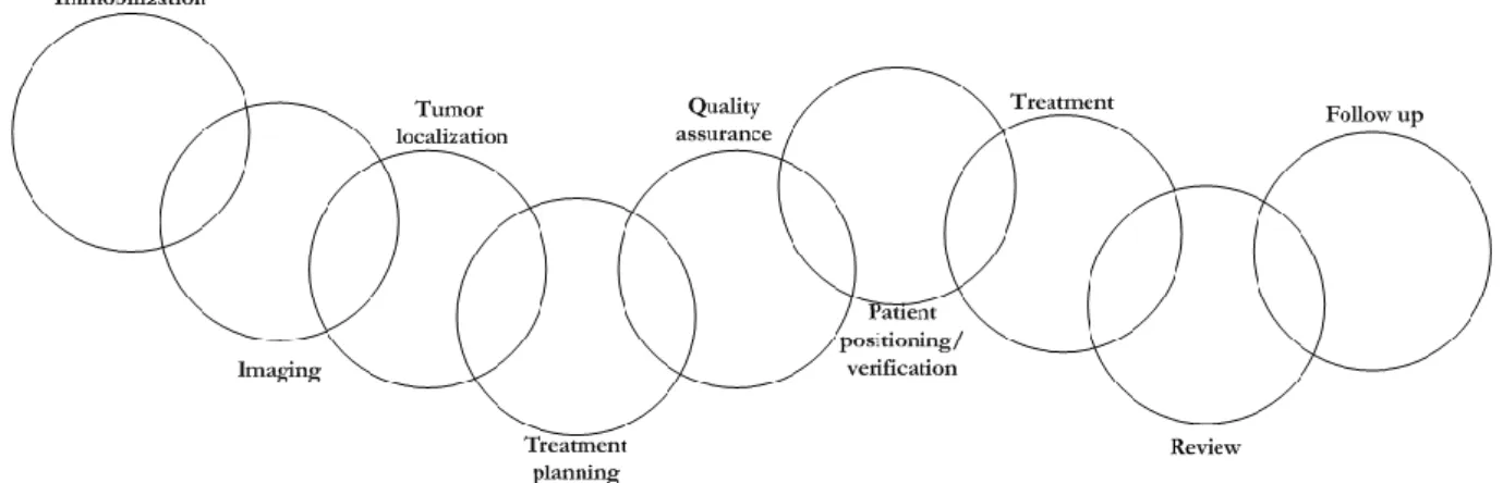
The basis of the RT covers different aspects of all links in the “chain of radiotherapy” procedure, as shown in figure 1. The link of treatment planning includes the following steps: delineation of the target volumes and organs at risk, definition of the treatment technique, dose calculation and evaluation of the 3-D dose distribution (Schlegel et al., 2006).
Figure 1. Chain of RT adapted from (Schlegel et al., 2006)
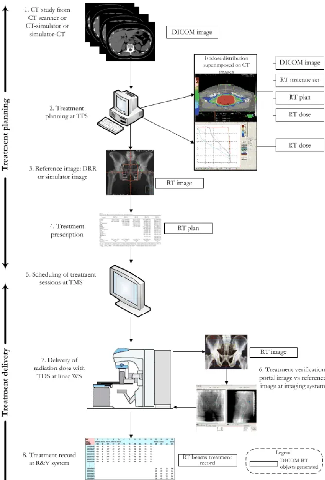
The authors describe some types of workflows in the external RT representing the flows depending the perspective, such as: clinical workflow (Law, 2005, Liu et al., 2007, Law and Liu, 2009), DICOM images flow (Law and Liu, 2009), DICOM-RT objects flow (Law, 2005, Law and Liu, 2009), RT dataflow (Law, 2005), etc. The RT workflow has two distinct phases: treatment planning and treatment delivery, and is described ahead with the identification of IS used and associated DICOM-RT objects. (see figure 2).
In the treatment planning the process starts with a CT image study of the anatomic region to localize the tumor volume, performed with either CT-simulator or CT scanner or simulator-CT. The radiation therapist sets the patient up in his treatment position for scanning in CT room. The DICOM images are associated to the patient‟s information and stored in the WS or in the picture archiving communication system (PACS) (step1). If the CT study is acquired at CT-simulator, a virtual simulation (VS) application retrieves the images and performs the simulation producing RT structure set and RT plan. The TPS reads the CT images (step2) and other previous available diagnostic images (diagnostic CT, MRI and positron emission tomography (PET) can be fused with the CT to aid in the delineation of tumor volume), the RT structure set, and the RT plan. At TPS, target volumes and organs at risk are delineated interactively by medical physicist, radiation oncologist or dosimetrist according to International Commission on Radiation Units and Measurements (ICRU) reports nº 50 (I.C.R.U., 1993) and 62 (I.C.R.U., 1999). After this procedure, the machine‟s parameters are determined (beam modifiers and beam geometries) and the radiation dose distribution within the body is calculated (dosimetry data). This planning requires a balance between accurate target volume coverage and sparing of neighboring normal tissue evaluated by dose-volume histogram (DVH) and isodoses distribution superimposed on CT images. From CT study projection X-ray image can be reconstructed named digitally reconstructed radiograph (DRR) creating RT image (step3). These images or the simulator images will serve as the reference images for treatment verification. At TPS new RT Plan object is created, and RT image is produced. After the evaluation and approval by radiation oncologist the treatment is prescribed (step4) and the treatment sessions are scheduled in the TMS (step5) and transferred to the treatment unit (linac) through the completed RT plan by network for the treatment delivery system (TDS) (Law and Liu, 2009, Huang, 2010).
In the treatment delivery the process starts with the verification of the treatment; a portal image is obtained at the linac to be compared with the reference image using imaging system, creating RT image (step6). After this procedure, the treatment is delivery (step7) if the portal images were found to match the reference images in aligning with the treatment field. At each session, every parameters of the treatment will be recorded at linac WS or (record and verify) R&V system, creating RT beams treatment record (step8). Usually, a treatment session is daily, five days a week, during 5 to 7 weeks, depending on the type of cancer. The radiation oncologist will review the patient‟s progress and a follow-up appointment will be made at the TMS (Law and Liu, 2009, Huang, 2010).
RT is prepared for the individual patient, so it is important use informatics to optimize workflow and patient treatment outcomes. Problems with the integration of the IS and equipments of the RT department compromise the efficiency of RT workflow (Huang, 2010).
2.1.3 Equipments
The kilovoltage X-rays has been used in RT from the earliest attempts at external beam treatments. The kilovoltage range covers X-ray beams generated between 10 kV and 400 kV and is usually subdivided into four categories according to beam penetration: grenz rays (10 kV to 20 kV), rarely used in modern RT; contact therapy (10 kV to 60 kV), provides a useful treatment depth up to several mm; superficial therapy (50 kV to 150 kV), affords treatment for many superficial lesions and an adequate alternative to electrons; orthovoltage therapy (150 kV to 400 kV), has the 90% dose in 1 cm to 2 cm beneath the incident skin surface at the usual treatment distance of 50 cm source to surface distance. Multi-energy units covering both the superficial and orthovoltage ranges are used in many RT departments (Podgoršak, 2005, Mayles et al., 2007).
The cobalt-60 introduced the early megavoltage RT generating γ-rays at two well-defined energies (1.173 MeV and 1.332 MeV). Additional Compton emission generated in the source results in the beam incident on the patient having a continuum of energies below this with a mean energy less than 1 MeV (Mayles et al., 2007).
The linacs produce more penetrating beams with the versatility of the choice of beam energy delivering in a higher dose rate. A 6 MV beam will have a mean X-ray energy of around 2 MeV, and the range of the secondary electrons produced by interaction in tissue will be about 16 mm. This machine can have MLC, dynamic wedges, and electron beam generation (Mayles et al., 2007).
The simulator was introduced as an additional device to help the preparation of external RT, by checking before the treatment if the plan can be delivered in practice and if the relationship of the beam setup to the patient‟s anatomical features is accurate. This equipment has similar mechanical and geometrical characteristics of the therapy machine and the treatment table, and fitted with a radiodiagnostic X-ray tube. Simulator provides the ability to mimic the treatment geometries and to visualize the resulting treatment fields on radiographs or under fluoroscopic examination of the patient. Modern simulators are universal and match the characteristics of most existing therapy machines. Some simulators have a special attachment that allows them to collect patient cross-sectional information similarly to a CT scanner being designated a simulator-CT (Boyer et al., 2001, Mayles et al., 2007).
Dedicated CT scanners used in treatment planning and simulation are known as CT-simulators. The components of a CT-simulator include: a large bore CT scanner (with a large opening for patient positions with treatment accessories), room lasers (for patient setup and marking), a flat table top (to closely reproduce RT treatment tables) and a powerful graphics WS (Podgoršak, 2005).
The portal imaging devices were introduced in RT to provide quality assurance of patient setup. In order to provide online verification electronic portal imaging device (EPID) has been developed to acquire and display portal images. This suitable radiation detector is attached to the gantry of the linac and is capable of transferring the detector information to a computer that will process and convert it to an image that can be analyzed instantly (Podgoršak, 2005, Mayles et al., 2007).
2.1.4 Information Systems
IS will never replace the RT professionals, but it helps in making repetitive and time-consuming tasks with perfection and in presenting the patient information clearly freeing the professionals to work in the clinical care efficiently (Ambider, 2000). In RT departments the following IS are used: TPS, TMS, TDS, and imaging systems (e.g. RT-PACS). The abbreviation “R&V” has been used to describe systems that receive data from the TPS and interact with the TDS having the function of RT process control through the network. Data integrity in the RT workflow must be preserved and available in real time at the treatment WS (Siochi et al., 2009).
Most IS in RT fit a client-server architectural data processing model, consisting of multiple computers that share the application processing. The components are often heterogeneous, provided by different hardware and software vendors, and they transfer data via messages across a network. The network, given the complexity and amount of data and images, usually is internal network to allow the total interconnection among all equipment. This network of RT can be connected to a pre-existing external network (e.g hospital information system - HIS), in order to share common data, including clinical, administrative and financial database of the patient (Brooks et al., 1997, Dyk, 1999, Huh et al., 2000).
Generally, treatment plans are stored at TPS, simulated fields are stored at simulator WS, and treatment records are stored at R&V systems. The treatment information for a patient whose treatment involves various IS will be stored in different places, normally being linked by paper record. If the paper record is lost, the patient‟s treatment information disintegrates (Law and Liu, 2009).
Treatment Planning System - TPS
The TPS is used in the treatment planning process, having the following steps: data acquisition and entry (characteristics of the linac and treatment field configurations); patient data acquisition through CT scan with possibility of additional radiological information (coregistered CT, MRI, ultrasound, PET, single-photon emission computed tomography (SPECT), etc) to help in geometric planning in order to obtain a theoretical distribution of doses within the body‟s patient; treatment plan generation with operator-specific information (tissue delineation, application of margins and treatment extent); and transfer of data to the TDS (via DICOM-RT format). The electron density information extracted from CT data set is vital for calculation of dose heterogeneities due to the differing composition of human tissues and the theoretical dose distributions depend on the dosimetry, calibration of the linac, checks of the treatment and in vivo dosimetry (Ebert et al., 2004, Podgoršak, 2005, Mayles et al., 2007).
Regarding software components, TPS contain dose calculation algorithms, beam modifiers, heterogeneity corrections, image display with DVH, and biological modeling. Dose calculation algorithms are the most critical software component being responsible for the correct representation of dose in the patient, from simple 2-D calculations to full 3-D dose models. There are numerous dose calculation algorithms and ICRU report nº 42 (I.C.R.U., 1987) lists the chronological development of these for photon and electron beams as: Monte Carlo techniques, pencil beam algorithms, etc. The TPS must be capable of handling the many diverse beam modifying devices found on linac models, the photon beam modifiers consisting of jaws (monitorized collimating devices), blocks (field shielding), MLC (beam shaping device), wedges (static or motorized or dynamic), custom compensator (compensating filter), bolus (tissue equivalent material placed in contact with the skin) and the electron beam modifiers consisting of cones (applicators for external collimation), etc. Most TPS algorithms apply either a correction factor approach or a model based approach to heterogeneity corrections. Concerning image display, TPS calculates DVH, Beam‟s Eye View (BEV) often used in conjunction with DRR to aid in assessing tumor coverage and for beam shaping, and Room‟s Eye View (REV) to give the user a perception of the gantry and table relation helping the virtual plan to be practicable in actual patient setup. Optimization routines including inverse planning are provided by modern TPS with varying degrees of complexity. Algorithms can modify beam weights and geometry or calculate beams with a modulated
beam intensity to satisfy the user criteria. Beam time and monitor unit (MU) calculation by TPS is optional, and total prescription dose and fractionation information can be incorporated. Automatic contouring for regions, image segmentation for the DVH evaluation and algorithms for the distributions modeled on biological effects can also be incorporated in the TPS (Podgoršak, 2005).
TPS can be dedicated for special techniques as stand-alone systems and there are clinical procedures that require careful consideration, owing to their inherent complexity. A list of techniques that require special consideration and that may result in dedicated TPS is shown in table 1.
Table 1. RT techniques which require dedicated TPS (Podgoršak, 2005) RT Techniques
RT with dynamic MLC Intraoperative radiotherapy
RT with micro MLC Stereotactic radiosurgery
Orthovoltage Electron beam arc therapy
IMRT Brachytherapy
Total body irradiation Tomotherapy
Total skin electron irradiation
The development of new techniques of RT involves the unconditional utilization of the TPS with successive improvements in treatment planning hardware and software that have been most notable in the graphics, calculation and optimization aspects. The acquisition of images with more detail of the anatomy and tumor location, and the systems encompassing the virtual patient and image registration routines leads to an increasing complexity in the data handling. Traditional forward treatment planning, based on a trial and error approach is giving way to inverse planning, that makes use of dose optimization techniques to satisfy the user specified criteria for dose in the target and critical structures (Podgoršak, 2005, Mayles et al., 2007, Song and Li, 2008).
The TPS are connected via networks to the other equipments to enable the sharing of data and images. If the TPS and the simulator are linked to the network, the patients are simulated with the same planned data without the need for typing in filed parameters. The TPS can also be connected to exterior systems, including imaging systems, for importation of images acquired by CT or by other imaging modalities to image registration routines. One existing problem is that treatment plans from an old TPS cannot be retrieved for review after a system upgrade. The exchange of RT data between various TPS and different institutions has become an important issue (Wang and Huang, 2006, Mayles et al., 2007).
Treatment Management Systems - TMS
The TMS, also called radiation oncology information management systems or RT specific ePR, facilitates the management of clinical tasks and technical information. It centralizes information, controls, records and checks all aspects of each individual treatment, facilitates access, provides data handling, and integrates imaging, planning, and therapy modalities linking them to the administrative services. The TMS is increasingly important because of the complexity of new treatment modalities that require different data sources and complex software tools (Huh et al., 2000, Boyer et al., 2001, Wu, 2008).
The system is designed in response to clinical operations requirements such as patient registration (demographics information and referring physician), patient consultation (physician, nursing and other clinical staff), departmental scheduling (patient, equipments and staff), patient tracking, departmental patient charting (shared electronic chart, chart checks), patient treatment simulation (resources scheduling, geometric field setup information), patient treatment with backup data (R&V system containing radiation field information for each patient controlling the use of assisted setup and automated treatments), and administrative services (billing, tumor registry, documentation)(Dyk, 1999).
A TMS should be nominated to manage the R&V systems, that checks the adequate dosing of patients at the linac by comparing the daily treatment setup parameters with the prescribed values and respective
tolerances, and at the end of the treatment session, it records a daily sheet of all treatments carried out including the accumulated dose, the treated fraction, and overrides along with the identification of the operator that performed them. In order to customize the system to the users‟ requirements, and help to provide maximum use of all the features of the system, different levels of authorization are password protected and different tolerance tables are customizable by the user to specify the range over which parameters can differ from the prescribed value before treatment is inhibited. Regarding backup and archiving, the large amount of data requires considerable storage capacity. It is useful if the backup system allows individual patient records to be retrieved as well as a full restore of the database. A daily backup of patients on treatment is usually performed. Old data are permanently removed from the system and transferred to long-term storage during the archival process. It is important that backups of archived data are also maintained. Data storage should comply with national regulations relating to patient privacy. Another important issue is the system resilience and the fault reporting. In summary, TMS supplies critical information for the control of treatment units and other extras functions such as user rights, networking, backup, archiving and security services (Podgoršak, 2005, Mayles et al., 2007, Siochi et al., 2009, Colonias et al., 2011).
The TMS may be provided by the TPS manufacturer, the linac manufacturer or third party software. It should drive the RT workflow of any linked equipment from any vendor using standards such as DICOM-RT. It is vital that the data in R&V systems be quality controlled using independent checking to verify the input, and that the network infrastructure can handle the transfer of the large amounts of data, for not compromise the patient treatment. The performance of the TMS should be included in an appropriate quality assurance program with specific tests according to specific system (Podgoršak, 2005, Siochi et al., 2009).
Treatment Delivery Systems - TDS
In order to deliver RT treatments, parameters are electronically parsed out from a TMS database to the TDS in the linac WS for cooperation with localization and treatment couch. Depending on the capabilities and vendor selection for a given treatment unit, it is usual for a single console area to have at least five computers (Siochi et al., 2009).
TDS systems beyond enabling a computer-controlled user interface with electronic monitoring circuits also provides beam shaping systems, image-guided setup systems, respiratory gating systems, among others. These systems improve the accuracy of the treatment delivery by eliminating as many errors as possible, ensuring that the machine settings will be repeated throughout the treatment course and that the programmed total dose will not be exceeded. These systems can be linked together by networking, and the high-speed connectivity between image acquisition, treatment planning and final treatment delivery improves the efficiency of the process (Mayles et al., 2007).
The normative requirements for the TDS system are: linac system, fully integrated MLC, shapes integrated in patient chart, auto-setup of MLC, integrated display of MLC shape, integrated system of MLC shape, in-room MLC display, pendant control of MLC, capability for pre-planning treatment field changes, advanced planning of treatment courses, easy implementation of hyperfractionation, field setup capabilities, user defined setup fields, setup photos, auto-sequencing of fields, in-room call-up of next field, port films integrated into treatment course, override of individual treatment parameters, demographic functions, schedule functions, treatments parameters, treatment parameters tolerances, dose information and emergency treatment provisions (Dyk, 1999).
If the TDS are networked, it is possible to switch patients from one treatment machine unit to another from a different vendor with the utilization of DICOM-RT without having to re-enter the treatment data reducing the errors in data entry (Mayles et al., 2007).
Imaging Systems
The RT process requires diagnostic images for identification, characterization, and localization of tumor, and image-based treatment workflow for planning, simulation, execution, and verification of treatment. The imaging systems handles with BEVs, REVs, DRR, simulation images, portal images, reconstructed images, images resultant of co-registration, and with segmentation, automatic contouring, VS, etc.
The normative requirements for the imaging systems are: WS functions, demographic data, setup parameters, data entry and edit capability, field capture, volumes delineation, image comparison, image approval, image enhancement tools, image review, image tasks tracking, etc (Dyk, 1999).
Systems used for the acquisition, archiving, communication, display, control, image database manager, and processing of RT images and related data are referred to as a radiotherapy picture archival and communication system (RT-PACS), also known as oncology-specific PACS. The practice of RT has specific functions that a conventional PACS cannot guarantee. For one patient, the images required include several simulation images, a set of initial portal images, and a set of on-treatment portal images. In addition to the images themselves, various other types of data also must be incorporated, like scalar data (geometric parameters), vector data (outlines of treatment portals on simulator images and target volumes on CT images), and character strings (annotations and messages). Different types of users are involved in the acquisition of images (simulator technologist, CT technologist, and treatment machine technologist) and in other tasks related to these images (dosimetrist, physician, and radiation oncologist). The PACS must differentiate among the various types of users providing a common platform for supporting the various user-specific tasks. It should integrates modalities and WS with scalable archive to meet departmental needs, having RT information online and accessible any time and from any location through its web-based application (Mayles et al., 2007).
The portal imaging application can be separated into offline and online analysis: offline analysis can be used to quantify uncertainties for individual patients and online imaging allows a quick decision about continuation of treatment by comparing the portal image with the reference image and looking for unacceptable discrepancies. The application includes features for image edition, image approval, image comparison, and measuring tools for field placement errors by using overlays of patient anatomy and of field shape. The values of the field placement errors need to be stored along with the images (Hendee and Ritenour, 2002, Podgoršak, 2005, Mayles et al., 2007).
As the amount of non-textual data generated in RT has radically increased image integration into the patient database is also a major condition in RT practice. The diagnosis and treatment decision are made from CT study becoming the burden of the RT department to archive this information for legal purposes (Starkschall, 1997, Huh et al., 2000, Podgoršak, 2005).
Standards
A result of the commercial development of IS for the RT is proprietary data formats (e.g. TMS export
format- Helax®, iCOM- Elekta®, RTP link- Impac®, Varis link- Varian®) that lead to problems in interface
between the IS. Unless all the equipment being used in a department is from a single software supplier, it is a requirement that image and other data should be transferable between different IS in such a way that it can be interpreted by different software. The need to improve the RT workflow, as well as efforts to analyze and collect the treatment information for clinical trials has motivated the development of standards on RT information sharing. Some attempts by collaborative groups for the standardization of RT data formats, resulted in two relevant formats: the Radiation Therapy Oncology Group (RTOG) format and DICOM-RT extension. Another standards used are: for images, standards like the DICOM and the American College of Radiology-National Electrical Manufacturers Association (ACR-NEMA) 2.0, and file formats like joint photographic experts group (JPEG) and tagged image file format (TIFF); for administrative date, standards such as health level 7 (HL7) and languages such as extensible markup language (XML) (Ebert et al., 2004, Bosch et al., 2010).
Since 1994 the North American Radiation Therapy Oncology Group – use the format RTOG based on report nr.10 of the AAPM (1982), designed for the purpose of transferring RT data to the Radiotherapy and Oncology Group data centre (now called the Image-Guided Therapy Centre) so that quality assurance of clinical trials could be performed. It is widely used in systems originating in the U.S.A providing a common format for CT data, structures, treatment data and dose distributions used in RT. A typical RTOG data set consists of a collection of data files with a common name. The files are numbered sequentially, „Patient0000, Patient0001, …‟ with file „0000‟ containing patient demographic information and a description of data contained in all other files and the subsequent files containing planning information. This format is limited, because there are no modules for brachytherapy treatments and for IMRT. It also does not present any record of actual treatment (i.e. data from R&V system) or images used for alignment (i.e. simulator images, DRR, portal images) (Ebert et al., 2004).
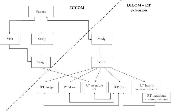
The RT extension of DICOM standard was created under the auspices of NEMA to support the RT data transferring between IS from within and outside RT department. It consists of a collection of DICOM-RT objects that can store information describing various components of a treatment plan (Dicom Standards Committee, 1999, Ebert et al., 2004). In next section, this standard will be present with more details.
2.2 DICOM-RT
The de facto standard for connectivity in the RT community has become with specifically designed objects of the DICOM standard. These have for been known as DICOM-RT, however, they form an integral part of the full DICOM standard being RT specific DICOM objects (Shakeshaft, 2010).
2.2.1 History
With the great development of medical images and the different ways to acquire, transfer and visualize images it became apparent the need for standardization in order to ensure connectivity and interoperability of all systems. In 1983, a committee formed by members of the medical community and of the medical equipment industry represented by the ACR and the NEMA respectively, introduced an industry standard to which all vendors of medical equipment could conform. Even though, the first versions of the standard, ACR-NEMA 1.0 (1985) and ACR-NEMA 2.0 (1988) never became popular among vendors, the later version DICOM 3.0 (1992) is by present-day standards ubiquitous. The main goals of the standard are to solve compatibility problems of digital image, concerning information exchange, interconnectivity and communications among IS to facilitate the expansion of PACS, and allow the creation of diagnostic information databases (Schlegel et al., 2006, Huang, 2010).
The first version specified standards in point-to-point message transmission, data formatting, and presentation and included a preliminary set of communication commands and a data format dictionary. The second version included hardware definitions and software protocols, as well as a standard data dictionary. Networking issues were not addressed sufficiently in either version. For this reason a new version aiming to include network protocols was released in 1992. Because of the magnitude of changes and additions it was given a new name: digital imaging and communications in medicine version 3.0 (DICOM 3.0). The two most distinguished new features in DICOM are adaptation of the object-oriented data model for message exchange and utilization of existing standard network communication protocols (Huang, 2010).
The DICOM was adopted by other standardization organizations including the European Committee for Standardization-Technical Committee (CEN/TC) 251, the Institute of Electrical and Electronics Engineers (IEEE), the HL7 and the American National Standards Institute (ANSI) and demonstrated at the Radiological Society of North America (RSNA) Annual Meetings. In 1994, during the RSNA meeting, the need for standardization of the way that RT data are transferred from one piece of equipment to