RESUMO.- [Progesterona e Hormônio Folículo-Estimu-lante interagem e promovem a sobrevivência e o de-senvolvimento in vitro de folículos pré-antrais
capri-nos.] Este trabalho veriicou os efeitos da progesterona e do hormônio folículo-estimulante (FSH) na sobrevivência e no crescimento de folículos pré-antrais caprinos.
Fragmen-Progesterone and Follicle Stimulating Hormone interact and promote
goat preantral follicles survival and development
in vitro
1Isabel B. Lima-Verde2*, Maria H.T. Matos2, Juliana J.H. Celestino2, Rafael Rossetto2,
Khesller P.O. Name3, Sônia N. Báo3, Cláudio C. Campello2 and José R. Figueiredo2
ABSTRACT.- Lima-Verde I.B., Matos M.H.T., Celestino J.J.H., Rossetto R., Name K.P.O., Báo S.N., Campello C.C. & Figueiredo J.R. 2012. Progesterone and Follicle Stimulating Hor-mone interact and promote goat preantral follicles survival and development in vi-tro. Pesquisa Veterinária Brasileira 32(4):361-367. Programa de Pós-Graduação em Ciências Veterinárias, Laboratório de Manipulação de Oócitos e Folículos Pré-Antrais, Universidade Estadual do Ceará, Av. Paranjana 1700, Campus do Itaperi, Fortaleza, CE 60740-000, Brazil. E-mail: isabel_limaverde@yahoo.com.br
We investigated the effects of progesterone and follicle stimulating hormone (FSH) on survival and growth of caprine preantral follicles. Pieces of ovarian tissue were cultured for 1 or 7 days in minimum essential medium (MEM) alone or containing progesterone (1, 2.5, 5, 10 or 20ng/mL), FSH (50ng/mL) or the interaction between progesterone and FSH. Fresh (non-cultured control) and cultured ovarian tissues were processed for histological and ultrastructural studies. After 7 days the addition of FSH to all progesterone concen-trations maintained the percentage of normal follicles similar to fresh control. At day 7 of culture, a higher percentage of developing follicles was observed only in 2.5ng/ml of progesterone associated with FSH or 10ng/ml of progesterone alone when compared with control. From day 1 to day 7 of culture, a signiicant increase in the percentage of develo-ping follicles was observed in MEM and 2.5ng/ml of progesterone + FSH. In addition, after 7 days, in all treatments, there was a signiicant increase in follicular diameter when com-pared with control, except for MEM alone and in 5ng/ml of progesterone + FSH or 10ng/ml of progesterone alone. Ultrastructural studies conirmed follicular integrity after 7 days of culture in 2.5ng/ml of progesterone with FSH. In conclusion, this study demonstrated that the interaction between progesterone and FSH maintains ultrastructural integrity, stimula-tes primordial follicles activation and further growth of cultured caprine preantral follicles.
INDEX TERMS: Progesterone, follicle stimulating hormone, FSH, preantral follicles, goats.
1 Received on July 18, 2011.
Accepted for publication on January 23, 2012.
2 Programa de Pós-Graduação em Ciências Veterinárias (PPGCV), Labo-ratório de Manipulação de Oócitos e Folículos Pré-Antrais (Lamofopa), Universidade Estadual do Ceará (UECE), Avenida Paranjana 1700, Cam-pus do Itaperi, Fortaleza, CE 60740-000, Brazil. *Corresponding author: isabel_limaverde@yahoo.com.br
3 Instituto de Biologia, Departamento de Biologia Celular, Laboratório de Microscopia Eletrônica, Universidade de Brasília (UnB), Brasília, DF 70910-900, Brazil.
em todos os tratamentos, houve um aumento signiicativo no diâmetro folicular em relação ao controle, exceto nos tratamentos com MEM sozinho, 5ng/ml de progesterona + FSH ou 10ng/ml de progesterona sozinha. A análise ultra--estrutural conirmou a integridade follicular após 7 dias de cultivo no tratamento com 2,5ng/ml de progesterona + FSH. Em conclusão, este estudo demonstrou que a intera-ção entre progesterona e FSH mantém a integridade ultra--estrutural, estimula a ativação de folículos primordiais e o posterior crescimento de folículos pré-antrais caprinos cultivados in vitro.
TERMOS DE INDEXAÇÃO: Progesterona, hormônio folículo-esti-mulante, FSH, folículo pré-antral, caprinos.
INTRODUCTION
In vitro follicle culture is an essential tool in understanding the mechanisms of oocyte growth and differentiation. Over recent years, various culture systems for preantral follicles have been developed in several species (Cecconi et al. 1999, Gutierrez et al. 2000, Matos et al. 2007a,b). Primordial folli-cles represent the earliest and the most abundant stage of ovarian follicles and consist of an oocyte surrounded by a single layer of lattened granulosa cells (Gougeon 1996). Mechanisms regulating the activation of follicles from the primordial through the primary stages of development are still limited. Because primordial follicles potentially repre-sent a large source of oocytes in humans and large animals, with several possible applications, such as infertility treat-ment in clinical medicine or improvetreat-ment of animal repro-ductive potential, efforts have been focused on developing culture systems for follicles at that stage. In addition, the in vitro culture of preantral follicles allows the evaluation of the effects of different substances (hormones, growth fac-tors, antibiotics, etc) on the ovarian physiology before their use in vivo in animals or humans.
Steroid hormones, such as progesterone, are important in the reproductive processes of mammalian female. The ovary synthesizes progesterone, and its secretion is depen-dent on gonadotrophin stimulation and physiological state of the ovary (Peluso 2006). This hormone acts in follicular growth, ovulation and luteinization (Peluso 2006), besides prevents apoptosis in granulosa cells in human (Makri-giannakis et al. 2000) and mouse (Shao et al. 2003). The effects of progesterone in the ovary are carried out directly by their receptors (PRA and PRB). In caprine, the plasma-tic physiological concentrations of progesterone alternate from 0.5 up to 13.3 ng/ml during estrous cycle (Mencha-ca and Rubianes 2002, Khanum et al. 2007). However, the effects of different physiological concentrations of proges-terone are not yet tested in the in vitro culture of caprine preantral follicles, being important to verify if this hormo-ne has any role in early folliculogehormo-nesis.
Some studies demonstrated that addition of FSH to the culture medium maintains viability and promotes in vi-tro growth of early caprine preantral follicles (Matos et al. 2007a), as well as antrum formation in different species after in vitro culture of large secondary follicles in mouse (Spears et al. 1998), murine (McGee et al. 1997), human (Wright et
al. 1999), ovine (Cecconi et al. 1999), bovine (Gutierrez et al. 2000) and swine (Mao et al. 2002). In addition, FSH stimula-tes granulosa cells proliferation in swine (Hirao et al. 1994), inhibits apoptosis of granulosa cells cultured in vitro and in-creases progesterone secretion by these cells (Yu et al. 2003). Although FSH receptors are expressed in granulosa cells (O´Shaughnessy et al. 1996, Ulloa-Aguirre et al. 2003) from primary follicles stage onward (Oktay et al. 1997), this hor-mone may act indirectly in the primordial follicles through paracrine factors secreted by larger follicles or stroma cells. However, there are no reports demonstrating that the inte-raction between progesterone and FSH is able to promote follicular activation and growth of caprine preantral follicles.
The aim of this work is to verify whether physiologi-cal concentrations of progesterone alone or in association with FSH have a beneicial effect on the survival, activation and growth of caprine preantral follicles cultured in vitro for 1 or 7 days.
MATERIALS AND METHODS
Unless mentioned otherwise, the culture media, progesterone and other chemicals used in the present study were purchased from Sigma Chemical Co. (St Louis, USA).
Source of ovaries. Ovarian cortical tissues (n=8 ovaries) were collected at a local slaughterhouse from four adult (1-3 years old), mixed-breed goats. Immediately postmortem, the ovaries were washed in 70% alcohol for 10 seconds followed by two times in Minimum Essential Medium (MEM) supplemented with 100 μg/ mL penicillin and 100 μg/mL streptomycin. The pairs of ovaries were transported within 1 hour to the laboratory in MEM at 33°C.
Experimental protocol. In the laboratory, the ovaries from each animal were stripped of surrounding fat tissue and liga-ments and then, cut in half. The medulla, large antral follicles, and corpora lutea were removed. Our organ culture system was described in detail earlier (Matos et al. 2007a,b). Ovarian tissue samples from each ovarian pair were cut in 25 slices (3mm x 3mm x 1mm) using a needle and scalpel under sterile conditions. The tissue pieces were then either directly ixed for histological and ultrastructural analysis (fresh control) or placed in culture for one or seven days. Caprine tissues were transferred to 24-well culture dishes containing 1ml of culture media. Culture was performed at 39°C in 5% CO2 in a humidiied incubator and all the media were incubated for 1 h prior to use. The basic culture medium (cultu-red control) consisted of MEM (pH 7.2-7.4) supplemented with ITS (insulin 6.25ng/mL, transferrin 6.25ng/mL and selenium 6.25ng/mL), 0.23mM pyruvate, 2mM glutamine, 2mM hypoxan-tine, 1.25mg/mL bovine serum albumin (BSA), which was called MEM+. Fragments were cultured in MEM+ alone or MEM+
contai-ning progeterone (1, 2.5, 5, 10 or 20ng/mL), FSH (50ng/ml) or the interaction between different concentrations of progesterone and FSH (50ng/ml) (porcine FSH, provided by Dr. J.F. Beckers, Liè-ge, Belgium), as shown in Table 1. Each treatment was repeated four times and the culture media was replenished every other day. The concentrations of progesterone were based on physiologic parameters for goats (Menchaca & Rubianes 2002, Khanum et al. 2007), whereas the concentration of FSH was based on prelimi-nary studies culturing caprine preantral follicles in our laboratory (Matos et al. 2007a).
(Synth, São Paulo, Brazil), the wax blocks containing the treatments were completely and serially sectioned (7µm sections), and every
section was mounted on glass slides and stained by Periodic Acid Schiff - hematoxylin. Follicle stage and survival were assessed mi-croscopically on serial sections. Coded anonymized slides were exa-mined on a microscopy (Nikon, Japan) under 400x magniication.
The developmental stages of follicles have been deined pre-viously (Silva et al. 2004) as primordial (one layer of lattened granulosa cells around the oocyte) or growing follicles (interme-diate: one layer of lattened to cuboidal granulosa cells; prima-ry: one layer of cuboidal granulosa cells, and secondaprima-ry: two or more layers of cuboidal granulosa cells around the oocyte). These follicles were still classiied individually as histologically normal when an intact oocyte was present, surrounded by granulosa cells which are well organized in one or more layers and that have no pyknotic nucleus. Atretic follicles were deined as those with a retracted oocyte, pyknotic nucleus, and disorganized granulosa cells detached from the basement membrane. Overall, 120 folli-cles were evaluated for each treatment (30 follifolli-cles per treatment in one repetition x 4 repetitions = 120 follicles).
To evaluate follicular activation, the percentages of healthy primordial and growing follicles were calculated before (fresh control) and after culture in each medium. In addition, follicle and oocyte diameters were measured only in healthy follicles. Folli-cle diameter was recorded from edge to edge of granulosa cell membrane, or from the outside edge of the theca cell layer when present. Oocyte diameter was recorded from edge to edge of the oocyte membrane. Two perpendicular diameters were recorded for each and the average of these two values was reported as folli-cle and oocyte diameters, respectively. Care was taken to count each follicle only once as we have also done in our earlier studies
(Matos et al. 2007a,b). Each follicle was examined in every section in which it appeared and matched with the same follicle on adja-cent sections to avoid double counting, thus ensuring that each follicle was only counted once, regardless of its size.
Ultrastructural analysis of preantral follicles. For better evaluation of the follicular morphology, ultrastructural studies were carried out on fragments (1 mm3) of fresh control and
treat-ments that maintained follicular morphology during the histologi-cal analysis. Briely, ovarian tissues were ixed in 2% paraformal-dehyde and 2.5% glutaralparaformal-dehyde in 0.1 M sodium cacodylate buffer (pH 7.2) for 4 h at room temperature. After ixation, fragments were post-ixed in 1% osmium tetroxide, 0.8% potassium ferricyanide and 5 mM calcium chloride in 0.1 M sodium cacodylate buffer for 1 h. Subsequently, the samples were dehydrated through a gradient of acetone solutions and the tissues were embedded in Spurr. Semi thin sections (3 µm) were cut on an ultramicrotome (Reichert
Su-pernova, Heidelberg, German) for light microscopy studies and stained with toluidine blue. The ultra-thin sections (60-70ηm)
were contrasted with uranyl acetate and lead citrate, and exami-ned under a Jeol 1011 (Jeol, Tokyo, Japan) transmission electron microscope operating at 80 kV. Parameters such as density and integrity of ooplasmic and granulosa cell organelles, vacuolization and basement membrane integrity were evaluated.
Statistical analysis. Data were analyzed statistically with Kol-mogorov-Smirnov and Bartlett’s tests, which were applied to con-irm normal distribution and homogeneity of variance, respectively. Analysis of variance was made using GLM procedure of SAS (1999) and Dunnett’s test was applied for comparison of control group against each treatment tested. Student Newman Keuls’ (SNK) test was used to compare percentages of surviving primordial or devel-oping follicles among treatments and days of culture. Because of the higher coeficient of variation observed in some comparisons, Duncan’s test was applied to compare treatments tested, whilst Stu-dent’s t-test was used to compare means between days of culture. Differences among groups were considered signiicant when P<0.05 and results were expressed as mean ± standard deviation (SD).
RESULTS
Caprine preantral follicle survival after in vitro culture A total of 3,000 preantral follicles were analyzed by classi-cal histology, which showed morphologiclassi-cally normal and de-generated follicles before (non-cultured control) and after in vitro culture for 1 or 7 days. In degenerated follicles, histolo-gical changes such as cytoplasmic retraction, picnotic nucleus and disorganized granulosa cells were observed (Fig.1).
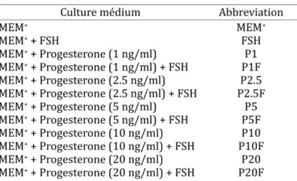
Table 1. Different media tested for the in vitro
culture of caprine preantral follicles
Culture médium Abbreviation
MEM+ MEM+
MEM+ + FSH FSH
MEM+ + Progesterone (1 ng/ml) P1 MEM+ + Progesterone (1 ng/ml) + FSH P1F MEM+ + Progesterone (2.5 ng/ml) P2.5 MEM+ + Progesterone (2.5 ng/ml) + FSH P2.5F MEM+ + Progesterone (5 ng/ml) P5 MEM+ + Progesterone (5 ng/ml) + FSH P5F MEM+ + Progesterone (10 ng/ml) P10 MEM+ + Progesterone (10 ng/ml) + FSH P10F MEM+ + Progesterone (20 ng/ml) P20 MEM+ + Progesterone (20 ng/ml) + FSH P20F
Fig.1. Histological section (400x) of preantral follicles cultured for 7 days with FSH plus progesterone (2.5ng/ml) (A) and MEM+ (B)
The percentages of histologically normal preantral folli-cles in control (fresh tissue) and after 1 or 7 days of cul-ture are shown in Table 2. After 1 day, it was observed a signiicant reduction (P<0.05) in the percentage of normal follicles after culture with 2.5 or 10ng/ml of progesterone alone, compared with fresh control. In the same period, the association of 2.5 ng/ml of progesterone + FSH was beneic to the culture, since it showed greater (P<0.05) percentage of normal follicles in relation to the same concentration of progesterone alone. At day 7 of culture, addition of FSH to all progesterone concentrations kept the percentage of his-tologically normal preantral follicles similar to fresh con-trol (P>0.05). It was not observed any signiicant difference in the percentage of normal follicles between other treat-ments and MEM+ at day 1 or 7 (P>0.05)
or 5 and 10ng /ml of progesterone alone, compared with fresh control (Fig.3). After 7 days, in all treatments, there was a signiicant increase (P<0.05) in follicular diameter in all treatments compared with fresh control, except in MEM+ and MEM+ when 5ng/ml of progesterone + FSH or 10ng/ ml of progesterone alone. When FSH was added to the me-dium containing progesterone (2.5, 10 and 20ng/ml), folli-cular diameter signiicantly increased (P<0.05) when com-pared with these same concentrations alone, after 1 day, while its addition decreased follicular diameter in 5 ng/ml of progesterone plus FSH (P<0.05). At day 7 of culture, the positive effect of FSH addition was veriied at the concen-tration of 10 ng/ml of progesterone (P<0.05). With the pro-gression of the culture period from 1 to 7 days, there was a signiicant increase (P<0.05) in follicular diameter in FSH,
Activation of caprine primordial follicles after in vitro
culture
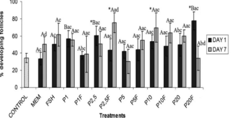
At day 1 of culture, 2.5ng/ml of progesterone alone or 20ng/ml of progesterone associated with FSH signiican-tly increased (P<0.05) the percentage of growing follicles when compared with fresh control (Fig.2). After 7 days, a signiicant increase (P<0.05) of growing follicles was ob-served with 2.5ng/ml of progesterone + FSH or 10ng/ml of progesterone alone, compared with fresh control. At day 1, the media containing 1 and 2.5ng/ml of progesterone alone and 20 ng/ml of progesterone + FSH showed signiicantly higher percentage of follicular activation than MEM+ alone (P<0.05). It is important to note that from days 1 to 7, the-re was a signiicant the-reduction (20ng/ml of progesterone + FSH) and, in some treatments, an increase (MEM+ alone and 2.5ng/ml of progesterone + FSH) in the percentage of growing follicles (P<0.05).
In vitro growth of caprine preantral follicles
After 1 day of culture, there was a signiicant increase (P<0.05) in follicular diameter in MEM+ or MEM+ supple-mented with 2.5, 10 and 20ng/ml of progesterone plus FSH,
Table 2. Percentages of morphologically normal caprine preantral follicles in control (non-cultured tissue) and
after 1 or 7 days of culture in medium containing progesterone and/or FSH
Control 85.02 + 4.30%
Day 1 (%) Day 7 (%) MEM 68.37 + 12.32c 59.17 + 6.88*c
FSH 69.20 + 11.98c 57.52 + 16.61*ac P1 70.87 + 12.87ac 56.70 + 14.40* ac P1F 69.20 + 9.17ac 68.37 + 10.38ac P2.5 49.17 + 13.44*ac 58.35 + 8.38*ac P2.5F 71.70 + 5.77bc 64.20 + 9.95ac P5 70.85 + 8.34ac 60.85 + 10.31*ac P5F 70.85 + 14.24ac 64.20 + 7.41ac P10 58.37 + 15.49*ac 58.35 + 6.95*ac P10F 70.85 + 15.50ac 63.37 + 6.65ac P20 69.20 + 9.57ac 56.70 + 16.77*ac P20F 79.20 + 4.19ac 62.52 + 15.22ac * Differs signiicantly from control follicles (P<0.05).
a,b Differs signiicantly from addition of FSH at the same concentra-tions and days of culture (P<0.05).
c,d Differs signiicantly between days of culture in the same treat-ment (P<0.05).
Fig.2. Percentages of developing preantral follicles in control (non-cultured tissue) and after 1 or 7 days of culture in me-dium containing progesterone and/or FSH.
Fig.3. Mean follicular diameter (µm) in the control (non-cultured
tissue) and after 1 or 7 days of culture in medium containing progesterone and/or FSH.
Fig.4. Mean oocyte diameter (µm) in the control (non-cultured
progesterone alone (1, 2.5 and 20ng/ml) and progesterone (1 and 20ng/ml) plus FSH, and a decrease in this diameter for culture performed with MEM+ or MEM+ supplemented with 5ng/ml of progesterone (P<0.05).
At day 1 of culture, MEM+ or MEM+ supplemented with 5ng/ml of progesterone promoted an increase (P<0.05) in oocyte diameter when compared to fresh control (Fig.4). However, after 7 days, only 20ng/ml of progesterone asso-ciated with FSH showed an oocyte diameter signiicantly higher than control or MEM+. Moreover, addition of FSH to 2.5ng/ml of progesterone was beneic to preantral follicles, since it was the only treatment that promoted an increase in oocyte diameter after 1 day of culture (P<0.05). After 7 days, the same effect was observed with 10 or 20ng/ml of progesterone. With the progression of the culture period from 1 to 7 days, it was observed a signiicant reduction (P<0.05) in the oocyte diameter with MEM+ or MEM+ sup-plemented with 5 or 10ng/ml of progesterone alone or in 10ng/ml of progesterone + FSH. In contrast, in such period, it was observed a signiicant increase in oocyte diameter in MEM+ supplemented with FSH, 2.5ng/ml of progesterone alone and progesterone (1 and 20ng/ml) + FSH.
Ultrastructural analysis of cultured caprine preantral follicles
For a better evaluation of follicular integrity, the ultras-tructural analysis was performed in tissues from fresh con-trol as well as in those cultured for 7 days with 2.5 ng/ml of progesterone, which showed satisfactory results at his-tological evaluation. Both treatments showed ultrastructu-rally normal follicles (Fig.5), with intact oocyte and nuclear membranes, nucleus with descondensed chromatin, some
vesicles and organelles uniformly distributed in the cyto-plasm. The granulosa cells were normal, with elongated nucleus and a high proportion nucleus-cytoplasm.
DISCUSSION
The present study showed the inluence of progesterone on the survival, activation and growth of caprine prean-tral follicles cultured in vitro. It is important to empha-size that the concentrations of progesterone used in this study were similar to the physiological levels reported in caprine specie (Menchaca & Rubianes 2002, Khanum et al. 2007).
After 7 days of culture, it was observed that addition of FSH to all concentrations of progesterone kept the per-centage of preantral follicles similar to fresh control, whi-ch demonstrate a positive interaction between these two hormones. Although there were few studies regarding the inluence of progesterone associated with FSH on prean-tral follicles culture, it is known that this steroid can pre-vent granulosa cells apoptosis in human (Makrigiannakis et al. 2000) and mouse (Shao et al. 2003). Furthermore, some studies demonstrated the importance of FSH for the maintenance of preantral follicle viability (Cortvrin-dt et al. 1997, Matos et al. 2007a), inhibition of apoptosis of caprine granulosa cells from antral follicles, as well as increasing progesterone secretion by these cells (Yu et al. 2003).
In this study, after 7 days of culture, it was veriied a gre-ater percentage of growing preantral follicles with 2.5ng/ ml of progesterone associated with FSH or 10ng/ml of pro-gesterone alone, compared to fresh tissue. Propro-gesterone is a steroid hormone synthesized by the ovary, and its
tion is dependent on gonadotrophin stimulation and phy-siological state of the ovary (Peluso et al. 2006). LH stimu-lates the conversion of cholesterol to progesterone in theca cells. Thus, progesterone will be converted to testosterone (still in theca cells), and the latter converted to estradiol in the granulosa cells under FSH inluence. Estradiol acts in-creasing progesterone production, until it reaches high se-ric levels and then decreases FSH action on granulosa cells and consequently estradiol production (Yarak et al. 2005). In this manner, we can suppose that the action of these two hormones is related and could have inluenced the results obtained in this work. Some studies demonstrated that progesterone acts in follicular growth, ovulation and lutei-nization (Peluso et al., 2006), besides promotes in vitro ma-turation of monkeys (Zheng et al. 2003) and bitches (Kim et al. 2005, Vannucchi et al. 2006) oocytes. Furthermore, FSH can promote in vitro follicular activation in ovine (Andrade et al. 2005) and bubaline (Santos et al. 2006), acting indi-rectly in the ovary, regulating growth factors that promote primordial follicle development (Thomas et al. 2005).
After 7 days, in all treatments tested, follicular diameter was larger than fresh control, except when 5ng/ml of pro-gesterone + FSH or 10ng/ml of propro-gesterone alone were used. In the same period, the concentration of 20 ng/ml of progesterone in association with FSH showed an oocyte diameter larger than control and MEM+ alone. The effect of progesterone in the ovary are exerted by their recep-tors (PRA and PRB) present in theca and granulosa cells of preantral and antral follicles in mouse (Gava et al. 2004), swine (Slomczynska et al. 2000) and bovine (D´Haeseleer et al. 2007) and these receptors increase progressively in conformity with follicular development. However, it was demonstrated that in mouse without progesterone recep-tors (PRA and PRB), follicular development can normally occurs (Lydon et al. 1996), suggesting that other receptors can be involved in progesterone action. Regarding FSH, their receptors are expressed in granulosa cells from pri-mary follicles stage onward (Xu et al. 1995, O´Shaughnessy et al. 1996), suggesting that this hormone have an impor-tant role in follicular growth.
In this study, TEM was performed in fresh control and in the treatment with 2.5 ng/ml of progesterone associated with FSH. Follicles were ultrastructurally normal with their featu-res similar to those observed previously in caprine preantral follicles cultured in vitro for up to 7 days (Matos et al. 2007a,b).
In conclusion, this study demonstrated that the asso-ciation of 2.5ng/ml of progesterone with 50ng/ml of FSH could promote caprine primordial follicles activation, as well as further in vitro follicular growth. In addition, the results showed that the interaction between these hormo-nes maintain ultrastructural integrity of caprine preantral follicles cultured in vitro for up to 7 days. This culture sys-tem should be useful for studying the regulation of early follicular growth and development, especially because the-se follicles reprethe-sent a large source of oocytes that could be used in vitro for embryo production.
Acknowledgements.- This work was supported by CNPq (Renorbio).
Isa-bel B. Lima-Verde is a recipient of a grant from Funcap/CAPES
(Fortale-za, Ceará, Brazil). We would like to acknowledge the generous donation of pFSH by Dr Jean-François Beckers of the University of Liége, Belgium, and the special contribution of Sarah Bezerra Honório and Jamily Bezerra Bruno.
REFERENCES
Andrade E.R, Seneda M.M., Alieri A.A., Oliveira J.A., Bracarense A.P.F.R.L., Figueiredo J.R. & Toniolli R. 2005. Interactions of indole acetic acid with EGF and FSH in the culture of ovine preantral follicles. Theriogenology 64:1104-1113.
Cecconi S., Barboni B., Coccia M. & Mattioli M. 1999. In vitro development of sheep preantral follicles. Biol. Reprod. 60:594-601.
Cortvrindt R., Smitz J. & Van Steirteghem A.C.1997. Assessment of the need for follicle stimulating hormone in early preantral mouse follicle culture in vitro. Hum. Reprod. 12:759-768.
D´Haeseleer M., Simoens P. & Van den Broeck W. 2007. Cell-speciic locali-zation of progesterone receptors in the bovine ovary at different stages of the oestrous cycle. Anim. Reprod. Sci. 98:271-281.
Gava N., Clarke C.L., Byth K., Arnett-Mansield R.L. & Fazio A. 2004. Expres-sion of progesterone receptors A and B in the mouse ovary during the estrous cycle. Endocrinology 145:3487-3494.
Gougeon A. 1996. Regulation of ovarian follicular development in prima-tes: Facts and hypothesis. Endocr. Rev. 17:121-154.
Gutierrez C.G., Ralph J.H., Telfer E.E., Wilmut I. & Webb R. 2000. Growth and antrum formation of bovine preantral follicles in long-term culture
in vitro. Biol. Reprod. 62:1322-1328.
Hirao Y., Nagai T., Kubo M., Miyano T., Miyake M. & Kato S. 1994. In vitro growth and maturation of pig oocytes. J. Reprod. Fertil.100:333-339. Khanum S.A., Hussain M. & Kausar R. 2007. Assessment of reproductive
parameters in female Dwarf goat (Capra hircus) on the basis of proges-terone proiles. Anim. Reprod. Sci. 102:267-275.
Kim M.K., Fibrianto Y.H., Oh H.J., Jang G., Kim H.J., Lee K.S., Kang S.K. & Hwang W.S. 2005. Effects of estradiol-17β and progesterone supple-mentation on in vitro nuclear maturation of canine oocytes. Therioge-nology 63:1342-1353.
Lydon J.P., DeMayo F.J., Conneely O.M. & O’Malley B.W. 1996. Reproductive phenotypes of the progesterone receptor null mutant mouse. J. Steroid. Biochem. Mol. Biol. 56:67-77.
Makrigiannakis A., Coukos G., Christoidou-Solomidou M., Montas S. & Coutifaris C. 2000. Progesterone is an autocrine/paracrine regulator of human granulosa cell survival in vitro. Ann. N.Y. Acad. Sci. 900:16-25. Mao J., Wu G., Smith M.F., McCauley T.C., Cantley T.C., Prather R.S., Didion
B.A. & Day B.N. 2002. Effects of culture medium, serum type and vari-ous concentrations of follicle-stimulating hormone on porcine prean-tral follicular development and antrum formation in vitro. Biol. Reprod. 67:1197-1203.
Matos M.H.T., Lima-Verde I.B., Luque M.C.A., Maia Jr J.E., Silva J.R.V., Celesti-no J.J.H., Martins F.S., Báo S.N., Lucci C.M. & Figueiredo JR. 2007a. Essen-tial role of follicle stimulating hormone in the maintenance of caprine preantral follicle viability in vitro. Zygote 15:173-182.
Matos M.H.T., Van den Hurk R., Lima-Verde I.B., Luque M.C.A., Santos K.D.B., Martins F.S., Báo S.N., Lucci C.M. & Figueiredo J.R. 2007b. Effects of ibroblast growth factor-2 on the in vitro culture of caprine preantral follicles. Cells Tissues Organs 186:112-120.
McGee E., Spears N., Minami S., Hsu S.Y., Chun S.Y., Billig H. & Hsueh A.J.W. 1997. Preantral ovarian follicles in serum-free culture: suppression of apoptosis after activation of the cyclic guanosine 3-5-monophosphate pathway and stimulation of growth and differentiation by follicle-stimu-lating hormone. Endocrinology 138:2417-2424.
Menchaca A. & Rubianes E. 2002. Relation between progesterone concen-trations during the early luteal phase and follicular dynamics in goats. Theriogenology 57:1411-1419.
O’Shaughnessy P.J., Dudley K. & Rajapaksha W.R. 1996. Expression of folli-cle stimulating hormone-receptor mRNA during gonadal development. Mol. Cell. Endocrinol. 125:169-175.
Peluso J.J. 2006. Multiplicity of progesterone´s actions and receptors in the mammalian ovary. Biol. Reprod. 75:2-8.
Santos S.S.D., Biondi F.C., Cordeiro M.S., Miranda M.S., Dantas J.K., Figuei-redo J.R. & Ohashi O.M. 2006. Isolation, follicular density, and culture of preantral follicles of buffalo fetuses of different ages. Anim. Reprod. Sci. 95:1-15.
Shao R., Markstrom E., Friberg P.A., Johansson M. & Billig H. 2003. Expres-sion of progesterone receptor (PR) A and B isoforms in mouse granulo-sa cells: stage-dependent PR-mediated regulation of apoptosis and cell proliferation. Biol. Reprod. 68:914-921.
Silva J.R.V., Van den Hurk R., Costa S.H.F., Andrade E.R., Nunes A.P.A., Fer-reira F.V.A., Lôbo R.N.B. & Figueiredo J.R. 2004. Survival and growth of goat primordial follicles after in vitro culture of ovarian cortical slices in media containing coconut water. Anim. Reprod. Sci. 81:273-286. Slomczynska M., Krok M. & Pierscinski A. 2000. Localization of the
proges-terone receptor in the porcine ovary. Acta Histochemica 102:183-191. Spears N., Murray A.A., Alisson V., Boland N.I. & Gosden R.G. 1998. Role of
gonadotro.ns and ovarian steroids in the development of mouse follicles in vitro. J. Reprod. Fertil. 113:19-26.
Thomas F.H., Ethier J.F., Shimasaki S. & Vanderhyden B.C. 2005. Follicle--Stimulating Hormone regulates oocyte growth by modulation of ex-pression of oocyte and granulosa cell factors. Endocrinology 146:941-949.
Ulloa-Aguirre A., Timossi C., Barrios-de-Tomasi J., Maldonado A. & Nayudu P. 2003. Impact of carbohydrate heterogeneity in function of FSH: Stud-ies derived from in vitro and in vivo models. Biol. Reprod. 69:379-389. Vannucchi C.I., Oliveira C.M., Marques M.G., Assumpção M.E.O.A. & Visitin
J.A. 2006. In vitro canine oocyte nuclear maturation in homologous ovi-ductal cell co-culture with hormone-supplemented media. Theriogenol-ogy 66:1677-1681.
Wright C.S., Hovatta O., Margara R., Trew G., Winston R.M.L., Franks S. & Hardy K.1999. Effects of follicle-stimulating hormone and serum subs-titution on the in-vitro growth of human ovarian follicles. Hum. Re-prod.14:1555-1562.
Xu Z., Garverick H.A., Smith G.W., Smith M.F., Hamilton S.A. & Youngquist R.S. 1995. Expression of follicle-stimulating hormone and luteinizing hormone receptor messenger ribonucleic acids in bovine follicles dur-ing the irst follicular wave. Biol. Reprod. 53:951-957.
Yarak S., Parada M.O.A.B., Bagatin E., Talarico-Filho S., Hassun K.M. 2005. Hiperandrogenismo e pele: síndrome do ovário policístico e resistência periférica à insulina. Anais Bras. Dermatol. 80:395-410.
Yu Y., Li W., Han Z., Luo M., Chang Z. & Tan J. 2003. The effect of follicle-sti-mulating hormone on follicular development, granulosa cell apoptosis and steroidogenesis an its mediation by insulin-like growth factor-I in the goat ovary. Theriogenology 60:1691-1704.