Valorisation of wild mushrooms as functional foods:
chemoinformatic studies
Hugo Jorge Calisto Froufe
Dissertação apresentada à Escola Superior Agrária de Bragança para obtenção do Grau de Mestre em Qualidade e Segurança
Alimentar
Orientado por
Isabel Cristina F.R. Ferreira Rui Miguel V. Abreu
AKNOWLEDGEMENTS
To all the people that were fundamental in providing me with help, direction and support throughout this journey and in particularly to:
Doctor Isabel Cristina F.R. Ferreira and Master Rui Miguel V. Abreu, my supervisors, thank you for an invaluable and rewarding experience, your support and encouragement has been a source of motivation.
ABSTRACT
Intermolecular interactions play essential roles in several life processes and understanding these interactions is critical for pharmaceutical and functional foods industries. Mushrooms represent an unlimited source of compounds with antitumor and immunostimulating properties and mushroom intake has been shown to reduce the risk of breast cancer. In this work, two in silico studies were performed in an attempt to
elucidate potential mechanisms of mushroom bioactivity. First, a QCAR (Quantitative Composition-Activity Relationships) modelling approach was used to study and predict mushroom antioxidant activity. Next, molecular docking and virtual ligand screening (VLS) studies were performed in an attempt to elucidate possible mechanisms of mushroom anti-breast cancer activity.
For the initial QCAR study a PLS (Partial Least Square) statistical technique was applied to evaluate the relationship between antioxidant potential (scavenging effect on free radicals and reducing power) and chemical composition of twenty three samples from seventeen Portuguese wild mushroom species. A wide range of analytical parameters including ash, carbohydrates, proteins, fat, monounsaturated fatty acids, polyunsaturated fatty acids, saturated fatty acids, phenolics, flavonoids, ascorbic acid
and β-carotene was studied and the data was analyzed by the PLS regression analysis to find correlations between all the parameters. Antioxidant activity correlated well with phenolic and flavonoid contents. A QCAR model was constructed, and its robustness and predictability was verified by internal and external cross-validation methods. This model proved to be a useful tool in the prediction of mushrooms reducing power.
For the VLS study, molecular docking software AutoDock 4 was used in order to evaluate which wild mushroom low molecular weight (LMW) compounds, including antioxidants, could be involved in anti-breast cancer activity. A representative dataset of 43 LMW compounds (individual phenolic acids, flavonoids, tocopherols, carotenoids, sugars and fatty acids) was selected and molecular docking was carried out against three known protein targets involved in breast cancer (Aromatase, Estrone Sulfatase and
proteins. 4-O-caffeoylquinic acid, naringin and lycopene stand out as the top ranked potential inhibitors for Aromatase, Estrone Sulfatase and 17β-HSD-1, respectively.
RESUMO
As interacções intermoleculares desempenham um papel essencial nos diversos processos biológicos, sendo fundamental a compreensão destas interacções nos Sectores das Indústrias Farmacêuticas e de Alimentos Funcionais. Os cogumelos representam uma fonte ilimitada de compostos com propriedades antitumorais e imunoestimulantes, e o seu consumo foi já relacionado com a redução do risco de cancro da mama. No presente trabalho, foram desenvolvidos dois estudos in silico com o intuito de melhor
compreender quais os mecanismos moleculares responsáveis por diferentes propriedades bioactivas dos cogumelos. Primeiro utilizou-se uma metodologia de modelação QCAR (Relações Quantitativas Composição – Actividade) para estudar e prever a actividade antioxidante de cogumelos. Num segundo estudo utilizaram-se
ferramentas de “docking” molecular e “virtual ligand screening” (VLS) para tentar elucidar possíveis mecanismos de actividade dos cogumelos contra o cancro da mama.
No estudo QCAR inicial foi utilizada a técnica estatística dos Mínimos Quadrados Parciais (PLS) para avaliar a relação entre o potencial antioxidante (efeitos bloqueadores de radicais livres e poder redutor) e a composição química de vinte e três amostras de dezassete espécies de cogumelos silvestres Portugueses. Estudaram-se vários parâmetros analíticos tais como cinzas, hidratos de carbono, proteínas, gorduras, ácidos gordos monoinsaturados, ácidos gordos polinsaturados, ácidos gordos saturados, fenóis, flavonóides, ácido ascórbico e β-caroteno, e os seus resultados foram analisados por PLS de forma a estabelecer correlações entre todos os parâmetros. A actividade antioxidante mostrou estar correlacionada com o teor em fenóis e flavonóides. Foi construído um modelo QCAR, cuja robustez e capacidade de previsão foram verificadas por métodos de validação cruzada internos e externos. Finalmente, este modelo provou ser uma ferramenta útil na previsão do poder redutor de cogumelos.
desidrogenase 1). Os compostos LMW foram classificados quanto à sua capacidade de inibição do cancro da mama. A informação obtida estabelece um bom ponto de partida para o desenvolvimento de inibidores das proteínas mencionadas. O ácido 4-o
-cafeoilquínico, a naringina e o licopeno revelaram-se, respectivamente, os melhores inibidores para Aromatase, Esterona Sulfatase e 17β-HSD1.
Os estudos de Química Computacional realizados permitiram a valorização dos cogumelos como alimentos funcionais, podendo ser muito úteis para Indústrias que visem o desenvolvimento de novos nutracêuticos ou alimentos funcionais.
TABLE OF CONTENTS
AKNOWLEDGEMENTS ABSTRACT
RESUMO
TABLE OF CONTENTS LIST OF TABLES LIST OF FIGURES ABBREVIATIONS
I. INTRODUCTION
1. Functional Foods
2. Mushrooms as Functional Foods 2.1 Nutritional Value
2.2 Antioxidant Activity
2.2.1 Oxidative stress
2.2.2 Contribution of mushrooms against oxidative tress
2.3 Anticancer Activity
2.3.1 Breast cancer
2.3.2 Contribution of mushrooms against cancer
3. Chemoinformatics methodologies
3.1 QCAR - Quantitative Composition-Activity Relationships 3.2 Molecular Docking and Virtual Ligand Screening
II RESULTS
1. A QCAR model for predicting antioxidant activity of wild mushrooms
1.1 Introduction 1.2. Methods
1.2.1 Data set
1.2.2 Statistical Analysis
1.2.3 QCAR model
1.3 Results and Discussion
1.3.1 Relationships between antioxidant activity and chemical
1.3.2 QCAR model
2. Insides on wild mushrooms anti-breast cancer activity by Virtual ligand screening of low molecular weight compounds
2.1 Introduction 2.2. Methods
2.2.1 Ligand dataset
2.2.2 Protein structure preparation
2.2.3 Virtual ligand screening using Autodock 4
2.3. Results and Discussion
2.3.1 Docking validation
2.3.2 Mushrooms LMW virtual ligand screening
2.3.3 Structure analysis of the best docked conformations
III CONCLUSIONS
IV. REFERENCES
27 32
32 34 34 35 36 36 36 38 43
46
LIST OF TABLES
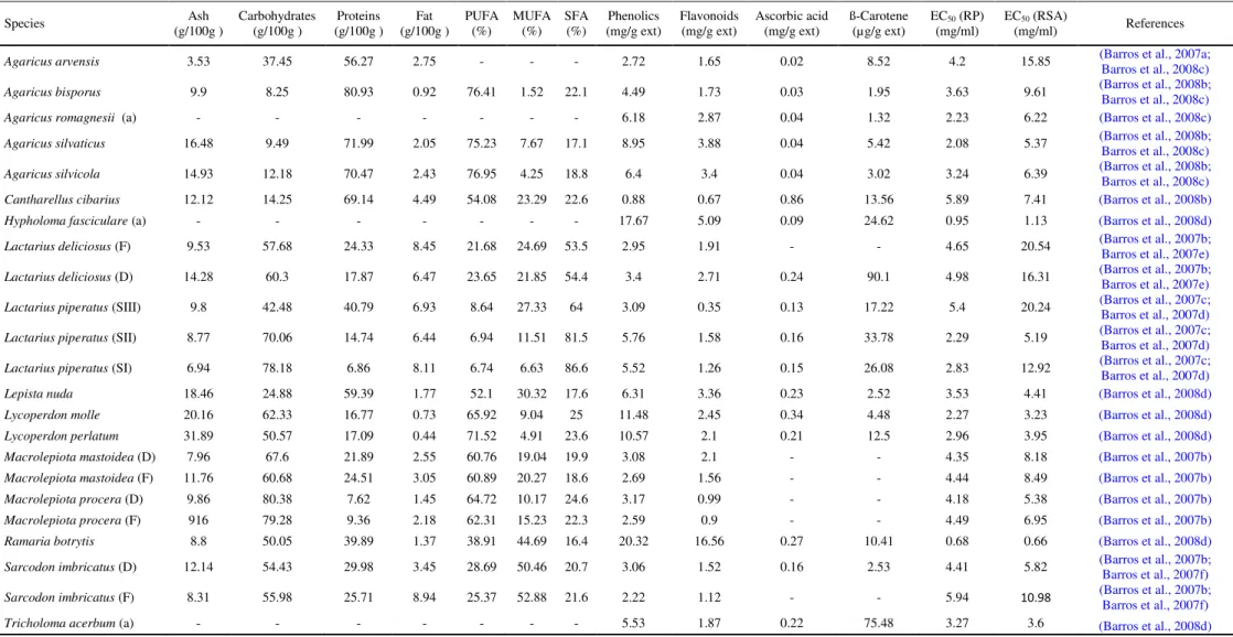
Table 1.
Chemical composition and antioxidant activity (reducing power, RP and radical scavenging activity, RSA) values of Portuguese wild mushrooms.
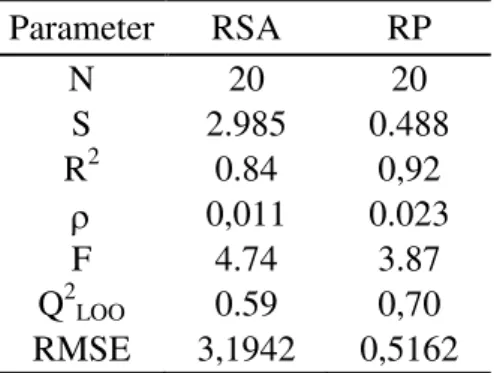
Table 2.
Statistical parameters of the models radical scavenging activity (RSA) and reducing power (RP) using PLS method.
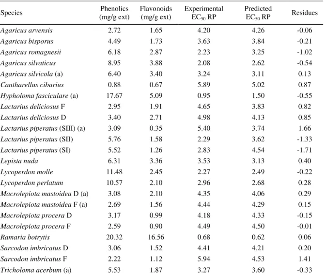
Table 3.
Phenolics, flavonoids, experimental and predicted EC50 reducing power
values of Portuguese wild mushrooms.
Table 4.
Comparison of estimated and experimental values of Km (ƞM) and ΔG
(Kcal/mol) values.
Table 5.
Docking studies with phenolic acids found in mushrooms as ligands.
Table 6.
Docking studies with flavonoids found in mushrooms as ligands.
Table 7.
Docking studies with vitamins and carotenoids found in mushrooms as ligands.
Table 8.
Docking studies with sugars and fatty acids found in mushrooms as ligands.
21
27
29
35
39
41
42
LIST OF FIGURES
Figure 1.
Major causes for over production of free radicals (oxidative stress), possible cellular targets and conditions associated to oxidative stress.
Figure 2.
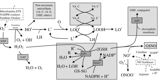
Overview of the main reactions involving reactive Oxygen species (ROS) / reactive Nitrogen species (RNS), and major endogenous enzymatic and non-enzymatic antioxidant defences in the cell.
Figure 3.
Estrogens pathway in breast cell.
Figure 4.
Estrone sulfatase X-ray protein (PDB: 1P49) represented in cartoon format; docked natural ligands in wire format.
Figure 5.
17β-HSD1 X-ray protein (PDB: 1FDT) represented in cartoon format; docked natural ligands in wire format.
Figure 6.
Aromatase (PDB: 3EQM) represented in cartoon format; docked natural ligands in wire format.
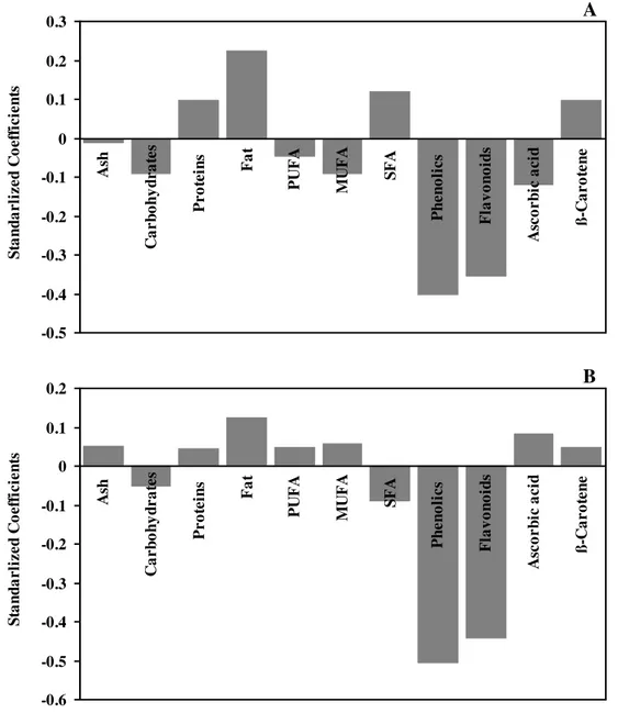
Figure 7.
Standardized coefficients of the chemicals compounds, Ash, Carbohydrates, Proteins, Fat, Polyunsaturated fatty acids, Monounsaturated fatty acids, Saturated fatty acids, Phenolics, Flavonoids, Ascorbic acid and β-Carotene, used in the in the approached model for RSA (A) and RP (B) .
Figure 8.
Distribution of reducing power (EC50 RP) versus number of samples for the
training set (black) and test set (grey) of the QCAR model.
Figure 9.
Results of the permutation test. The R2 and Q2 values were obtained from 100
permutations for the four developed PLS models.
Figure 10.
Predicted versus experimental EC50 RP for the training (●) and test sets (○).
Figure 11.
Residual versus experimental log EC50 RP, for the training (●) and test sets
(○).
5
6
10
11
12
13
25
29
30
31
Figure 12.
General view of estrogen formation and transformation of estrogens in human breast cancer: aromatase and sulfatase pathways.
Figure 13.
Superimposition of X-ray (sticks and balls) and docked configurations (wire) for: (A) Androstenedione in Aromatase and (B) Estrone in 17β-HSD1.
Figure 14.
Colour coded representation of the best results obtained by virtual ligand screening of the LMW dataset against the 3 protein targets.
Figure 15.
Docking of the top ranked inhibitor for each of the studied protein targets. Figure shows (A) Aromatase, (B) Estrone Sulfatase and (C) 17β-HSD1 docked with 4-O-caffeoylquinic, naringin and lycopene, respectively.
33
37
40
ABREVIATIONS
17β-HSD-1 17-β-hydroxysteroid dehydrogenase 1.
ΔG Gibbs free energy.
ρ Significance.
Ǻ Ångströms. A Androstenedione. CAT Catalase.
CYP19A1 Aromatase gene. DNA Deoxyribonucleic acid.
DPPH 1,1-Diphenyl-2-picrylhydrazyl. E1 Estrone.
E2 Estradiol.
E1S Estrone sulfate. E2S Estradiol sulfate.
ETS Electron transport system. F Fisher ratio value.
FuFoSE European Commission‟s Concerted Action on Functional Food Science in Europe.
GC-FID Gas Chromatography-Flame Ionization Detector. GPx Glutathione peroxidases.
Gred Glutathione redutase. GSH Glutathione.
GSNO S-nitrosoglutathione. GST Glutathione-S-transferases. GS-SG Glutathione disulphide
HPLC High Performance Liquid Chromatography
HPLC/DAD-ESI/MS HPLC/Diode Array Detector- Electron Spray Ionization/Mass Spectrometry.
HPLC-RI HPLC-Refraction Index Detector. HTS High-Throughput Screening.
ILSI International Life Science Institute Ki Inhibition constant.
LMW Low Molecular Weight. M Molar
MUFA Monounsaturated Fatty Acids.
NADP+ Nicotidamide adenine dinucleotide phosphate: oxidized. NADPH Nicotidamide adenine dinucleotide phosphate reduced. NOS Nitric oxide synthase.
PDB Protein Data Bank. PLS Partial Least Square.
PRESS Predictive Error Sum of Squares PSK Polysaccharide Krestin.
PUFA Polyunsaturated Fatty Acids.
Q2Chi-square test. Q2LOO Q2 Leave One Out
QCAR Quantitative Composition-Activity Relationships QSAR Quantitative Structure-Activity Relationships
R2 Squared correlation coefficient. Rcal Gas constant for calories/K/mol. RMSE Root Mean Squared Errors. RNS Reactive Nitrogen Species. ROS Reactive Oxygen Species. RP Reducing Power.
RSA Radical Scavenging Activity. S Standard deviation of regression. SFA Saturated Fatty Acids.
SOD Superoxide dismutase. STS Estrone sulfatase. T Testosterone.
I. INTRODUCTION
1. Functional foods
In the last decades consumer demands in the field of food production has changed considerably. Consumers more and more believe that foods contribute directly to their health. Today foods are not intended to only satisfy hunger and to provide necessary nutrients for humans but also to prevent nutrition-related diseases and improve physical and mental well-being of the consumers. In this regard, functional foods play an outstanding role. The increasing demand on such foods can be explained by the rapid advances in science and technology, increasing cost of healthcare, changes in food laws affecting label and product claims, the steady increase in life expectancy, the desire of older people for improved quality of their later years, and rising interest in attaining wellness through diet (Siro et al., 2008).
According to the Institute of Medicine‟s Food and Nutrition Board “Functional Foods” are foods or dietary components that may provide a health benefit beyond basic
nutrition. We can take greater control of our health through the food choices we make, knowing that some food can provide specific health benefits (Hasler, 1998). The
European Commission‟s Concerted Action on Functional Food Science in Europe
(FuFoSE), coordinated by International Life Science Institute (ILSI) Europe defined
functional food as follows: „„a food product can only be considered functional if
together with the basic nutritional impact it has beneficial effects on one or more functions of the human organism thus either improving the general and physical conditions or/and decreasing the risk of the evolution of diseases. The amount of intake and form of the functional food should be as it is normally expected for dietary purposes. Therefore, it could not be in the form of pill or capsule just as normal food
form‟‟ (Siro et al., 2008).
Functional food could not exist without nutraceutical compounds, the bioactive compounds that give functional properties to food. A nutraceutical can be defined as a substance that may be considered a food or part of a food and provides medical or health benefits like the prevention and treatment of disease. Nutraceuticals may range
foods, herbal products and processed products such as cereals, soups and beverages (Andlauer and Furst, 2002).
The term „„functional food‟‟ itself was first used in Japan, in the 80s, for food
products fortified with special constituents that possess advantageous physiological effects (Hasler, 1998; Siro et al., 2008). Functional foods may improve the general conditions of the body (e.g. pre- and probiotics), decrease the risk of some diseases (e.g. cholesterol-lowering products), and could even be used for curing some illnesses. It was recognized that there is a demand for these products as different demographical studies revealed that the medical service of the aging population is rather expensive. The European market for functional foods was estimated to be between 4 and 8 billion US$ in 2003 depending which foods are regarded as functional. This value has increased to around 15 billion US$ by 2006. The current market share of functional food is still below 1% of the total food and drink market. Germany, France, the United Kingdom and the Netherlands represent the most important countries within the functional food market in Europe (Siro et al., 2008).
The design and development of functional foods is a scientific challenge that should rely on a stepwise process. The process begins with basic scientific knowledge relevant to functions that are sensitive to modulation by food components, which are pivotal to maintenance of well-being and health, and that, when altered, may be linked to a change in the risk of a disease. The exploitation of this knowledge in the development of markers that can be shown to be relevant to the key functions is the second step. Next is a new generation of hypothesis-driven human intervention studies that will include the use of these validated, relevant markers and allow the establishment of effective and safe intakes. Last is the development of advanced techniques for human studies that, preferably, are minimally invasive and applicable on a large scale. The targets for functional food science may include: Gastrointestinal functions; Redox and antioxidant systems; Metabolism of the macronutrients; Development in fetal and early life; Xenobiotic metabolism and its modulation by non-nutritive dietary components; Mood and behaviour or cognition and physical performance (Roberfroid, 2000).
2. Mushrooms as Functional Foods
Mushrooms are something special in the living world, being neither plant nor animal. They have been placed in a kingdom, called Myceteaea. The word mushroom may mean different things to different people and countries. In a broad definition
“mushrooms are macrofungus with a distinctive fruiting body, which can be either epigeous or hypogeous and large enough to be seen with naked eye and to be picked by
hand”. Thus, mushrooms can be Ascomycetes that can grow underground and have a
non-fleshy texture and need not be edible (Miles and Chang, 1997).
Edible mushrooms have been widely used as human food for centuries and have been appreciated for texture and flavours as well as some medicinal and tonic attributes. However, the awareness of mushrooms as a healthy food and as an important source of biological active substances with medicinal value has only recently emerged. Various activities of mushrooms have been studied which includes antibacterial, antifungal, antioxidant, antiviral, antitumor, cytostatic, immunosuppressive, antiallergic, antiatherogenic hypoglycaemic, anti-inflammatory and hepatoprotective activities (Lindequist et al., 2005). In the present work we will focus on antioxidant and antitumor activities.
2.1 Nutritional Value
Mushrooms are considered a healthy food because they are low in calories and fat but rich in protein and dietary fibers (Cheung et al., 2003; Manzi et al., 1999).
aspartic acid, proline and serine. They are important in providing structure to cells, tissues and organs and therefore essential for growth and repair (Diez and Alvarez, 2001; Kalac, 2009; Manzi et al., 1999).
Mushrooms are recognized as an excellent choice for low energy diets, as they have high water and low fat content (average of 2–6% of dry weight). Fat in mushrooms contains all classes of lipid compounds including free fatty acids, mono-, di-, and triglycerides, sterols, sterol esters and phospholipids.. Within fatty acid composition, polyunsaturated linoleic acid (C18:2n-6), monounsaturated oleic acid (C18:1n-9) and nutritionally undesirable saturated palmitic acid (C16:0) prevail. The proportions of
nutritionally neutral saturated stearic acid (C18:0), and especially of desirable α -linolenic acid (C18:3n-3), are low. Other fatty acids are present at only low levels (Heleno et al., 2009a). Contents of odd- and branched-chain acids and hydroxy fatty acids are negligible. The occurrence of trans fatty acids in mushrooms has not been
reported and it is not expected. Phosphatidylcholine was the major phospholipid present in 55 of 58 wild growing mushroom species of several families. The nutritional value of wild growing mushroom lipids is thus limited, due to low total lipid content and a low proportion of desirable n-3 fatty acids (Kalac, 2009).
Cultivated mushrooms are a good source of several vitamins, such as riboflavin, niacin, and folates, with concentrations that vary within the range of 1.8-5.1, 31-65, and 0.30-0.64 mg/100g dry weight, respectively, depending on the species. The vitamin B2 content in mushrooms is higher than than generally founds in vegetables, and in some varieties even at a level found in egg and cheese. Mushrooms contain moderately high amounts of folates at concentrations that are of the same magnitude as is generally found in vegetables. In addition to riboflavin, niacin and folates, cultivated mushrooms also contain small amounts of vitamin C and vitamin B1 and traces of vitamins B12 and D2 (Clifford et al., 1991; Mattila et al., 2001).
The carbohydrate content of edible mushrooms varies with species and ranges from 3 to 65% dry weight. Glucose, mannitol and trehalose are the main representatives of monosaccharides, their derivatives and oligosaccharide groups, respectively. Usual contents of glucose and trehalose are low; the content of mannitol, which participates in volume growth and firmness of fruiting bodies, differs widely. Reducing sugars are only a small part of carbohydrates content since wild edible mushrooms are rich in non-starch polysaccharides (dietary fiber, 3–32% dry weight), such as glycogen (animal and
mushrooms are believed to contain a high levels of oligosaccharides and only a low levels of total soluble sugars (Bano and Rajarathnam, 1988; Kalac, 2009).
2.2 Antioxidant activity
2.2.1 Oxidative stress
Free radicals are produced in the normal natural metabolism of aerobic cells,
mostly in the form of reactive oxygen species (ROS). Once produced, most of the free
radicals are neutralized by cellular antioxidant defences (enzymes and non-enzymatic
molecules). Beneficial effects of ROS occur at low or moderate concentrations and
involve cellular physiological roles of signalization and regulation. Nevertheless, the
equilibrium between ROS production and antioxidant defences might be displaced
either by the overproduction of ROS or by the loss of the cell antioxidant defences. This
disequilibrium is known as oxidative stress (Ferreira et al., 2009).
Oxidative stress might have natural causes such as extreme exercise or
inflammation processes, or non-natural causes such as the presence of xenobiotics in the
organism or situations related to several diseases (Figure 1).
Figure 1. Major causes for over production of free radicals (oxidative stress), possible cellular targets and
conditions associated to oxidative stress. Source: Ferreira et al., 2009.
Cancer
Cardiovascular diseases
Neurological disorders
Pulmonary diseases
Diabetes
Arthritis
Aging process Consequences
Membrane lipids
Proteins
Nucleic Acids
Carbohydrates Targets
Free radicals Causes
Environmental
Drugs Metallic ions
Radiation Extreme exercise
In fact, the non-controlled production of free radicals has been related to more
than one hundred diseases including several kinds of cancer, diabetes, cirrhoses,
cardiovascular diseases, neurological disorders, among others. The overproduction of
ROS has also been related to the aging process.
Several ROS production pathways and the main endogenous antioxidant
defences of the cell are described in Figure 2.
Figure 2. Overview of the main reactions involving reactive Oxygen species (ROS) / reactive Nitrogen species
(RNS), and major endogenous enzymatic and non-enzymatic antioxidant defences in the cell. The most representative endogenous sources (traced rectangles) of ROS/RNS are presented and include: Mitochondrial ETS (Electron transport system), NADPH oxidases, Xanthine oxidase for ROS and NO synthases for RNS. The main antioxidant defences are presented in shaded rectangles and the enzymes involved are presented in italic. Molecular Oxygen (O2), superoxide anion (O2•−), hydrogen peroxide (H2O2), hydroxyl radical (HO•), hydroxide ion (HO-)
membrane lipids (LH), lipid radical (L•), peroxyl radical (LOO•), hydroperoxide lipid (LOOH), lipid alkoxyl radical (LO•), nitric oxide (NO•), radicals (R•), non-radicals (R), alcohols (LOH), glutathione (GSH), glutathione disulphide
(GS-SG), α-tocopherol or vitamin E (vit. E), vitamin E radical (vit. E•), vitamin C (vit. C), vitamin C radical (vit.
C•), S-nitrosoglutathione (GSNO), nicotidamide adenine dinucleotide phosphate: oxidized (NADP+), reduced
(NADPH). Enzymes: Superoxide dismutase (SOD), catalase (CAT), glutathione peroxidase (GPx), glutathione redutase (Gred), glutathione-S-transferases (GST), Mitochondrial ETS (electron transport system), nitric oxide synthase (NOS). Adapted from Ferreira et al., 2009.
Transition metals
Non-enzymatic antioxidants (vit. C, vit. E, GSH, others)
O O2•− HO•
L•
LOO• LOOH LO•
O2•
−
H2O + O2
CAT
H2O+ LOH GS-SG 2GSH GSH- conjugated electrophilic xenobiotic GSNO GST O2 LH L• GPx Gred
H2O Mitochondria ETS
NADPH oxidases Xanthine Oxidase
Arginine LH
SOD O2 + OH-
Vit. C •
Vit. C
Vit. E Vit. E •
NADPH + H+
NADP+ NO•
ONOO− H2O2
NADP+
NADPH + H+
Citrulline
NO Synthases R•
Superoxide anion (O2•− - “primary” ROS) is mostly produced in mitochondria,
due to a small but continuous “leak” of the electrons in the mitochondrial electron
transport system (ETS). Superoxide anion can also be produced by different
endogenous enzymatic systems present in the cell like NADPH oxidases and xanthine
oxidase. Even though O2•− is not a very active radical, it can interact with other
molecules generating what are considered as “secondary” ROS, such as hydrogen
peroxide (H2O2) and hydroxyl radical (OH•). Hydroxyl radical has a very short life time
but is considered to be the most toxic among all ROS, being responsible for the attack
to DNA molecules, damaging purins and pyrimidines and the structure of desoxyribose
DNA. Mitochondria are the most important source of ROS, but they are also the first
targets of these radicals because ROS have an easy access to the membrane lipids,
which are susceptible to free radicals attack. This attack is called lipid peroxidation and
promotes the production of different types of ROS (Figure 2). The lipid peroxidation
usually begins with the extraction of a hydrogen atom from a polyunsaturated lipid
(LH) chain, through the action of reactive species such as HO•. This generates a highly
reactive lipid radical (L•) that can react with O2 to form a peroxyl radical (LOO•). If not
neutralized by antioxidants defences, the peroxyl radical will react with other adjacent
lipids producing hydroperoxides lipids (LOOH) that can easily be decomposed to form
new L• radicals, initiating a process that is known as chain propagation reactions. This
process when not stopped, can lead to much superior damage than the ROS that started
the reaction. It is also important to notice the existence of radicals with nitrogen called
Reactive Nitrogen species (RNS). The principal RNS is nitric oxide (NO•) and it is
generated in biological tissues by specific nitric oxide synthases (NOS), which
metabolise arginine to citrulline (Figure 2) (Ferreira et al., 2009).
Exposure to free radicals from a variety of sources has led organisms to the development of a series of defence mechanisms (Figure 2). These defences were the evolution response to the inevitability of the existence of oxygen radicals in aerobic life conditions, and can be classified into enzymatic and non-enzymatic. There are many different endogenous enzymatic antioxidant defences in the organism, either in intracellular or extracellular medium. Examples of these defences include superoxide dismutase (SOD), catalase (CAT), glutathione peroxidases (GPx), and glutathione redutase (Gred) among others. The endogenous non-enzymatic antioxidant defences
The implication of oxidative and nitrosative stress in the etiology and progression of several acute and chronic clinical disorders has led to the suggestion that antioxidants can have health benefits as prophylactic agents. This suggests that changes in dietary behaviour, increasing consumption of plant-based foods, which contain significant amounts of bioactive phytochemicals, may provide desirable health benefits, beyond basic nutrition, to reduce the risk of chronic diseases.
2.2.2. Contribution of mushrooms against oxidative stress
Natural products with antioxidant activity may help the endogenous defence system. In this perspective, the antioxidants present in the diet assume a major importance as possible protector agents reducing oxidative damage.
Many studies have concluded that edible mushrooms possess potent antioxidants. Research conducted in Japan showed the antioxidant activity of the crude ethanol extract of 150 Japanese mushrooms using the peroxide value in the methyl linoleate system (Cheung, 2009). It showed that many mushrooms, especially those belonging to the Suillus genus, had a peroxide value 80% lower than the control. A
study of methanol extracts from black (Auricularia mensenterica), red (Auricularia polytricha) and snow (Auricularia fuscosuccinea) ear mushrooms found that they had
an inhibitory effect on lipid peroxidation, 1,1-diphenyl-2-picrylhydrazyl (DPPH) radical scavenging, hydroxyl radical scavenging, strong reducing power and ability to chelate ferrous ions. Similar studies of other mushrooms including, Dictyophora indusiata, Grifola frondosa, Hericium erinaceus , Tricholoma giganteum, Lentinula edodes, Pleurotus cystidiosus, and Pleurotus ostreatus, showed that these mushrooms also
possess the afore mentioned antioxidant properties. It is therefore likely that most mushrooms possess hydroxyl and DPPH radical scavenging effects, inhibit lipid peroxidation, chelate metals, and has a strong reducing effect (Mau et al., 2001; Mau et al., 2002; Yang et al., 2002). Similar antioxidant properties have also been reported for other edible mushrooms, including Agrocybe cylindracea and Hypsizigus marmoreus,
both of which belong to the Tricholomataceae family (Lee et al., 2007; Tsai et al., 2006).
Furthermore, several other mushrooms from Portugal (Lactarius deliciosus, Lactarius piperatus, Macrolepiota mastoidea, Macrolepiota procera, Sarcodon
imbricatus, Agaricus arvensis, Agaricus bisporus, Agaricus silvicola, Agaricus
Cantharellus cibarius, Hypholoma fasciculare, Lepista nuda, Lycoperdon molle,
Lycoperdon perlatum, Ramaria botrytis and Tricholoma acerbum), Korea (Grifola frondosa and Lentinus edodes), China (Lentinus edodes, Volvariella volvacea and Agrocybe aegerita), Taiwan (Grifola frondosa, Morchella esculenta, Termitomyces albuminosus, Dictophora indusiata, Grifola frondosa, Hericium erinaceus, Trichloma
giganteum, Ganoderma lucidum, Ganoderma tsugae, Coriolus versicolor, Armillariella
mellea, from India Termitomyces heimii, Helvella crispa, Termitomyces tylerance,
Lactarius sanguifluus, Morchella conica, Termitomyces mummiformis, Pleurotus
sajor-caju, Termitomyces shimperi, Lentinus squarrulosus, Boletus edulis, Pleurotus djamor,
Macrolepiota procera, Cantharellus clavatus, Morchella angusticeps, Termitomyces
microcarpus, Lactarius deliciosus, Geastrum arinarius, Hydnum repandum, Lentius
sajor-caju, Sparassis crispa, Russula brevepis, Auricularia polytricha and Cantharellus cibarius), Turkey (Agaricus bisporus, Polyporus squamosus, Pleurotus ostreatus, Lepista nuda, Russula delica, Boletus badius, Verpa conica and Lactarius deterrimus)
and Brazil (Lentinula edodes and Agaricus blazei) were also reported to have
antioxidant activity, which was mainly related to their phenolic content (Barros et al., 2007b; Barros et al., 2007d; Barros et al., 2008c; Barros et al., 2007f; Barros et al., 2008d; Cheung and Cheung, 2005; Cheung et al., 2003; Choi et al., 2006; Elmastas et al., 2007; Ferreira et al., 2007; Kitzberger et al., 2007; Lee et al., 2008; Lo and Cheung, 2005; Mau et al., 2004; Mau et al., 2002; Ng et al., 2007; Puttaraju et al., 2006; Sarikurkcu et al., 2008; Soares et al., 2009; Song et al., 2003; Tsai et al., 2007; Turkoglu et al., 2007; Yang et al., 2002).
The antioxidants found in mushrooms are mainly phenolic compounds (phenolic acids and flavonoids), followed by tocopherols, ascorbic acid and carotenoids. These molecules were quantified in tens of different species mainly from Finland, India, Korea, Poland, Portugal, Taiwan and Turkey. The values are available in literature, but expressed in different basis (dry weight, fresh weight and extract) (Ferreira et al., 2009).
2.3 Anticancer activity
2.3.1. Breast Cancer
Breast cancer is the most common type of cancer among women worldwide and its rate is increasing in both developed and developing countries. The burden is not evenly distributed and there are large variations in the incidence rates of breast cancer between different countries (Parkin et al., 2005).
Most breast cancers (about 95%), whether in pre- or postmenopausal women, are initially hormone-dependent (Pasqualini and Chetrite, 2005). The majority of breast cancers occur during the postmenopausal period when the ovaries have ceased to be functional. Despite the low levels of circulating estrogens, the tissular concentrations of estrone (E1), estradiol (E2) and their sulfates (E1S, E2S) in breast tumours are several times higher than those found in the plasma or in the area of the breast considered as normal tissue, suggesting a specific tumoral biosynthesis and accumulation of these hormones (Pasqualini, 2004).
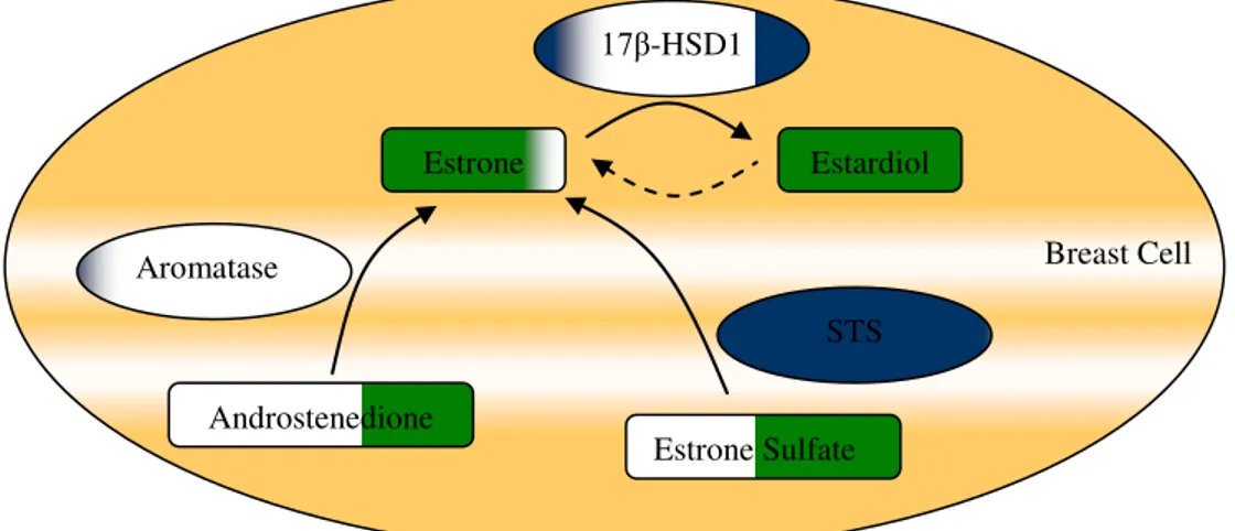
Figure 3 shows the biosynthesis of estrogens from steroid precursors via the aromatization of androstenedione to estrone by aromatase or via the hydrolysis of estrone sulfate by Estrone sulfatase (STS).
Figure 3. Estrogens pathway in breast cell. Estrone sulfatase (STS), 17-β-hydroxysteroid dehydrogenase
type 1 (17β-HSD1) and Aromatase.
Androstenedione
Estrone Estardiol
Estrone Sulfate Aromatase
17β-HSD1
STS
There are three enzymes that are directly involved in the production of estrone and estradiol in the breast cell, that are Estrone sulfatase (STS), 17-β-hydroxysteroid
dehydrogenase type 1 (17β-HSD1) and Aromatase. These proteins were used as targets in the present work.
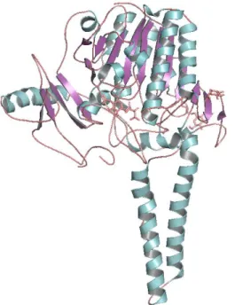
Estrone sulfatase is a microsomal enzyme and is an integral membrane protein of the Endoplasmic Reticulum (Figure 4). It is most active at or near neutral pH and can be solubilised only in the presence of detergents (Ghosh, 2007).
Figure 4. Estrone sulfatase X-ray protein structure (PDB: 1P49) represented in cartoon format; docked
natural ligands are represented in wire format (pink).
STS is an alternative source of sex-steroid precursors for the local biosynthesis of active estrogens and androgens. STS catalyzes the hydrolysis of E1S to unconjugated E1, which is subsequently reduced to estradiol (E2) by 17-β-hydroxysteroid
There are fifteen discovered 17β-HSD enzymes in mammals (Jansson, 2009). The common feature for this enzymes is the possibility to catalyze oxidation or reduce the carbon at position 17 in steroids. The enzymes have different preferences for substrates such as estrone, estradiol, testosterone, androstenedione, and dihydrotestosterone, are expressed in different parts of the cell, and in diverse tissues. This shows that the enzymes have separate physiological functions (Jansson, 2009).
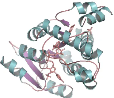
17β-HSD1 (Figure 5) known to be of main importance in breast tissue (Pasqualini, 2004).
Figure 5. 17β-HSD1 X-ray protein structure (PDB: 1FDT) represented in cartoon format; docked natural
ligands represented in wire format (pink).
Aromatase (Figure 6), an enzyme of the cytochrome P450 (CYP450) subfamily and the product of the CYP19A1 gene, is highly expressed in the placenta and in the granulose cells of ovarian follicles in premenopausal women. In menopause, androstenedione produced in the adrenals and, to a small extent, testosterone produced in the ovaries are released to the circulation and then sequestered to nonglandular tissues (e.g., liver and breast cells), where they are converted to estrone and estradiol, respectively, by aromatase located in these tissues (Desta et al., 2009).
Figure 6. Aromatase (PDB: 3EQM) represented in cartoon format; docked natural ligands in wire format
(pink).
Drugs that effectively inhibit the aromatase-mediated synthesis of estrogens in peripheral tissues including the breast, thus depriving the system of estrogens, are widely used in the treatment of breast cancer. These drugs include the nonsteroidal triazole derivatives anastrozole and letrozole and the steroidal exemestane (Desta et al., 2009). Aromatase inhibitors are used widely as second-line therapy in breast cancer; and there is now evidence for a chemopreventive role for these agents (Zaidman et al., 2005).
The present work explores mushrooms as sources of potential inhibitors of the
2.3.2 Contribution of mushrooms against cancer
The mushroom Cordyceps militaris has been use for a long time in eastern Asia
as a nutraceutical and in traditional Chinese medicine as a treatment for cancer patients.
Cordyceps militaris proteins exerted strong antifungal effect against the growth of the
fungus Fusarium oxysporum, and exhibited cytotoxicity against human breast and
bladder cancer cells. New discoveries in molecular oncology along with rapid expansion of our knowledge concerning the processes that govern differentiation, apoptosis, immune surveillance, angiogenesis, metastasis, cell cycle, and signal transduction control have unveiled an abundance of specific molecular targets for cancer therapy, including a variety of small-molecule compounds that inhibit or stimulate these molecular targets (Park et al., 2009).
In a recent study, it was found that Ganoderma lucidum, Phellinus rimosus, Pleurotus florida and Pleurotus pulmonaris possessed profound antioxidant and
antitumor activities. This indicated that these mushrooms would be valuable sources of antitumor and antioxidant compounds. Extracts of fruiting bodies of Boletus edulis and
other Basidiomycetes also revealed antitumor activity against Sarcoma 180 line in mice. In the 1960s, calvacin was the most commonly cited natural product isolated from the medicinal mushroom Calvatia gigantean and was broadly used in many laboratories as
an antitumor agent (Ajith and Janardhanan, 2007; Lucas et al., 1957).
In Eastern Europe, the fruiting bodies of Ionotus obliquus have been used as a
folk medicine for cancer and stomach diseases since the 16th or 17th century. antitumor effects of several extracts and isolated compounds from mushrooms could be demonstrated in tumour cell systems and in animal assays (Burczyk et al., 1996; Molitoris, 1994).
Several phytochemicals have been isolated from medicinal mushrooms and three of these, which are carcinostatic polysaccharide drugs, have been developed from
mushrooms in Japan. These are “Krestin” (PSK), from the cultured mycelium of
Kawaratake (Trametes versicolor), “Lentinan” from the fruiting bodies of Shiitake
(Lentinus edodes) and “Schizophyllan” (Sonifilan) from the culture fluid of Suehirotake
(Schizophyllum commune)(Mizuno, 1993). Lentinan and schizophyllan are pure β
-glucans, whereas PSK is a protein bound polysaccharide (Larone, 2002). The biological activity of these three products is related to their immunomodulating properties, which
immunopotentiators, or immunoinitiators, are also referred as “biological response
modifiers” (Zaidman et al., 2005; Zjawiony, 2004).
A recent clinical study suggests that higher dietary intakes of fresh and dried mushrooms are associated with a reduced breast cancer risk with a dose–response relationship in both pre- and postmenopausal Chinese women. The combination of dietary intake of mushrooms and green tea drinking decreased breast cancer risk with an additional reduced effect on the malignance (Zhang et al., 2009)
3. Chemoinformatics methodologies
The use of bioinformatic tools is widespread in all areas of basic and applied scientific knowledge. These tools include chemoinformatics methods that are used in the design, creation, organization, management, retrieval, analysis, dissemination, visualization and use of chemical information. In this work, we selected the appropriate chemoinformatic tool for each study performed: QCAR for mushroom antioxidant activity prediction and molecular docking for potential mushroom anti-breast cancer activity against selected protein targets.
3.1 QCAR - Quantitative Composition-Activity Relationships
Because most sets of biological observations, particularly those produced by testing different chemical structures in the same biological system, cannot be adequately described by existing theory, researchers often seek semi empirical models in which the changes in observed values are predicted as a mathematical function of properties which are better understood (Cramer, 1993).
In modern pharmaceutical industry, computer-aided drug design methods, such as quantitative structure–activity relationship (QSAR) study has greatly accelerated the pace of drug discovery in recent decade. The underlying assumption behind QSAR analysis is that the variation of biologic activity within a group of compounds can be correlated with the variation of their respective structural and chemical features (Wang et al., 2006).
To develop the QCAR model, the PLS (Partial Least Squares) statistical method was used. PLS is an important technique for producing a linear equation to describe or predict differences in the values of one or more properties from differences in the values of other properties (Cramer, 1993). In composition–bioactivity studies, such a linear equation is usually called a QCAR model. The described or predicted properties (in this case, antioxidant activity) are called the 'dependent variables' or, in the PLS literature, the 'Y-block'. The describing or predicting properties (chemical composition) are called the 'independent variables' or the 'X-block'. To implement PLS technique and build a QCAR model we used SIMCA-P software a user-friendly software program (Fernandez et al., 2005).
By quantitatively analyzing the chemical composition–bioactivity relationship, a QCAR mathematical model was established that is able to successfully predict antioxidant activity of mushrooms. The QCAR approach is very recent and, when used to study complex biological matrixes (like mushrooms), is a very promising methodology, better suitable for biological activity predictions than the more widely used QSAR methods.
3.2 Molecular Docking and Virtual Ligand Screening
Structure based drug design is now an established approach in drug discovery. Computational methodologies are used to facilitate structure based drug design at various stages of the process. One of the most important and routinely adopted methods is molecular docking (or just docking) and refers to the prediction of the binding mode of a specified compound within the active site of the protein target of interest (Congreve et al., 2005; Verdonk et al., 2007).
Selecting (or designing) compounds in silico that bind to a protein active site is
difficult. First, the in silico method must solve the docking problem by finding the
conformation of the protein. Currently, state-of-the-art docking programs correctly dock ~70–80% of ligands when tested on large sets of protein–ligand complexes (Congreve et al., 2005).
The second challenge is that the in silico method must score the compound so
that its relative affinity can be judged versus other compounds.
The use of molecular docking to search large databases of compounds for possible ligands of a protein receptor is usually termed virtual ligand screening (VLS) and has been successfully applied in several therapeutic programs at the lead discovery stage (Ghosh et al., 2006). Rapid accumulation of high-resolution three-dimensional structures, further accelerated by the structural proteomics initiative and the improvements of docking and scoring technology, are making VLS an attractive alternative to the traditional methods of lead discovery. VLS can sample a virtually infinite chemical diversity of drug-like molecules without synthesizing and experimentally testing every screened molecule. Typically, a corporate high-throughput screening (HTS)-ready compound library ranges from 200,000 to 1,000,000 molecules. The high cost of such massive experimental testing and its technical complexity are further motivation for the theoretical alternative. Finally, the virtual experiment, as opposed to a high-throughput assay, can be easily designed to select for a particular binding site or receptor specificity (Abagyan and Totrov, 2001).
II RESULTS
1. A QCAR model for predicting antioxidant activity of wild
mushrooms
1.1 Introduction
Free radicals play important roles in many physiological and pathological conditions (Valko et al., 2007). In general, excess of free radicals caused by the imbalance between free radical generation and scavenging may contribute to disease development. Free radicals can damage membranes, proteins, enzymes and DNA,
increasing the risk of diseases such as cancer, Alzheimer‟s, Parkinson‟s,
angiocardiopathy, arthritis, asthma, diabetes, and degenerative eye disease (Machlin and Bendich, 1987; Valko et al., 2007).
Mushrooms have become attractive as functional foods and as a source of physiologically beneficial compounds including antioxidants (Lindequist et al., 2005; Wasser, 1999). Different wild mushroom species were reported to have antioxidant activity, which was mainly related to their phenolic content (Cheung et al., 2003; Elmastas et al., 2007; Kim et al., 2008; Lee et al., 2008; Mau et al., 2004; Soares et al., 2009; Tsai et al., 2007). Nevertheless, none of the available reports present a quantitative study to obtain a predict model for antioxidant potential. Furthermore, other chemical substances in mushrooms including proteins, carbohydrates, vitamins and fibers could also contribute to the antioxidant capacity (Maisuthisakul et al., 2008).
coupled to a diode array detector and mass spectrometry (HPLC/DAD-ESI/MS), carotenoids and ascorbic acid, by spectrophotometric techniques. The antioxidant activity was screened through chemical and biochemical assays (Barros et al., 2007b; Barros et al., 2007d; Barros et al., 2007f; Barros et al., 2008c; Barros et al., 2008d). Numerous tests have been used for measuring the antioxidant capacity of food and biological samples. However, there is no universal method that can measure the antioxidant capacity of all samples accurately and quantitatively (Prior et al., 2005).
DPPH radical scavenging activity (RSA) and reducing power (RP) assays are two of the most widely used methods for antioxidant activity screening.
The relationship between chemical substances including phenolic compounds and antioxidant properties may be complex, and there is very little data to elucidate the relationship between chemical composition and antioxidant capacity of wild mushrooms. Herein, the antioxidant potential (RSA and RP) and chemical composition of some Portuguese wild mushrooms were evaluated using linear regression analysis (Partial Least Square, PLS), in order to find possible relationships between those parameters. Furthermore, a Quantitative Composition-Activity Relationships (QCAR) model was constructed in order to predict the reducing power of mushrooms.
1.2. Methods
1.2.1. Data set
A total of twenty three samples from seventeen Portuguese wild mushroom species were used in this study (Table 1). The samples were selected using the following criteria: wild mushrooms studied by our research group using the same methodologies; results of chemical composition and antioxidant activity available on the same sample. In some species, results considering different stages of fruiting body growth (SI, SII and SIII) and different conservation conditions (frozen and dehydrated) were available, and therefore used in this study.
Table 1. Chemical composition and antioxidant activity (reducing power, RP and radical scavenging activity, RSA) values of Portuguese wild mushrooms. Species (g/100g ) Ash Carbohydrates (g/100g ) (g/100g ) Proteins (g/100g ) Fat PUFA (%) MUFA (%) SFA (%) (mg/g ext) Phenolics Flavonoids (mg/g ext) Ascorbic acid (mg/g ext) ß-Carotene (µg/g ext) EC50 (RP)
(mg/ml) EC(mg/ml) 50 (RSA) References
Agaricus arvensis 3.53 37.45 56.27 2.75 - - - 2.72 1.65 0.02 8.52 4.2 15.85 (Barros et al., 2007a; Barros et al., 2008c)
Agaricus bisporus 9.9 8.25 80.93 0.92 76.41 1.52 22.1 4.49 1.73 0.03 1.95 3.63 9.61 (Barros et al., 2008b; Barros et al., 2008c)
Agaricus romagnesii (a) - - - 6.18 2.87 0.04 1.32 2.23 6.22 (Barros et al., 2008c)
Agaricus silvaticus 16.48 9.49 71.99 2.05 75.23 7.67 17.1 8.95 3.88 0.04 5.42 2.08 5.37 (Barros et al., 2008b; Barros et al., 2008c)
Agaricus silvicola 14.93 12.18 70.47 2.43 76.95 4.25 18.8 6.4 3.4 0.04 3.02 3.24 6.39 (Barros et al., 2008b; Barros et al., 2008c)
Cantharellus cibarius 12.12 14.25 69.14 4.49 54.08 23.29 22.6 0.88 0.67 0.86 13.56 5.89 7.41 (Barros et al., 2008b)
Hypholoma fasciculare (a) - - - 17.67 5.09 0.09 24.62 0.95 1.13 (Barros et al., 2008d)
Lactarius deliciosus (F) 9.53 57.68 24.33 8.45 21.68 24.69 53.5 2.95 1.91 - - 4.65 20.54 (Barros et al., 2007b; Barros et al., 2007e)
Lactarius deliciosus (D) 14.28 60.3 17.87 6.47 23.65 21.85 54.4 3.4 2.71 0.24 90.1 4.98 16.31 (Barros et al., 2007b; Barros et al., 2007e)
Lactarius piperatus (SIII) 9.8 42.48 40.79 6.93 8.64 27.33 64 3.09 0.35 0.13 17.22 5.4 20.24 (Barros et al., 2007c; Barros et al., 2007d)
Lactarius piperatus (SII) 8.77 70.06 14.74 6.44 6.94 11.51 81.5 5.76 1.58 0.16 33.78 2.29 5.19 (Barros et al., 2007c; Barros et al., 2007d)
Lactarius piperatus (SI) 6.94 78.18 6.86 8.11 6.74 6.63 86.6 5.52 1.26 0.15 26.08 2.83 12.92 (Barros et al., 2007c; Barros et al., 2007d)
Lepista nuda 18.46 24.88 59.39 1.77 52.1 30.32 17.6 6.31 3.36 0.23 2.52 3.53 4.41 (Barros et al., 2008d)
Lycoperdon molle 20.16 62.33 16.77 0.73 65.92 9.04 25 11.48 2.45 0.34 4.48 2.27 3.23 (Barros et al., 2008d)
Lycoperdon perlatum 31.89 50.57 17.09 0.44 71.52 4.91 23.6 10.57 2.1 0.21 12.5 2.96 3.95 (Barros et al., 2008d)
Macrolepiota mastoidea (D) 7.96 67.6 21.89 2.55 60.76 19.04 19.9 3.08 2.1 - - 4.35 8.18 (Barros et al., 2007b)
Macrolepiota mastoidea (F) 11.76 60.68 24.51 3.05 60.89 20.27 18.6 2.69 1.56 - - 4.44 8.49 (Barros et al., 2007b)
Macrolepiota procera (D) 9.86 80.38 7.62 1.45 64.72 10.17 24.6 3.17 0.99 - - 4.18 5.38 (Barros et al., 2007b)
Macrolepiota procera (F) 916 79.28 9.36 2.18 62.31 15.23 22.3 2.59 0.9 - - 4.49 6.95 (Barros et al., 2007b)
Ramaria botrytis 8.8 50.05 39.89 1.37 38.91 44.69 16.4 20.32 16.56 0.27 10.41 0.68 0.66 (Barros et al., 2008d)
Sarcodon imbricatus (D) 12.14 54.43 29.98 3.45 28.69 50.46 20.7 3.06 1.52 0.16 2.53 4.41 5.82 (Barros et al., 2007b; Barros et al., 2007f)
Sarcodon imbricatus (F) 8.31 55.98 25.71 8.94 25.37 52.88 21.6 2.22 1.12 - - 5.94 10.98 (Barros et al., 2007b;
Barros et al., 2007f)
Tricholoma acerbum (a) - - - 5.53 1.87 0.22 75.48 3.27 3.6 (Barros et al., 2008d)
The samples were collected in Bragança (Northeast of Portugal), in autumns of 2005 and 2006. Different chemical parameters (ash, carbohydrates, proteins, fat, monounsaturated fatty acids (MUFA), polyunsaturated fatty acids (PUFA), saturated fatty acids (SFA),
phenolics, flavonoids, ascorbic acid and β-carotene), radical scavenging activity (RSA) and reducing power (RP) values were obtained from previous reports of our research group (Barros et al., 2007a; Barros et al., 2007b; Barros et al., 2007c; Barros et al., 2007d; Barros et al., 2007e; Barros et al., 2007f; Barros et al., 2008b; Barros et al., 2008c; Barros et al., 2008d).
Ash was determined by incineration at 600±15 ºC; the proteins content (N 4.38) was estimated by the macroKjeldahl method; fat was determined by extracting a known weight of powdered mushroom sample with petroleum ether, using a Soxhlet apparatus; carbohydrates were calculated by difference; saturated fatty acids (SFA), monounsaturated fatty acids (MUFA) and polyunsaturated fatty acids (PUFA) were determined by GC-FID after a
trans-esterification procedure. Phenolics, flavonoids, ascorbic acid and β-carotene were determined by spectrophotometer assays. These phytochemicals are frequently analyzed in mushrooms and reported in literature.
For antioxidant activity data, we used the results of two in vitro chemical assays,
To make the RSA and RP data homogenous and directly comparable, all the values were reported as EC50, (expressed in mg/mL, concentration required to achieve 50% of RSA or 0.5 of absorbance in RP).
1.2.2. Statistical Analysis
The relationships between antioxidant activity (RP and RSA) and the different chemical composition parameters were studied using PLS (Wold et al., 2001) method implemented in SIMCA-P v12 statistics software (Fernandez et al., 2005), and using NIPALS algorithm for missing data (A.B. Umetrics, 2008). Because Agaricus romagnesii, Hypholoma fasciculare and Tricholoma acerbum had more than 50% of missing data, these observations
were not used in the models.
The goodness of fit of the model was evaluated using the following statistical parameters: squared correlation coefficient (R2), standard deviation of regression (S),
significance of the model (ρ) and Fisher ratio value (F).
The predictive stability and robustness of the model was first verified by internal cross-validation calculating the following parameters: Q2LOO (“Leave-One-Out”; 1 -PRESS/TSS were PRESS is the Predictive Error Sum of Squares and TSS the Total Sum of Squares) and RMSE (training set) (Root Mean Squared Errors for the training set) (Gramatica, 2007; Gramatica and Papa, 2005).
1.2.3. QCAR model
To build the QCAR model the complete data set was used (table 1) and the Partial Least Square (PLS) method implemented in SIMCA-P v12 statistics software was used. The twenty three samples were first divided in two groups: training and test sets. The training set, representing about 3/4 of the total number of samples (17 samples), was used to build the QCAR model. The remaining 1/4 (6 samples) was assigned to the test set and used to validate the model. The division was made to cover all the antioxidant activity scale (Farkas et al., 2004; Saiz-Urra et al., 2007) and the samples included on the training set were randomly selected within each group (Durand et al., 2007).
The goodness of fit of the models was evaluated using the following statistical parameters: squared correlation coefficient (R2), standard deviation of regression (S),
The predictive stability and robustness of the model was first verified by internal cross-validation calculating the following parameters: Q2LOO (“Leave-One-Out”; 1 -PRESS/TSS were PRESS is the Predictive Error Sum of Squares and TSS the Total Sum of Squares), permutation test of SIMCa-p software and RMSE (training set) (Root Mean Squared Errors for the training set) (Eriksson et al., 1997; Gramatica, 2007; Gramatica and Papa, 2005). Using the test set, the model was further checked by external cross-validation by calculating parameters: Q2ext (External, 1-PRESS/SD) and RMSE (test set) (Root Mean Squared Errors for the test set). PRESS is defined as the sum of the squared difference between the observed value and the predicted value for each compound in the test set, and SD is defined as the sum of the squared deviation between the observed value and the mean measured value of the training test (Gramatica, 2007).
1.3. Results and Discussion
1.3.1. Relationships between antioxidant activity and chemical composition
Figure 7. Standardized coefficients of the chemicals compounds, Ash, Carbohydrates, Proteins, Fat,
Polyunsaturated fatty acids (PUFA), Monounsaturated fatty acids (MUFA), Saturated fatty acids (SFA), Phenolics, Flavonoids, Ascorbic acid and β-Carotene, used in the in the approached model for RSA (A) and RP (B) .
This analysis presented good statistical parameters for both RP and RSA, as summarized in Table 2. Information about some chemical compounds was not available (Table 1). To overcome this lack of information, we used a specific statistic algorithm available on SIMCA-P (A.B. Umetrics, 2008). This algorithm dynamically selects samples with more than 50% of the chemical compound results. From the data set available four mushroom species did not meet this criterion and were not considered in this initial analysis.
Table 2. Statistical parameters of the models radical scavenging activity (RSA) and reducing power (RP) using
PLS method.
N- number of samples, S- standard deviation, R2- squared correlation coefficient, ρ- significance, F- Fisher ratio,
Q2
LOO- “Leave-One-Out” correlation coefficient and RMSE(training set)- Root Mean Squared Errors for the training
set.
A similar relationship between chemical composition and antioxidant activity was observed for RSA (Figure 7A) and RP (Figure 7B), with the exception of fatty acids and ascorbic acid. A close observation of the standarlized coefficients of the analysed chemical parameters shows that antioxidant activity is strongly positively related to phenolics and flavonoids contents (Figure 7). This is in agreement with several manuscripts reporting phenolic compounds as the main antioxidant substances in mushrooms, particularly phenolic acids and flavonoids (Ferreira et al., 2009). Phenolic substances (ArOH) serve as oxidation terminators by scavenging radicals to form resonance stabilized radicals (Rice-Evans et al., 1997), according to:
Ascorbic acid is a well-known powerful antioxidant, reported in mushrooms in lower amounts than phenolics (Ferreira et al., 2009). Accordingly, it seems to contribute positively to the RSA but with less significance. Otherwise, it gives a negative contribution to the reducing power of the samples. In fact, ascorbic acid mechanism of action has been related to free radicals scavenging effects and not to reducing processes with electrons transference (which is present in RP assay- reducing Fe3+ to Fe2+); ascorbic acid protect biomembranes against lipid peroxidation damage by eliminating peroxyl radicals in the aqueous phase before the latter can initiate lipid peroxidation (Davey et al., 2000). Carbohydrates gave a small positive contribution possible due to the presence of mannitol (reducing sugar), a very
Parameter RSA RP
N 20 20
S 2.985 0.488
R2 0.84 0,92
0,011 0.023
F 4.74 3.87
Q2
LOO 0.59 0,70
RMSE 3,1942 0,5162
abundant sugar in mushrooms which functions to provide support and expansion of the fruit body (Barros et al., 2007a; Barros et al., 2007b; Barros et al., 2007c; Barros et al., 2008b; Barros et al., 2008d). Unsaturated fatty acids (MUFA and PUFA) show also a small positive contribution to RSA but a negative contribution to RP; the presence of double bonds makes them susceptible to oxidation; they can react with free radicals and become radicals themselves. Therefore, they act mostly as free radical scavengers. Particularly, oleic (C18:1) and linoleic acid (C18:2), abundant in mushrooms (Barros et al., 2007a; Barros et al., 2007b; Barros et al., 2007c; Barros et al., 2008b; Barros et al., 2008d) proved to have more than 80% of RSA (Henry et al., 2002). Nevertheless, it should be noticed that fatty acids exist in mushrooms in a very low concentration.
Surprisingly, β-carotene gave a small negative contribution to the antioxidant activity. This can be explained by the fact that this compound is present is mushrooms only in vestigial amounts (Ferreira et al., 2009). The negative correlation between the fat and the antioxidant properties was expectable since total fat obtained by soxhlet extraction include linked compounds, not free fatty acids; to obtain fatty acids, a derivatization process should be done. Saturated fatty acids (SFA) are not antioxidants and therefore, seem not to contribute to the scavenging effects. Particularly, stearic acid (C18:0), a fatty acid abundant in mushrooms proved to have less than 20% of RSA (Henry et al., 2002). Surprisingly, SFA show a slightly positive contribution to the RP of samples, probably through the reducing properties of the carboxylic moiety. The biosynthesis of phenolic compounds is derived from some amino acids, including tyrosine and tryptophan, in the shikimic acid pathway. A possible explanation for the negative contribution of proteins to the antioxidant properties is that they might be used as a source of amino acids to obtain phenolics, decreasing proteins content. The negative correlation between the ash and the reducing properties can also be explained as ash contains minerals and heavy metals (including iron) which can act as pro-oxidants (Maisuthisakul et al., 2008).
1.3.2. QCAR model
RSA analysis (Table 2) and thus we selected RP values to build the predictive QCAR model (Table 3).
Table 3. Phenolics, flavonoids, experimental and predicted EC50 reducing power values of Portuguese wild
mushrooms.
Species (mg/g ext) Phenolics Flavonoids (mg/g ext) Experimental EC
50 RP
Predicted
EC50 RP Residues
Agaricus arvensis 2.72 1.65 4.20 4.26 -0.06
Agaricus bisporus 4.49 1.73 3.63 3.84 -0.21
Agaricus romagnesii 6.18 2.87 2.23 3.25 -1.02
Agaricus silvaticus 8.95 3.88 2.08 2.62 -0.54
Agaricus silvicola (a) 6.40 3.40 3.24 3.11 0.13
Cantharellus cibarius 0.88 0.67 5.89 5.02 0.87
Hypholoma fasciculare (a) 17.67 5.09 0.95 1.50 -0.55
Lactarius deliciosus F 2.95 1.91 4.65 3.83 0.82
Lactarius deliciosus D 3.40 2.71 4.98 4.13 0.85
Lactarius piperatus (SIII) (a) 3.09 0.35 5.40 3.74 1.66 Lactarius piperatus (SII) 5.76 1.58 2.29 3.62 -1.33 Lactarius piperatus (SI) 5.52 1.26 2.83 4.54 -1.71
Lepista nuda 6.31 3.36 3.53 3.13 0.40
Lycoperdon molle 11.48 2.45 2.27 2.49 -0.22
Lycoperdon perlatum 10.57 2.10 2.96 2.68 0.28
Macrolepiota mastoidea D (a) 3.08 2.10 4.35 4.06 0.29 Macrolepiota mastoidea F (a) 2.69 1.56 4.44 4.29 0.15 Macrolepiota procera D 3.17 0.99 4.18 4.33 -0.15 Macrolepiota procera F 2.59 0.90 4.49 4.50 -0.01
Ramaria botrytis 20.32 16.56 0.68 0.62 0.06
Sarcodon imbricatus D 3.06 1.52 4.41 4.21 0.20
Sarcodon imbricatus F 2.22 1.12 5.94 4.53 1.41
Tricholoma acerbum (a) 5.53 1.87 3.27 3.60 -0.33
(a) test set observations; F- frozen mushrooms; D- dehydrated mushrooms; SI immature (cap diameter less than 4.5 cm); SII mature (cap diameter between 4.5 and 7 cm) with immature spores; SIII mature (cap diameter higher than 7 cm) with mature spores. All the other samples were lyophilized and in SII.
Figure 8A shows the number of samples assigned to the three groups of antioxidant activity: 0–3, 3–4.5 and 4.5–6 according to EC50 values.
The QCAR model equation obtained and the statistical parameters were the following: EC50 RP = 10(-0.0239517 xPhenolics (mg/g ext) - 0.027891 xFlavonoids (mg/g ext) + 0.740363)
N=17; R2=0,84; F=37.53 ρ=2.37x10⁶; S=0.8114 ; Q2
were N is the number of samples used, R2 is the squared correlation coefficient, ρ is the
significance of the model, F is the Fisher ratio, Q2LOO is the “Leave-One-Out” correlation coefficient and RMSE(training set) and RMSE(test set) are Root Mean Squared Errors for the training and test sets, respectively.
6
7
4
2
3
1
0 2 4 6 8 10
0 - 3 3 - 4.5 4.5 - 6
N
u
m
b
e
r
o
f
sa
m
p
le
s
Reducing power
Figure 8. Distribution of reducing power (EC50 RP) versus number of samples for the training set (black) and
test set (grey) of the QCAR model.
The model was evaluated for its robustness and predictive power by internal leave-one-out (LOO) validation, as demonstrated by R2 and Q2 values, and by external validation as demonstrated by Q2ext value. Also RMSE values, for both the training and test sets, validate the model by presenting low and similar values. External validation is acknowledged to be the best method to validate a model as it is usually immune to overfitness and overprediction.