Differentiation of Human Pluripotent Stem Cells to the
Neuronal Dopaminergic Fate
by
Ana Rita Ferreira Martins
Master's Dissertation
Supervisor:
Gustavo Tiscornia, PhD
Mestrado em Ciências Biomédicas 2013
Declaração de Autoria do Trabalho
Declaro ser a autora deste trabalho, que é original e inédito. Autores e trabalhos consultados estão devidamente citados no texto e constam da listagem de referências incluída.
Ana Rita Ferreira Martins
_________________________________________________
Copyright, Ana Rita Ferreira Martins
A Universidade do Algarve tem o direito, perpétuo e sem limites geográficos, de arquivar e publicitar este trabalho através de exemplares impressos reproduzidos em papel ou de forma digital, ou por qualquer outro meio conhecido ou que venha a ser inventado, de o divulgar através de repositórios científicos e de admitir a sua cópia e distribuição com objetivos educacionais ou de investigação, não comerciais, desde que seja dado crédito ao autor e editor.
i
Abstract
Gaucher’s disease (GD) is a rare, autosomal and an inherited lysosomal storage disorder due to a deficiency of glucocerebrosidase (GBA or acid β-glucosidase). This leads to excessive storage of glucosylceramide in the liver, spleen, bone, and bone marrow. In addition, GD patients can present neurological involvement. The most consistent neuropathological finding in neuronophatic forms of GD is the periadvential accumulation of glucosylceramide along with neuroinflammation and neuronal loss. Neurons from GD patients are difficult to procure; therefore, alternative sources are needed for study of basic pathogenic mechanisms and development of therapies. Once approach is to differentiate pluripotent cells to neurons, as a first step, we attempted to establish a practical, reproducible and efficient protocol for differentiation of human embryonic stem cells (hESc) into dopaminergic (DA) neurons. We attempted several protocols with different approaches: 1) overexpression of specific transcription factors, 2) co-culture with PA6 cells and 3) embryoid body formation. In addition, we tested a number of different parameters, including platting substrates, cell densities, multiplicity of infection (MOI) and neuronal inducing media. We were able to obtain differentiated cells, including mature neurons and they were analyzed by immunofluorescence to identify neuronal markers – β-tubulin III (TUJ1) and tyrosine hydroxylase (TH). Subsequently, we attempted to purify mature neurons by fluorescence activated cell sorting (FACS). By adapting this protocol to Induced pluripotent stem cells (iPSc) derived from GD patients we can obtain neurons with GD mutations that can provide new opportunities for basic research into GD and the development of novel therapeutic compounds.
ii
Resumo
A doença de Gaucher foi descrita pela primeira vez por Phillipe Gaucher em 1882.
É uma doença rara e hereditária que se deve à deficiência da enzima glucocerebrosidade. Esta deficiência leva ao armazenamento excessivo de glucosilceramida no fígado, baço, osso e medula óssea. Além disso, pacientes com doença de Gaucher podem apresentar, também, manifestações neurológicas. A apresentação clínica desta doença envolve, então, um fenótipo sistémico e um fenótipo neurológico. Ao longo dos anos, tem vindo a ser descrito três tipos principais da doença de Gaucher, com base na presença (tipo 2 e tipo 3) ou ausência (tipo 1) e da severidade das características neurológicas (Elstein, Abrahamov et al. 2001). O tipo 1 é o mais comum; é particularmente prevalente entre os judeus de Ashkenazi e é caracterizado como a variante não-neurpática. O tipo 2 e 3 são caracterizados como variantes neuropáticas, porque, para além da apresentação sistémica, o sistema nervoso central é afectado e são distinguidos pela idade de início e progressão da doença. (Grabowski, Barnes et al. 2011). A marca do tipo 2 é a grave neurodegeneração. O tipo 3 tem manifestações neurológicas mais atenuadas. (Grabowski, Barnes et al. 2.011, B. e N. 2013) A descoberta neuropatológica mais consistente nas formas neuropáticas da doença de Gaucher é o acúmulo periadventicial de glucosilceramida juntamente com neuroinflamação e perda neuronal.
Actualmente, tratamentos para doença de Gaucher, clinicamente usados para o tipo sistémico da doença, incluem a terapia de reposição enzimática e terapia de redução de substrato. A terapia de reposição enzimática envolve perfusão regular de glucocerebrosidase recombinante para a corrente sanguínea dos pacientes. A enzima recombinante é sujeita a endocitose por macrófagos e a função lisossómica é parcialmente restabelecida (& B. N., 2013; Elstein, Abrahamov, Hadas-Halpern, & Zimran, 2001). A terapia de resução de susbstrato envolve a administração oral de um inibidor de glucosilceramida sintetase, por conseguinte, impedindo a síntese do substrato. Significativamente, não existem actualmente tratamentos disponíveis para os aspectos neurológicos da GD, sendo portanto, necessário a procura por novas abordagens.
Neurónios de pacientes com este distúrbio são difíceis de encontrar; por conseguinte, são necessárias fontes alternativas para o estudo dos mecanismos patogénicos básicos e o desenvolvimento de terapias. Uma vez que a nossa abordagem é diferenciar células pluripotentes para os neurónios, inicialmente, tentou-se estabelecer um
iii
protocolo prático, reprodutível e eficiente para a diferenciação de células estaminais embrionárias humanas em neurónios dopaminérgicos. As células estaminais embrionárias humanas têm a capacidade de se diferenciar nas três camadas germinativas (ectoderme, mesoderme e endoderme) tanto in vitro como in vivo. (Reubinoff, Pera, Fong, Trounson, & Bongso, 2000; Thomson, 1998) A pluripotência in vitro é muitas vezes demonstrada pela capacidade para formar corpos embrióides, que consistem numa massa multicelular, exibindo células das três camadas germinativas. A pluripotência in vivo pode ser confirmada pela capacidade para formar teratomas, após injecção de células num animal hospedeiro, normalmente esse animal é caracterizado por imunodeficiência combinada grave (SCID). (Heins et al., 2004; Reubinoff et al., 2000)
Realizámos vários protocolos com diferentes abordagens: 1) sobre-expressão de fatores de transcrição específicos, 2) co-cultura com células PA6 e 3) formação de corpos embrióides. Além disso, foram testados diferentes parâmetros, incluindo diversos substratos (gelatina, matrigel, fibroblastos e células estromais), densidades celulares, multiplicidade de infecção e meios de indução neuronal. No final de algumas experiências realizadas foi possível observar células diferenciadas, incluindo neurónios maduros e estas células foram analisadas pela técnica de imunofluorescência para identificar marcadores neuronais - β-tubulina III (TUJ1) e tirosina hidroxilase (TH). Posteriormente, efectuou-se uma tentativa de purificar neurónios maduros por citometria de fluxo. Ao adaptar este protocolo para células estaminais pluripotentes induzidas derivadas de fibroblastos de pacientes com a doença de Gaucher, podemos obter neurónios com mutações específicas da doneça, que pode proporcionar novas oportunidades para a pesquisa básica da mesma, e no desenvolvimento de novos compostos terapêuticos.
iv
List of Figures
1.1 Formation of Neural Tube by neurulation
3.1 Schematic representation of the differentiation protocol
3.2 Differentiation of hESCs into DA neurons after ANL overexpression 3.3 Schematic representation of the differentiation protocol
3.4 Differentiation of hESc on PA6 for 15 days 3.5 Differentiation of hESc into DA neurons
3.6 Differentiation of hESc on PA6 for 21 and 29 days 3.7 Analysis by immunofluorescence of differentiated cells 3.8 Schematic representation of the differentiation protocol
3.9 Differentiation of hESc into DA neurons by overexpression of specific transcription factors and co-culture with PA6 cells
3.10 Immunofluorescence analysis of differentiated cells 3.11 Schematic representation of the differentiation protocol 3.12 EBs after the transfer to a low adherence dish in culture 3.13 Immunofluorescence analysis of differentiated EBs 3.14 Schematic representation of the differentiation protocol 3.15 Differentiation of EBs for 21 days
3.16 Immunofluorescence analysis of differentiated cells from EBs formation 3.17 Schematic representation of the differentiation protocol
3.18 Differentiation of EBs for 21 days in culture
3.19 Analysis of immunofluorescence for GFP and TUJ1 3.20 Purification of SYN-GFP cells by FACS
3.21 SYN-GFP neurons
v
List of Tables
vi
Contents
1. Introduction ... 1
1.1 Aim ... 1
1.2 Gaucher’s Disease ... 1
1.3 Embryonic Stem Cells ... 4
1.4 Differentiation of hESc to dopaminergic neurons... 5
1.5 Development of the neural tube ... 5
1.6 Specification of midbrain neuronal field ... 6
1.7 Development of DA neurons in midbrain ... 7
1.8 Differentiation of hESC to mdDA neurons ... 7
2. Materials and Methods ... 9
2.1 H9 human embryonic stem cells ... 9
2.2 Human Foreskin Fibroblasts (HFF) and PA6 stromal cell line culture ... 9
2.3 Preparation of gelatin-coated culture dishes ... 10
2.4 Preparation of matrigel-coated culture dishes ... 10
2.5 Preparation of laminin-coated culture dishes ... 10
2.6 Immunocytochemistry ... 10
2.7 Preparation of Lentivirus ... 11
2.8 Fluorescence-activated cell sorting of hESc ... 11
3. Results and Discussion ... 13
3.1 Differentiation of hESc into dopaminergic neurons by overexpression of specific transcription factors ... 13
3.2 Differentiation of hESc into DA neurons by co-culture with stromal cell populations... 16
3.3 Differentiation of hESc into DA neurons by overexpression of specific transcription factors and co-culture with PA6 cells ... 19
3.4 Differentiation of hESc into DA neurons by embryoid body formation ... 22
3.5 Differentiation of hESc into DA neurons by embryoid body formation and co-culture with PA6 cells ... 24
3.6 Differentiation of hESc into DA neurons by embryoid body formation, co-culture with PA6 cells and overexpression of specific transcription factors ... 26
3.7 Purification of dopaminergic neurons ... 28
4. Final Commentary ... 33
1
1. Introduction
1.1 Aim
As part of a larger project of development of an in vitro human cell culture model of Gaucher’s Disease (GD), the specific aim of this thesis was to establish a protocol for differentiation of human embryonic stem cells (hESc) to dopaminergic (DA) neurons.
1.2 Gaucher’s Disease
Gaucher’s disease (GD) is a rare, autosomal-recessive inherited lysosomal storage disorder, resulting from reduced catalytic activity of the enzyme glucocerebrosidase (GBA or acid β-glucosidase) due to mutations in GBA gene, located on chromosome 1 (1q21) (Grabowski, Barnes et al. 2011). This enzyme is required for the degradation of its substrates, glucosylceramide and its deacetylated form, glucosylsphingosine within the lysosomes of macrophages of the reticuloendothelial system, predominantly in bone, bone marrow, liver, and spleen (Elstein, Abrahamov et al. 2001). It is the most prevalent of approximately 40 hereditary lysosomal storage disorders (LSDs) affecting 1:100 000 in worldwide. Its prevalence is greatly increased in certain ethnic groups, such as Ashkenazi Jewish (1:800) (Grabowski, Barnes et al. 2011).
The clinical presentation of GD involves a systemic phenotype and a neurological phenotype. Three main types of GD have been described based upon the presence (GD type 2 and GD type 3) or absence (GD type 1) and severity of neurological features (Elstein, Abrahamov et al. 2001). GD type 1 is the most common, is especially prevalent among Ashkenazi Jews and is characterized as the non-neunoropathic variant. It mainly affects the visceral organs, such as liver, spleen, lungs, bone and bone marrow. The manifestations can occur from early childhood to adulthood although is often referred to as the “adult type”. The typical clinical presentation includes hepatosplenomegaly, peripheral blood cytopenias and skeletal disease; atypical symptoms include pulmonary disease and portal hypertension secondary to cirrhosis (Katz, Booth et al. 2011). Type 2 and 3 are characterized as neuronopathic variants, because in addition to the systemic presentation, the central nervous system is affected and are distinguished by age of onset
2
and disease progression. Type 2 GD is an infantile onset, severe, rapidly progressive neurological disorder and visceral manifestations and with a life expectancy of ≤ 2 years. (Grabowski, Barnes et al. 2011). The hallmark of GD type 2 is severe neurodegeneration. GD type 3 has more attenuated neurological manifestations such as incoordination, mental deterioration, and myoclonic seizures and variable degrees of visceral involvement. (Grabowski, Barnes et al. 2011, B. and N. 2013) The most consistent neuropathological finding in neuronophatic forms of GD is the periadvential accumulation of glucosylceramide along with neuroinflammation and neuronal loss. Although type 1 GD does not show neurological manifestations, various studies suggest a genetic connection between the GD and Parkinson's disease (PD) making dopaminergic neurons a cell type of great interest for this disease (Giraldo, Capablo et al. 2011).
More than 300 mutations have been identified in the β-glucocerebrosidase gene, including point mutations, insertions, deletions and recombinations with a highly homologous GBA pseudogene, but genotype-phenotype correlations are poor. Four mutations are responsible for more than 90% of disease alleles in Ashkenazi Jewish patients: N370S, 84GG, L444P and IVS2+1G. (Thomas, Mehta et al. 2014) Non-Jewish patients exhibit a much wider range of genotypes, although two mutations (N370S and L444P) are common in both populations. Homozygosity for the L444P mutation is found almost exclusively in patients with neuropathic forms of the disease, whereas presence of at least one N370S allele is considered protective against the neurological manifestations seen in Type 3 GD.
The mechanisms underlying the disease are broadly understood. Mutations in GBA cause missfolding of the enzyme, making it unstable and prone to premature turnover in the ER as it is trafficked to the lysosome. As a consequence, glucocerebrosidase activity is reduced, causing accumulation of glucosylceramide, the enzyme’s substrate, in the lysosome. The lysosome becomes dysfunctional and affects mainly macrophages and certain types of neurons. Although the details are not well knwn, macrophages normally perfuse into a number of organs, explaining the multisystemic presentation of the disease.
Although the disease was first described more than 125 years ago, it has taken more than 100 years to find reasonably effective, if partial, therapies. Treatments for GD currently in clinical use for the systemic aspects of GD include enzyme replacement therapy (ERT) and substrate reduction therapy (SRT). ERT involves regular perfusion of
3
recombinant glucocerebrosidase into the patients bloodstream. The recombinant enzyme is endocytosed by macrophages and lysosomal function is partially restored (B. & N., 2013; Elstein, Abrahamov, Hadas-Halpern, & Zimran, 2001). SRT involves the oral administration of an inhibitor of glucosyceramide synthetase, therefore blocking synthesis of the substrate. Significantly, there are currently no available treatments for the neurological aspects of GD. Although ERT is safe and efficacious on non-neurological manifestations it has no demonstrable effect on neurological abnormalities since the recombinant enzyme does not cross the blood-brain barrier. Newer treatments are available or under investigation including cell and gene therapy as well as chemical chaperones (Vitner and Futerman 2013).
In the context of this thesis, two points are worth making. One is that the mechanisms underlying neurodegeneration in GD type II and type III are poorly understood (Tiscornia, Vivas, & Izpisua Belmonte, 2011; Tiscornia et al., 2013); the second is that we do not yet have a convenient human model for research. A number of mouse models of GD have been developed and have provided us with valuable insights into the mechanisms underlying GD (Tiscornia et al., 2011). However, while mouse models of GD have provided much information, none of them faithfully represent all aspects of the human disease. Furthermore, animal models are by definition not human, and even mice, which are relatively close to humans in phylogenetic terms can have species specific aspects which influence the phenotypes (Farfel-Becker, Vitner, & Futerman, 2011; Tiscornia et al., 2011) . Until recently, human models have been restricted to human patient fibroblasts, as other cell types, particularly neurons, are difficult to obtain.
The discovery of human induced pluripotent stem cells in 2007 offers a system where neurons carrying GD mutations can be studied (Tiscornia et al., 2011). Briefly, fibroblasts from GD patients are reprogrammed to the pluripotent state by forced expression of 4 transcription factors (Oct4, Sox2, Klf4 and c-Myc). These cells are similar to human embryonic stem cells and can potentially be differentiated to all cell types in the body, including neurons offering new opportunities for basic research into GD and the development of novel therapeutic compounds.
4
1.3 Embryonic Stem Cells
Embryonic stem cells are characterized by two properties: pluripotency and self-renewal. Pluripotency is the capability to differentiate into all cell types of the organism. Self-renewal is the ability to proliferate indefinitely while maintaining the property of pluripotency (Thomson, 1998). The first hESc were derived in 1998 (Thomson, 1998). Thomson and colleagues were able to derive hESc lines via removing the outer trophectodermal layer with immunosurgery, subsequently isolating the ICM that were plated on a monolayer of mitotically inactivated mouse embryonic fibroblasts (MEFs) which, with the addition of growth factors bFGF (basic Fibroblast Growth Factor) and Activin / Nodal / TGF-beta (Tumor Growth Factor beta) allowed activation of signaling pathways necessary for self-renewal in culture (Thomson et al. 1998; Reubinoff et al. 2000; Sidhu et al. 2008; Sidhu et al. 2010).
hESc can be characterized by morphology, marker expression, telomerase activity and pluripotency in vitro and in vivo. hESc in culture the distinctive morphological characteristics of undifferentiated cells. Cells have prominent nucleoli with a high ratio of nucleus to cytoplasm; usually colonies are round/circular; morphology of colony should be compact and have defined edges (Heins et al., 2004; Thomson, 1998). In addition to these morphological characteristics, there are several pluripotent transcriptional factors which undifferentiated hESc express, such as Oct4, Nanog and Sox2 as well as high levels of alkaline phosphatase activity. These cells also express cell surface markers including stage-specific embryonic antigen-3 (SSEA-3), stage specific embryonic antigen-4 (SSEA-4), tissue rejection antigen 1-160 (TRA1-60) and tissue rejection antigen 1-81 (TRA1-81). Furthermore, hESCs express high levels of telomerase activity which is associated with their ability to divide indefinitely in culture. (Heins et al., 2004)
In addition, pluripotency of hESc is demonstrated differentiating the cells into the three embryonic germ layers (ectoderm, mesoderm and endoderm) both in vitro and in
vivo. In vitro pluripotency is often demonstrated by the ability to form embryoid bodies
(EB) which consist of the cell types derived from of the three germ layers. In vivo pluripotency can be confirmed by assessing the ability to form teratomas, following injection of cells into a host animal, frequently severe combined immunodeficiency (SCID) mice (Heins et al., 2004; Reubinoff et al., 2000; Thomson, 1998).
5
1.4 Differentiation of hESc to dopaminergic neurons
In general, in vitro differentiation protocols seek to reproduce patterning events that occur during development. Therefore, it is useful to consider the events that take place during development of the central nervous system (CNS), with particular focus on the main stages of neuronal differentiation, in particular to the dopaminergic fate.
1.5 Development of the neural tube
After gastrulation, a portion of the dorsal ectoderm forms the neural ectoderm and its cells become distinguishable by their columnar appearance; this region of the embryo is called the neural plate, which ultimately forms the central nervous system (CNS) (Alves dos Santos & Smidt, 2011; Gilbert, 2010). Neurulation is the process by which the neural tube is formed. The developing neuroectoderm is positioned along the midline of anterior-posterior (AP) axis and there are two main ways of converting the neural plate into a neural tube: primary neurulation (giving rise to the anterior portion of the neural tube) and secondary neurulation (giving rise to the posterior portion of the neural tube). Eventually, the neural plate subdivides into 4 morphogenetic domains along the AP axis; forebrain (prosencephalon); midbrain (mesencephalon); hindbrain (rhombencephalon) and spinal cord, whilst the dorsal-ventral (DV) axis is subdivided crosswise into floor plate, basal plate, alar plate and roof plate. The neural plate then elongates, thickens and folds in upon itself to form a neural tube.(Alves dos Santos & Smidt, 2011; Chizhikov & Millen, 2005)
The floor plate and roof plate, along with the notochord, secrete differential secretory factors that determine the successful regionalization of the early CNS and subsequent correct commitment and positioning of mesodiencephalic dopaminergic (mdDA) neurons later on in development (Alves dos Santos & Smidt, 2011).The specification of the axis is initiated by two major paracrine factors. Sonic hedgehog (SHH), a signaling molecule secreted by the notochord, leads to the formation of the floor plate and the establishment of a SHH gradient from ventral to dorsal side within the neural tube. The cells of the roof plate secrete bone morphogenetic proteins (BMP) that subsequently induce expression of Transforming growth factor-beta (TGF-β) in the dorsal ectoderm (Roussa & Krieglstein, 2004).
6
In the dorsal region of the neural tube spinal neurons receive input from sensory neurons, whereas the ventral region is populated by motor neurons reside; connecting both regions are numerous interneurons. Several additional signaling pathways such as fibroblast growth factor (FGF2), FGF8, retinoid acid (RA), and wingless-related MMTV integration site family (Wnt-1), generate and define positions of the specific neuronal subtypes along the AP axis of the neural tube (Gilbert, 2010; Roussa & Krieglstein, 2004).
1.6 Specification of midbrain neuronal field
The midbrain neuronal field originates from the isthmus, a neuroepithelial signalling centre which is localised at the midbrain-hindbrain boundary (MHB). Here, mesencephalic cell fates are induced by FGF8 acting as an AP morphogen.The isthmus is established by the opposing expression domains of two transcriptional repressors: orthodenticle homeobox 2 (OTX2) and gastrulation brain homeobox 2 (GBX2). OTX2 is
Figure 1.1 Formation of Neural Tube by neurulation. Adapted from Nature Reviews Neuroscience 6, 945-954 (December 2005) doi:10.1038/nrn1805
7
involved in the regional patterning of the midbrain and forebrain whereas GBX2 is required in order for the anterior hindbrain precursors survive and form correctly.Other regulatory molecules involved in the induction and maintenance of the midbrain and hindbrain are engrailed 1/2 (EN1/2), LIM homeobox transcription factor 1 (LMX1) A/B, paired box gene (PAX) 2/5/8 and the signalling molecules FGF, Wnt and RA, which ultimately is important for maintaining the isthmic region (Alves dos Santos & Smidt, 2011).
1.7 Development of DA neurons in midbrain
In the adult CNS, almost 75% of all DA neurons reside in the ventral midbrain (VM) and these DA neurons are generated in the floor plate region of the mesencephalon. After the formation and patterning of the ventral midbrain (VM) region has occurred, the identity of VM DA neural precursors is established by a sequential pattern of gene expression, which eventually give rise VM DA neurons. These gene expressions include proteins such as TH, AADC, VMAT2 and DAT which are involved in DA synthesis. In addition to these essential proteins for DA synthesis and transport, numerous TFs such as nuclear receptor related 1 (NURR1), PITX3, LMX1B are essential for the specification of neuronal fate, maturation and survival of post-mitotic mdDA neurons.
Early mdDA neurogenesis has been described in mice that demonstrated that around mouse embryonic day (E) 10, proliferative mdDA precursors which are in the VM migrate ventrally along radial glia towards the pial surface. These progenitors express several genes; EN1/2, LMX1A/B, FOXA1/2 and AADC, followed by expression of NURR1 at E10. After this stage, the cells are considered as post-mitotic mdDA progenitors. The cells that express the enzyme TH, were first reported to appear in the mouse VM between E9.5 and E11.5. At this time-point, the mdDA precursors become mdDA neurons as they co-express PITX3 (Smidt et al. 2000). The neurogenesis of mdDA peaks around day E12.5 and then decreased (Bayer et al. 1995). Hereafter, the axons of mdDA neurons start extending towards their target projection areas within the striatum and cortex (Alves Dos Santos and Smidt 2011).
8
Many groups have invested much effort in development of methods for the differentiation of hESC to ectoderm and the neuronal lineage, and in particular, to the dopaminergic fate. From the methodological point of view, these efforts involve three main approaches: 1) co-culturing hESc on neurogenic stromal feeder layers (possibly including an embryoid body formation step) 2) genetic modification of hESC to over-express the mdDA neuronal-related genes and 3) addition of soluble growth factors, neutrophic factors or chemicals to hESc. It is not unusual to find reports in the literature where these approaches are combined; additionally, a wide range of secondary parameters have also been tested, although not systematically.
9
2. Materials and Methods
2.1 H9 human embryonic stem cells
Gelatin coated culture dishes (Sarstedt) were seeded with a feeder layer of 250,000 mitomycin-C inactivated human foreskin fibroblasts (HFF). The cells were maintained in a pluripotent state in CDM medium [IMDM/F12 (Life Technologies) containing 5 mg/ml Bovine Serum Albumin (Sigma), 1x Lipid (Life Technologies), 450 µM Monothioglycerol (Sigma), 7 µg/ml Insulin (Sigma), 15 µg/ml Transferin (Sigma), 12 ng/ml bFGF (Peprotech) and 10 ng/ml Activin A (Peprotech) at 5% CO2 and 37ºC and
the medium was changed daily. Cells were passaged mechanically every 4-5 days. If necessary, differentiated cells were dissected out to ensure that cells remained in a pluripotent state.
2.2 Human Foreskin Fibroblasts (HFF) and PA6 stromal cell line culture
Human Foreskin Fibroblasts and PA6 cells (a mouse stromal cell line) were cultured in DMEM (Life Technologies) supplemented with 10% FBS (Life Technologies), 1x Glutamax (Life Technologies), 1x Penicillin/Streptomycin (Life Technologies) at 37°C with 5% CO2 (medium was changed every 3 days). The cells were regularly passaged every 5-6 days by dissociation in 0.05% trypsin for 5 min at 37ºC. The trypsin was neutralized by adding 4 volumes of pre-warmed DMEM supplemented with 10% FBS and fraction of cells were passaged to a new culture dish.
Cryopreservation:
During the periods in which cells were not required, they were cryogenically preserved. Cell suspensions were counted and centrifuged at 800 rpm for 10 min. The supernatant was discarded and the pellet was resuspended in cryopreservation medium (DMEM supplemented with 10% FBS and 10% DMSO), aliquoted into cryogenic vials (approximately 1x10⁶ cells/vial) and stored in a freezing container (-80°C) overnight. Next day, the vials were then transferred to a -150ºC freezer on the next day for long term storage. To thaw cells, vials of PA6 cells from box at -150ºC were quickly thawed at 37°C. Cells were immediately resuspended in the appropriate medium), centrifuged at
10
800 rpm for 10 mi and the supernatant discarded. The pellet was resuspended in fresh culture medium, transferred onto a culture dish and incubated at 37°C with 5% CO2.
2.3 Preparation of gelatin-coated culture dishes
To coat tissue culture dishes, a 0,1% gelatin solution was overlayed on the culture vessel and incubated 1 hr at 37ºC.
2.4 Preparation of matrigel-coated culture dishes
Matrigel (BD Biosciences) was thawed at 4°C overnight and diluted (1:50) in Knockout DMEM (Life Technologies) overlayed on the culture vessels and incubated at 4˚C overnight.
2.5 Preparation of laminin-coated culture dishes
Laminin (0,5mg/ml) was thawed at 4ºC overnight, diluted in PBS for a final concentration of 2ug/cm2, overlayed on the culture vessel and incubated for at least 1 hr at 37ºC or at 4ºC overnight.
2.6 Immunocytochemistry
Expression of neuronal markers was examined by immunocytochemistry. Cell cultures were fixed in 4% paraformaldehyde for 20 min at room temperature (RT), washed three times with 1x DPBS, permeablised with 1% Triton X-100 in DPBS for 5 min at RT, washed 3x in DPBS and blocked with 6% Donkey Serum and 0.2% Tween-20 in DPBS (Blocking solution) for 1h30 at RT. Cells were incubated with appropriate primary antibodies in Blocking solution, overnight at 4°C. Cultures were washed 3x with 1x DPBS. AlexoFluor® conjugated secondary antibodies were employed for 1h30 min at RT, then were washed three times with 1x DPBS and nuclear staining was performed with Hoechst 33342 or DAPI (1:1000) for 5 min. Fluorescence images were captured by Axioimager Z2 microscope using AxionVision software.
11
Table 2.2 Antibodies used in this study:
Antibody Supplier Catalogue # ICC
TUBB3 Covance MMS-435P 1:1000
TH Sigma T8700 1:200 / 1:1000
Alexa Fluor Donkey anti-mouse 594 nm Invitrogen A21203 1:200 Alexa Fluor Goat anti-rabbit 488 nm Invitrogen A11008 1:200
2.7
Preparation of LentivirusTet Inducible lentiviral vectors (TET-ON) expressing the dopaminergic transcription factors ASCL1, NURR1, and LMX1A were purchased from Addgene. Lentivirus were packaged by co-transfection of the lentiviral vector with 3 helper plasmids (3rd generation lentiviral vector system) into HEK 293T cultured in DMEM supplied with 10% FBS, 1x Pen/Strep and 1x Glutmax (Life Technologies). For the transfection, 4,5 x 10⁶ HEK 293T were seeded on 10 cm dishes and incubated for 24h. The following mix of plasmids was prepared for the transfection: 10 µg of DNA vector plasmid, 6,5 µg of pMDL, 2,5 µg of pREV and 3,5 µg of VSV-G in 970 µl of Sodium Chloride (NaCl) 150 mM. After vortex, 9 µl of poliethylenimide (PEI) 10 mg/ml was added, mixed vigorously, incubate for 5 min and dropped over the cells. Medium was changed at 16 hrs (range 10-20 hrs) post-transfection and the viral supernatant was collected 48 hrs after post-transfection, filtered through a 0.22 µm filter, aliquoted and stored at -80ºC. To estimate titer of the lentiviral preparations, a lentiviral vector expressing GFP was produced in parallel and viral supernatant determined by serial dilution of the preparation, infection of HEK293T cells and determining % of GFP cells by FACS.
2.8 Fluorescence-activated cell sorting of hESc
The hESc colonies were picked manually and then treated with trypsin/EDTA for 30 min. The enzymatic reaction was stopped with GMEM supplied with 10% FBS (Life Technologies), cells dissociated by repeated flushing through a pipette and then the cell suspension was filtered through a 70 µm strainer. The cells were collected by centrifugation at 800 rpm for 5 min and resuspended in 1x DPBS supplied with 5% FBS.
12
Cells were sorted using a BD FACS Aria II (BD Biosciences, Erembodegem, Belgium) and FACS Diva software (Version 6.1.3). Cells excitation was performed using the blue laser (488 nm) and the violet laser (405 nm), whereas the emission signal was registered in FITC channel centered at 530/30 nm for the GFP and in DAPI channel centered at 450/40 nm for the DAPI fluorescence. A 100-um nozzle with 20 PSI was chosen and proper electronic gates of side scatter and forward scatter parameters were set in order to exclude cell debris and dead cells. Sorted cells were collected into collecting tubes containing 2 ml of GMEM supplied with 2x Pen/Strep.
13
3. Results and Discussion
Differentiation of hESCs into neurons is a central step in many cell therapy strategies aimed at treating neurological disease. In general, these strategies involve cell culture steps that attempt to mimic normal differentiation during development. A number of strategies have been described in the literature, such as a) culturing with signaling molecules, b) co-culture with stromal cell populations, c) embryoid body formation or d) overexpression of specific transcription factors. However many of the currently used methods for neuronal in vitro differentiation are laborious, time-consuming, inconsistent and result in heterogeneous populations.
We set out to establish a protocol that would allow us to differentiate human embryonic stem cells to dopaminergic neuronal cells. Ideally, the protocol would be straightforward in terms of procedure, efficient in terms of yield and reproducible from experiment to experiment. To do this, we started by testing two published methods.
3.1 Differentiation of hESc into dopaminergic neurons by overexpression of specific transcription factors
One particular approach was recently described by Theka et al (Theka et al., 2013) in order to differentiate human iPSc (hiPSc), which were generated by reprogramming IMR90 fetal fibroblasts, into DA neurons. hiPSc were infected with a lentiviral cocktail expressing the developmental TFs Ascl1, Lmx1a and Nurr1 and seeded on mouse embryonic fibroblasts (MEFs). After 2 weeks in neuronal inducing medium plus doxycycline, 51% of all the ANL-infected hiPSc were differentiated into β-III-tubulin neurons and 65% expressed the marker TH+. These cells expressed all the central molecular markers of the DA pathway and exhibited sophisticated functional features including spontaneous electrical activity and dopamine release.
We decide to reproduce the Theka approach in H9 hESc, with some modifications. Briefly, hESc were disaggregated into single cells and infected with a lentivirus expressing the three transcription factors Ascl1, Lmx1a and Nurr1 (as a polycistronic construct, TetO-ALN) and the doxycycline-inducible reverse transactivator (FUW-rtTA)
14
with the multiplicity of infection (MOI) of 10. Infected hESCs were then seeded on several different substrates: matrigel alone, gelatin alone, gelatin with PA6 feeders and gelatin with MEFs. 24 hs after seeding, cells were switched to neuronal inducing medium (containing 25 µg/ml insulin, 50 µg/ml transferrin, 30 nM sodium selenite, 20 nM progesterone, 100 nM putrescine and 1x Pen/Strep) for 21 days. The negative control consisted of mock-infected hESc; the positive control were hESCs infected with green fluorescent protein (GFP)-expressing lentivirus (L-PGK-GFP) and then cultured in identical conditions to those of infected cells. From day 9 was added B27 supplement to help in the growth and maintenance of neurons. A timeline of the experiment is shown in Figure 3.1.
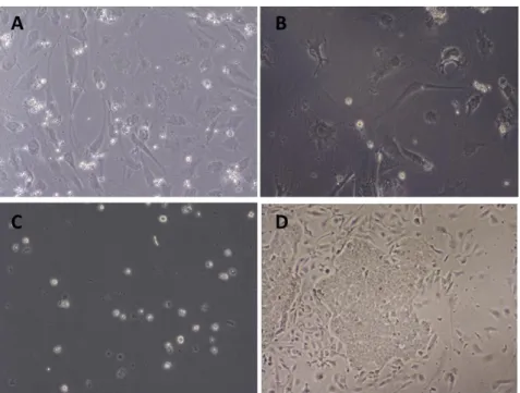
The Theka protocol was reported to generate mature and functional dopaminergic neurons from hiPSc in 21 days, skipping all the intermediate steps for inducting and selecting embryoid bodies or rosette-neural precursors required by other methods. However, both infected hESc and controls failed to attach to the any of the substrates tested (Figure 3.2 Panels A-C) and died after a few days in culture. The fact that control (untreated with virus) cells died suggested that our initial manipulation (making single cell suspensions followed by mock infection) might be toxic to the cells. Therefore we repeated the control procedure but seeding the cells in CDM rather than neuronal inducing medium. In this case, cells survived, attached to the substrate and formed colonies (Figure 3.2, Panel D).
15
Therefore, the published protocol of Theka et al did not work in our hands. It is possible that the viral vectors we were using were not expressing properly (although they were exactly the same vectors used by Theka, procured from Addgene). Alternatively, Theka et al used induced pluripotent stem cells instead of human embryonic stem cells as we did, although given the high similarity between these types of cells, one would not expect the protocol to work for iPSc and fail for hESc. Another reason could be that after seeding the single cell suspension on to the substrate, we switched the cells from ESc media to neuronal inducing medium too quickly (after 24 hs), but this was the procedure indicated in the original Theka protocol. We did not investigate the causes of the lack of success of this protocol and decided to try a different approach.
Figure 2.2 Differentiation of hESCs into DA neurons after ANL overexpression. (A) Negative control seeded on PA6. (B) Negative control seeded on MEFs. (C) Negative control seeded on gelatin. (D) Negative control seeded on MEFs and feeded with CDM media.
16
3.2 Differentiation of hESc into DA neurons by co-culture with stromal cell populations
Another approach that has been described is the differentiation of hESc into dopaminergic neurons using PA6 cells as feeder layer by Zeng et al (Zeng, Cai, & Chen, 2004). When seeded on this substrate and cultured in Glasgow Minimum Essential Media (GMEM) for 3 weeks, hESc formed colonies, approximately 70% of which were positive for TuJ1; furthermore 87% of these colonies Tuj1 contained TH-positive post-mitotic cells. Differentiated cells also expressed other dopaminergic markers, receptors and transcriptions factors such as Sox1, Nurr1, Lmx1b, VMAT and DAT. Neurons that had been differentiated on PA6 cells were able to release dopamine and 3,4-dihydroxphe-hylacetic acid (DOPAC) but not noradrenalin. In addition, PA6-induced dopaminergic cells were transplanted into the striatum of rats that had received unilateral lesions with 6-hydroxydopamine 4 weeks before transplantation. After 5 weeks of transplantation, they observed a small number of cells expressing TH. Therefore, differentiated hESc by co-culture with PA6 cells exhibit biochemical and functional features expected in mature dopaminergic neurons.
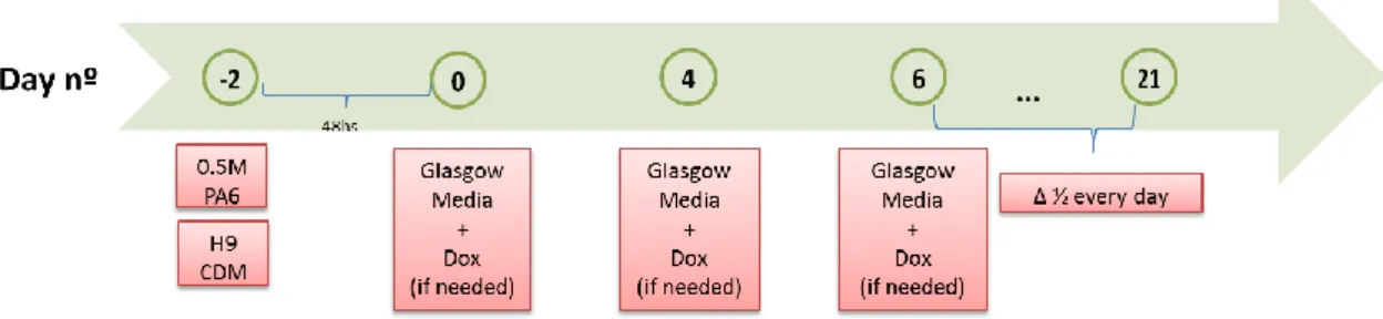
We decided to reproduce the Zeng approach, with some modifications. Neural differentiation of H9 hESc was induced by the mouse stromal cell line PA6. H9 hESc were disaggregated into single cells and cultured on PA6 feeder cells in GMEM supplemented with 10% KSR, 1 mM pyruvate, 0.1 mM nonessential amino acids (NEAA), and 0.1 mM β-mercaptoethanol in slide flasks for between 15 and 29 days. Medium was changed on day 4, 6 and every day thereafter. A timeline of the experiment is shown in Figure 3.3.
17
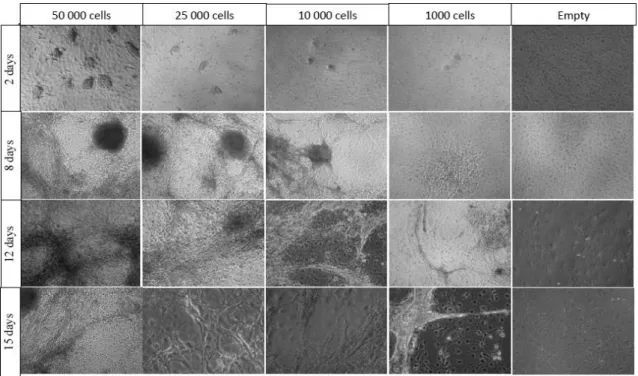
In our first attempt to reproduce this protocol, we tested different cell numbers (50000, 25000, 10000 and 1000 cells per flask). These cells were able to attach to the substrate, and after 2 days, small colonies could be seen (Figure 3.4, panel 1). The plating efficiency was approximately 5%. This low plating efficiency is normally observed when plating hESc. As expected, the number of colonies in each flask clearly reflected the number of cells initially seeded. The colonies grew and quickly started to differentiate, primarily at the edge of the colony. After 12 days in culture, it became clear that growth was excessive in the flasks that had received 50000, 25000 and 10000 cells (Figure 3.4 panel 3). Growth in the flask that had received 1000 cells was reasonable. After 15 days in culture, we performed immunofluorescence analysis on all cultures for a marker of neurons (Tuj1). Signal was weak or absent in flask that had received 50000, 25000 and 10000, but Tuj1 positive cells could clearly be seen in the flask that received 1000 cells (data not shown).
The next step was to perform the same experiment with only 1000 cells per slide flask for 21 days. The cells were seeded on PA6 feeders and had the same development as in the previous experiment. Then we analyzed them by immunofluorescence for both neural (β-tubulin III) and dopaminergic (TH) markers. After 21 days in culture, many colonies contained large numbers of TUJ1-positive cells (of 32 colonies examined, 28
Figure 3.4 Differentiation of hESc on PA6 for 15 days. In this experiment, several cell densities were tested (50 000, 25 000, 10 000 and 1000)
18
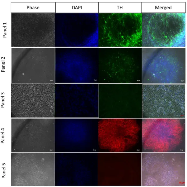
expressed TUJ1; in 24 of them, expression was high in both cell numbers and signal intensity); In most TUJ1 positive colonies, only a few cells were TH-positive, but few colonies we observed large numbers of TH positive cells with strong signal (Figure 3.5).
Figure 3.5 Differentiation of hESc into DA neurons. Analysis by immunofluorescence was performed. Panel 1 and 2) Cells were marked for TH and DAPI. Panel 3) Negative Control for TH. Panel 4) Cells were marked for TUJ1 and DAPI. Panel 5) Negative control for TUJ1.
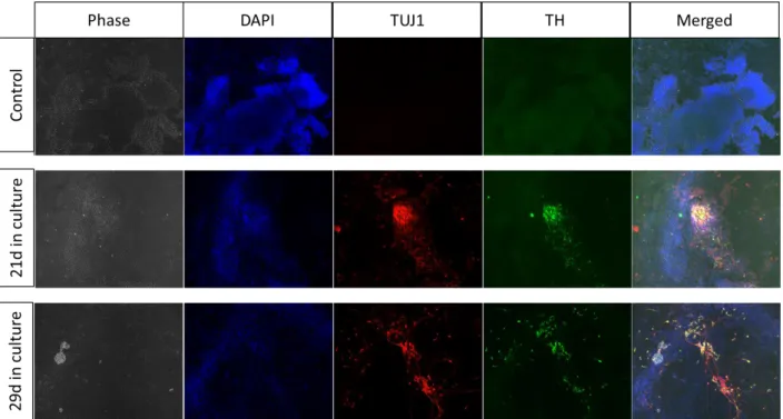
To test whether time in culture would affect neuronal differentiation, we repeated the experiment testing different times of culture (21 and 29 days). The cells (1000) were seeded on PA6 feeders in supplemented GMEM as mentioned above. No obvious morphological differences were apparent between the 21 d and 29 time-points (Figure 3.6). Analysis by immunofluorescence did not reveal any differences in yield of TuJ1 and TH positive cells between both time-points of differentiation Figure 3.7).
19
3.3 Differentiation of hESc into DA neurons by overexpression of specific transcription factors and co-culture with PA6 cells
Figure 3.6 Differentiation of hESc on PA6 for 21 and 29 days.
Figure 3.7 Analysis by immunofluorescence of differentiated cells. Both cell cultures (21d and 29d) were marked for neuronal markers (TUJ1 and TH). One Negative control for both markers was performed.
20
As explained above, our initial attempt at the Theka protocol did not succeed. Our hypothesis was that seeding in Theka neuronal medium after 24 hs was impeded cell attachment. We therefore re-attempted the Theka protocol with modifications: First, we seeded the single cell suspension in ESc media (CDM) and cultured the cells in CDM media for 48hs (as opposed to 24hs); second, after 48 hs we switched the cells to supplemented GMEM (the medium used to induce neuronal differentiation in the Zeng protocol), as opposed to Theka’s neuronal inducing medium.
As before, hESc were disaggregated into single cells and infected with a lentivirus expressing the three transcription factors Ascl1, Lmx1a and Nurr1 (as a polycistronic construct, TetO-ALN) and the doxycycline-inducible reverse transactivator (FUW-rtTA) with the multiplicity of infection (MOI) of 10 or 100. Infected hESc (1000 cells) were then seeded on gelatin with PA6 feeders in CDM for 48 hs, after which the medium was switched to the supplemented GMEM for 21 days. One negative control consisted of mock-infected hESc; a second negative control consisted of infected cells cultured without DOX. The positive control were hESc infected with green fluorescent protein (GFP)-expressing lentivirus (L-PGK-GFP). Doxycycline (Dox) was added to treatments which needed it with every medium change. A timeline of the experiment is shown in Figure 3.8.
The plating efficiency was approximately 2,5%. The cells formed colonies and by day 8 differentiation was clearly visible (Figure 3.9). In the positive control, GFP was only visible at MOI =100. After 21 days in culture, no morphological difference was observed between the ALN infected cells in which Dox was added, ALN infected cells without Dox and the mock infected negative control. Immunofluorescence analysis for TUJ1 and
21
TH, revealed differentiation to both TUJ+ and TH+ cells with a yield similar to that obtained with the Zeng protocol and no obvious differences between treatments: the results of ALN infected cells with Dox, the ALN infected cells without Dox and both positive and negative controls were overall not significantly different. Most of colonies were TUJ1-positive and some colonies expressed TH (Figure 3.10). During the 21 days, the expression of GFP in the positive control was only visible for MOI=100 (Figure 3.9); while the lack of success for MOI =10 could be attributed to insufficient transduction, this argument cannot justify the results at MOI = 100. Again, the Theka protocol did not give positive results in our hands.
Figure 3.9 Differentiation of hESc into DA neurons by overexpression of specific transcription factors and co-culture with PA6 cells. A similar morphology in the different cell cultures can be observed.
22
3.4 Differentiation of hESc into DA neurons by embryoid body formation
Another strategy that has been reported is the differentiation of hESc into dopaminergic neurons using embryoid body formation. These differentiation protocols start with the generation of embryoid bodies (EBs) (three-dimensional multicellular aggregates of pluripotent stem cells (PSc)). There are numerous methods for inducing EB formation: (i) the hanging drop (HD) method - is a classic method widely used to form EBs from mouse ES cell lines, (ii) culture in methylcellulose semisolid media, (iii) suspension culture in bacterial-grade dishes and (iv) centrifugation in conical-bottom 96 well plates followed by culture of clusters in low-adherence vessels. (Kurosawa, 2007; Murashov, Pak, & Katwa, 2005)
Although PSCs seeded/plated in suspension culture form EBs spontaneously, the efficiency of this process varies, is not reproducible and the homogeneity of EBs is low.
Figure 3.10 Immunofluorescence analysis of differentiated cells. The cells were marked for TUJ1 (red) and TH (green); Not significantly differences were observed in the infected H9 that received Dox and the infected H9 without Dox, either for MOI=10 or MOI=100. A negative control was performed for both markers.
23
Most current methods generate EBs of different size and shapes, which in turn affects their differentiation capabilities. The size of EBs is one of the key factors effecting differentiation of PSCs and should be controlled.
In order to develop a consistent and reproducible protocol to generated EBs of an adequate size, H9 hESc were disaggregated into single cells and seeded at densities of 1000 and 5000 cells per well of a conical 96-well plate, centrifuged at 230g for 5 min. and cultured for 48h. Subsequently, the EBs were transferred to a low adherence dish and cultured in N2B27 media for more 72h. After these 72hs, the EBs were seeded onto MEFs for 21 days. Media was changed daily. A timeline of the experiment is shown in Figure 3.11.
The EBs formed in conical-bottom 96-well plates were very homogeneous in size (Figure 3.12). When seeded on MEFs, the EBs were able to attach to the substrate and proliferated to form large cell masses, particularly when EBs formed by 5000 cells were plated. The plating efficiency was approximately 100%. The colonies started to differentiate, primarily at the edge of the colony as observed in previous experiments. After 10 days in culture, it became clear that growth was excessive in the slide flasks that had received EBs formed by 5000 cells, while growth of EBs with 1000 cells was adequate for the dimensions of the flask (data not shown). After 21 days we performed immunofluorescence analysis for neuronal markers TUJ1 and TH. Most of the colonies
24
expressed TUJ1 and TH-positive cells, and the yield in this experiment was noticeable greater than in our previous approaches (Figure 3.13).
3.5 Differentiation of hESc into DA neurons by embryoid body formation and co-culture with PA6 cells
Figure 3.12 EBs after the transfer to a low adherence dish in culture.
Figure 3.13 Immunofluorescence analysis of differentiated EBs. A and B) the cells were marked for TUJ1 and TH. Most of the colonies expressed TUJ1 and TH-positive cells.
25
The positive results obtained by introducing an EB formation step prompted us to introduce this step into the Zeng protocol. We therefore tested the effect seeding EBs on PA6 feeders followed by culture in supplemented GMEM. The H9 hESc were disaggregated into single cells in CDM medium, were seeded at density of 1000 cells per well in a conical-bottom 96-well plate, centrifuged at 230g for 5 min. and incubated for 48hs. Then, the EBs were transferred to a bacterial dish to grow in suspension in GMEM media for 72hs. Then EBs (15 EBs) were seeded onto PA6 feeders for 21 days and the media changed daily. The timeline of the experiment is shown in Figure 3.14.
The EBs were able to attach to substrate and proliferated into large cell masses. The plating efficiency was approximately 100%. The differentiation is clearly visible by day 4 (Figure 3.15), especially at the edges of the colony.
Figure 3.14 Schematic representation of the differentiation protocol
26
After 21 days of culture an analysis by immunofluorescence was performed for neuronal markers TUJ1 and TH. Our analysis revealed high levels of differentiation to both TUJ+ and TH+ cells (Figure 3.16). In three experiments, we obtained 48%, 71% and 100% of colonies with high number of TUJ1 positive cells with strong signal. If colonies with less number of positive cells (or having cells with weaker signal) were also included, the percentage of TUJ1 positive colonies was 84%, 100% and 97%. For TH marker we obtained 24%, 50% and 29% of colonies with high number/strong signal, and 48%, 78% and 41% if all colonies showing signal were considered. Therefore this protocol has a high yield of TUJ1 positive cells, of which up to 35% are also TH positive. However, although all experiments resulted in differentiation of hESc to TUJ1 and TH positive cells, yield showed considerable variability.
3.6 Differentiation of hESc into DA neurons by embryoid body formation, co-culture with PA6 cells and overexpression of specific transcription factors
As mentioned above, our previous attempts at using the Theka approach (first overexpression of ALN and seeding several substrates in Theka medium, and second by ALN overexpression followed by seeding on PA6 in GMEM medium) failed to give positive results. We asked whether introducing an EB formation step would improve results, based on the rationale that transcription factor overexpression in the context of a differentiating EB might drive the differentiation process forward more efficiently. Therefore, we infected single cells, generated EBs and seeded them on PA6 feeders in supplemented GMEM medium.
Figure 3.163 Immunofluorescence analysis of differentiated cells from EBs formation. The cells were marked for TUJ1 and TH. The differentiation of hESc to DA neurons in this experiment was noticeable.
27
As previously, H9 hESc were disaggregated into single cells and infected with a lentivirus expressing the three transcription factors Ascl1, Lmx1a and Nurr1 (as a polycistronic construct, TetO-ALN) and the doxycycline-inducible reverse transactivator (FUW-rtTA) with the multiplicity of infection (MOI) of 10 or 100. After infection the cells were seeded at density of 1000 cells per well in a conical-bottom 96-well plate for 48hs. Then, the EBs were transferred to a bacterial dish to grow in suspension in GMEM media for 72hs. After these 72hs, the EBs were seeded onto PA6 feeders for 21 days. The timeline of the experiment is shown in Figure 18. One negative control consisted of mock-infected EBs; a second negative control consisted of mock-infected EBs cultured without DOX. The positive control were EBs infected with green fluorescent protein (GFP)-expressing lentivirus (L-PGK-GFP). Doxycycline (Dox) was added to treatments which needed it with every medium change. The timeline of the experiment is shown in Figure 3.17.
The EBs were able to attach to substrate and they formed large cell masses. The plating efficiency was approximately 100%. The colonies grew and quickly started to differentiate by day 5, primarily at the edge of the huge colony. In the positive control, GFP was clearly visible at MOI=10 and MOI=100 (data not shown). After 21 days in culture, once again, no morphological difference was observed between the ALN infected cells in which Dox was added, ALN infected cells without Dox and the mock infected negative control. Immunofluorescence analysis for TUJ1 and TH, revealed high differentiation to both TUJ+ and TH+ cells (data not shown) with a yield similar to that
28
obtained with the EBs without infection seeded on PA6 feeders. Therefore, after multiple attempts, we concluded that overexpression of ALN transcription factors in hESc is not a reproducible approach in our hands and discontinued this strategy. We cannot able to explain the why the Theka protocol failed in our hands. Of note, we did not test whether the vectors expressed the transcription factors appropriately. While we could have performed this experiment, we decided not to because if the system expressed appropriately, we would still have a negative result; on the other hand, if the system did not express appropriately, we would have an explanation for the lack of success, but then would have needed to invest time and resources in making it work. Given that other approaches were giving satisfactory results, we did not pursue this path.
3.7 Purification of dopaminergic neurons
After various attempts to differentiate hESc into dopaminergic neurons, we conclude that the most efficient approach, was to use EBs in co-culture with PA6 cells. However, this protocol resulted in a heterogeneous population. Future experiments planed in our laboratory would benefit from homogeneous populations of dopaminergic neurons. We therefore considered the possibility of using fluorescence activated cell sorting (FACS) to purify neurons from our heterogeneous cultures. The strategy would involve, differentiation followed by disaggregation of cultures into single cell suspensions and FACS using a fluorescent reporter or appropriate surface cell markers. In any case, a FACS step would be needed. Ultimately, we are interested in dopaminergic neurons, but as a first approximation we decide to test whether we could infect hESc with a lentiviral vector expressing GFP from a synapsin promoter and use GFP fluorescence to FACS neurons. Synapsin expression occurs late during differentiation, so successful sorting of GFP positive cells could be expected to result in a homogeneous populations of neurons, most of which would be TUJ1 positive and an unknown proportion of which would be TH positive. Therefore, we infected the cells with a lentiviral vector with a neuron-specific promotor (synapsin) driving the expression of green fluorescent protein (GFP) (SYN-GFP) with the MOI of 10, 50 and 100. The negative control consisted of mock-infected hESc; the positive control were hESc mock-infected with green fluorescent protein (GFP)-expressing lentivirus (L-PGK-GFP). Subsequently, the cells were seeded at density of 1000 cells per well in a conical-bottom 96-well plate for 48hs. The EBs were
29
transferred to a bacterial dish to grow in suspension in GMEM media for 72hs. After these 72hs, the EBs (15 EBs) were seeded onto PA6 feeders for 21 days.
The expression of GFP in the EBs infected with Lenti-SYN-GFP with a MOI of 50 and 100 was visible by day 9. After 21 days of culture, the expression of GFP in the positive control was visible for all MOI and the mock-infected cells did not express GFP (Figure 3.18).
After 21 days, immunofluorescence analysis was performed for TUJ1 and GFP. Most colonies were TUJ1-positive. However, the GFP antibody we used gave us high background even in absence of transduction with Lenti-SYN-GFP, so we could not verify the co-localization with TUJ1 (Figure 3.19).
Figure 3.18 Differentiation of EBs for 21 days in culture. After these 21 days the expression of GFP in the positive control was visible for all MOI and the mock-infected cells did not express GFP. The expression of GFP was possible observe in the EBs infected with Lenti-SYN-GFP with a MOI of 50 and 100 by day 9.
30
Even without confirmation that cells expressing synapsin were TUJ1 positive neurons, we attempted to purify the SYN-GFP cells by Fluorescence-Activated Cell Sorting. In our first attempt, the EBs were disaggregated with 0.25% Trypsin. After cell sorting, we collected approximately 50,000 GFP-positive and Dapi-negative (ie, alive) cells (Figure 3.20) and the approximately 15,000 cells were seeded in dishes coated with laminin, gelatin or PA6 feeders. The cells did not attach and we could not able to see GFP-positive cells (data not shown).
Figure 3.19 Analysis of immunofluorescence for GFP and TUJ1. The GFP antibody we used gave us high background even in absence of transduction with Lenti-SYN-GFP (control), so we could not verify the co-localization with TUJ1.
Figure 3.20 Purification of SYN-GFP cells by FACS. A) In the gate P2, was selected the GFP-positive cells whereas B) in the gate P1 was selected the DAPI-negative cells. Most of the cells were DAPI-negative.
31
In second attempt we collected approximately 32,000 GFP-positive and Dapi-negative cells and seeded them at higher density on laminin coated dishes (10,000 and 22,000 cells in wells form a 12 well plate. Few cells attached to the plate and some showed neuronal like processes; however GFP expression was not evident (Figure 3.21).
In a final attempt to purify neurons, we tested different ways to disaggregate the EBs with 1) 0.25% Trypsin, 2) Accutase and 3) 0.25% Trypsin plus collagenase (1mg/ml). Cell survival and yield seemed best for treatment 1). After cell sorting, we collected approximately 25,000 GFP-positive and Dapi-negative cells and seeded them at high density (25,000 per 12-well) on laminin. Few cells attach to substrate and we able to see clear (albeit weak) GFP signal in some of the attached cells (Figure 3.22).
32
In general, these preliminary attempts did not yield positive results. Although by adjusting some of the parameters in our purification protocol such as disaggregation conditions, seeding density or plating substrate we were able to obtain some GFP+ cells attached to the substrate, the yield was too low to be practical. More work will need to be done to determine whether a purification protocol based on disaggregation of mature neurons from a differentiated EB followed by FACs is a viable purification strategy.
Figura 3.22 SYN-GFP neurons. After a third attempt to purify neurons, few cells attached to substrate and GFP signal is weak.
33
4.
Final Commentary
In sum, we have tested a number of methods and parameters with the goal of obtaining a robust, reproducible and practical protocol to differentiate human embryonic stem cells to the dopaminergic neuronal fate. Based on our experimental results, we can present the following conclusions:
1) The strategy of inducing differentiation by overexpression of specific transcription factors ALN (Theka et al) either in single cells or embryoid bodies did not work in our hands, despite the fact that a number of different parameters were tested, including MOI, plating substrate, and neuronal induction media.
2) Neuronal differentiation by co-culture of PA6 cells using
supplemented GMEM (Zeng et al) either in single cells or embryoid bodies gave us satisfactory results, albeit with considerable variability.
3) Embryoid body formation showed to be a better approach than seeding single cells. Neuronal differentiation (TUJ1 and TH) was more noticeable in the embryoid body protocols.
4) Purification of mature neurons by FACS did not yield positive results, despite the fact that several cell densities and substrates were tested. More work will need to be done to ensure that this protocol is practical.
34
5. References
Alves dos Santos, M. T. M., & Smidt, M. P. (2011). En1 and Wnt signaling in midbrain dopaminergic neuronal development. Neural Development, 6(1), 23. doi: 10.1186/1749-8104-6-23
B., R., & N., W. (2013). Gaucher Disease: A Comprehensive Review. Critical Reviews in
Oncogenesis.
Chizhikov, V. V., & Millen, K. J. (2005). Roof plate-dependent patterning of the vertebrate dorsal central nervous system. Dev Biol, 277(2), 287-295. doi:
10.1016/j.ydbio.2004.10.011
Elstein, D., Abrahamov, A., Hadas-Halpern, I., & Zimran, A. (2001). Gaucher's disease. The
Lancet, 358(9278), 324-327. doi: 10.1016/s0140-6736(01)05490-3
Farfel-Becker, T., Vitner, E. B., & Futerman, A. H. (2011). Animal models for Gaucher disease research. Dis Model Mech, 4(6), 746-752. doi: 10.1242/dmm.008185
Gilbert, S. F. (2010). Developmental Biology (Ninth ed.).
Heins, N., Englund, M. C. O., Bergh, C., Sjoblom, C., Lindahl, A., Dahl, U., . . . Semb, H. (2004). Derivation, Characterization and Differentiation of Human Embryonic Stem Cells. Stem
Cells.
Kurosawa, H. (2007). Methods for inducing embryoid body formation: in vitro differentiation system of embryonic stem cells. J Biosci Bioeng, 103(5), 389-398. doi:
10.1263/jbb.103.389
Murashov, A. K., Pak, E. S., & Katwa, L. C. (2005). Parallel development of cardiomyocytes and neurons in embryonic stem cell culture. Biochem Biophys Res Commun, 332(3), 653-656. doi: 10.1016/j.bbrc.2005.04.167
Reubinoff, B. E., Pera, M. F., Fong, C.-Y., Trounson, A., & Bongso, A. (2000). Embryonic stem cell lines from human blastocysts: somatic differentiation in vitro. Nat Biotechnol.
Roussa, E., & Krieglstein, K. (2004). Induction and specification of midbrain dopaminergic cells: focus on SHH, FGF8, and TGF-beta. Cell Tissue Res, 318(1), 23-33. doi: 10.1007/s00441-004-0916-4
Theka, I., Caiazzo, M., Dvoretskova, E., Leo, D., Ungaro, F., Curreli, S., . . . Broccoli, V. (2013). Rapid generation of functional dopaminergic neurons from human induced pluripotent stem cells through a single-step procedure using cell lineage transcription factors.
Stem Cells Transl Med, 2(6), 473-479. doi: 10.5966/sctm.2012-0133
Thomson, J. A. (1998). Embryonic Stem Cell Lines Derived from Human Blastocysts. Science,
282(5391), 1145-1147. doi: 10.1126/science.282.5391.1145
Tiscornia, G., Vivas, E. L., & Izpisua Belmonte, J. C. (2011). Diseases in a dish: modeling human genetic disorders using induced pluripotent cells. Nat Med, 17(12), 1570-1576. doi: 10.1038/nm.2504
Tiscornia, G., Vivas, E. L., Matalonga, L., Berniakovich, I., Barragan Monasterio, M., Eguizabal, C., . . . Izpisua Belmonte, J. C. (2013). Neuronopathic Gaucher's disease: induced pluripotent stem cells for disease modelling and testing chaperone activity of small compounds. Hum Mol Genet, 22(4), 633-645. doi: 10.1093/hmg/dds471
Zeng, X., Cai, J., & Chen, J. (2004). Dopaminergic Differentiation of Human Embryonic Stem Cells. Stem Cells.