Diferentiation Potential of O Bombay Human-Induced
Pluripotent Stem Cells and Human Embryonic Stem
Cells into Fetal Erythroid-Like Cells
Fatemeh Ganji, M.Sc.1, Saeid Abroun, Ph.D.1, 2*, Hossein Baharvand, Ph.D.1, Nasser Aghdami,
Ph.D.1, 3, Marzieh Ebrahimi, Ph.D.1, 3
1. Department of Stem Cells and Developmental Biology at Cell Science Research Center, Royan Institute for Stem Cell Biology and Technology, ACECR, Tehran, Iran
2. Department of Hematology, School of Medical Sciences, Tarbiat Modares University, Tehran, Iran 3. Department of Regenerative Biomedicine at Cell Science Research Center, Royan Institute for Stem Cell Biology
and Technology, ACECR, Tehran, Iran
*Corresponding Address: P.O. Box: 16635-148, Department of Stem Cells and Developmental Biology at Cell Science Research Center, Royan Institute for Stem Cell Biology and Technology, ACECR, Tehran, Iran
Email: abroun@modares.ac.ir
Received: 8/Sep/2013, Accepted: 6/Nov/2013
Abstract
Objective: There is constant dificulty in obtaining adequate supplies of blood compo
-nents, as well as disappointing performance of "universal" red blood cells. Advances in somatic cell reprogramming of human-induced pluripotent stem cells (hiPSCs) have pro
-vided a valuable alternative source to differentiate into any desired cell type as a therapeu
-tic promise to cure many human disease.
Materials and Methods:In this experimental study, we examined the erythroid dif
-ferentiation potential of normal Bombay hiPSCs (B-hiPSCs) and compared results to human embryonic stem cell (hESC) lines. Because of lacking ABO blood group expression in B-hiPSCs, it has been highlighted as a valuable source to produce any cell type in vitro.
Results:Similar to hESC lines, hemangioblasts derived from B-hiPSCs expressed ap
-proximately 9% KDR+CD31+ and approximately 5% CD31+CD34+. In semisolid media,
iPSC and hESC-derived hemangioblast formed mixed type of hematopoietic colony. In mixed colonies, erythroid progenitors were capable to express CD71+GPA+HbF+ and
ac-companied by endothelial cells differentiation.
Conclusion: Finally, iPS and ES cells have been directly induced to erythropoiesis with
-out hemangioblast formation that produced CD71+HbF+erythroid cells. Although we observed
some variations in the eficiency of hematopoietic differentiation between iPSC and ES cells, the pattern of differentiation was similar among all three tested lines.
Keywords: Induced Pluripotent Stem Cells, Differentiation, Hemangioblasts, Erythroid Cells
Cell Journal(Yakht eh), Vol 16, No 4, Wint er 2015, Pages: 426- 439
Citation: Ganji F, Abroun S, Baharvand H, Aghdami N, Ebrahimi M. Differentiation potential of o Bombay human-induced pluripotent stem cells and human embryonic stem cells into fetal erythroid-like cells. Cell J. 2015; 16(4): 426-439.
Introduction
Blood transfusions are universally used to treat various types of hematological diseases, such as hemoglobin abnormalities (sickle cell disease, thalassemia and methemoglobinemia) and abnor-malities in the red blood cell (RBC) membrane or metabolism, as well astraumatic injury, surgery, treatment for burn victims and organ transplant
occur with transfusion of RBCs. Moreover, ABO incompatible transfusion due to clinical or labora-tory error remains the most widespread cause of transfusion related morbidity and mortality. Sev-eral studies have demonstrated that it is possible to enzymatically cleave A and B antigens to produce 'universal' RBCs, but to date this has not found widespread clinical application (1).
Thus, there is an emphasis on the need for better treatment methods, including hematopoietic cell re-placement strategies as an alternative source for blood cells. Studies pioneered by Douay and Andreu (2) have demonstrated the feasibility of in vitro produc-tion of RBCs from CD34+ hematopoietic stem cells
and progenitors that have been isolated from cord blood, bone marrow or peripheral blood. However, bone marrow or peripheral derived hematopoietic
stem cells are dificult to expand and the possibility of
using these cells for high scale industrial production of major blood components remains unresolved.
Pluripotent stem cells such as embryonic stem cells (ESC) and induced pluripotent stem cells (iP-SCs) have been introduced as the best candidates to substitute for blood production in vitro. Human ESC
(hESC) possess indeinite proliferative capacity in vitro, and have been shown to differentiate into all three germ layers that give rise to all type of somatic cells, including blood cells (3). In a comparison be-tween ESCs and iPSCs, ethical issues do not avoid of iPSCs because they do not need embryonic or fetal material (4, 5) and they are more compatible because of autologous terminally differentiated somatic cells
in mice and humans. Also iPSCs exhibit high similar
-ity to ESCs due to effective proliferation and eficient
differentiation into several cell types (6-8).
Previously, human iPSCs (hiPSCs) from donor
ibroblasts derived from a Bombay phenotype have been established in our institute by ectopic expres -sion of transcription factors that played a fundamen-tal role in hESCs (9). The established cell line has been named Bombay hiPSCs (B-hiPSCs). According to mutational analysis, the Bombay phenotype fails
to express the FUT1 and FUT2 genes by sequence
analyses of ibroblasts and iPSCs which lead to lack of ABH antigen expression on blood cells, related to
the ABO blood group system. The discovery of the Bombay phenotype, as a rare blood group, is an
im-portant discovery for the ield of immunohematology.
B-hiPSC-derived RBCs can be introduced as a his-tocompatible erythroid crucial for future cell therapy applications.
We sought to determine if differentiation of iP-SCs into erythroid cells would follow the same patterns as that observed for hES cells. To achieve this goal we have used B-hiPSCs and two hES cell lines of various genomic sources, Royan H5 and 6 (RH5, RH6) and induced their differentiation into erythrocytes (10). The results revealed that cells
produced in all lines were similar in the expression
pattern of hemangioblast and erythroid progenitors regardless of their genomic diversity. Importantly, we observed in this system that hESCs differentia-tion closely resembled early human erythropoiesis development. In other word, sequential
differentia-tion has been identiied by formadifferentia-tion of hemangio -blast colonies. Afterwards, these colonies
differen-tiated to erythroid cells that expressed hemoglobin
F (α2γ2), however, they could not produce adult hemoglobin or hemoglobin A (α2β2).
Materials and Methods
Cell lines
In this experimental study, RH6 (44+XY), RH5 (44+XX) and BhiPSCs-11 (44+XY) with normal
karyotype were used and cell passage number was between 30 and 40. B-hiPSCs-11 have been shown
to be deicient in FUT1 and FUT2 genes expres -sion was established at Royan Institute and main-tained as undifferentiated cells in a feeder-free cul-ture established previously by Larijani et al. (11).
Adherent feeder-free and suspension culture of hiPS and hES cells
hiPS and ES cells were cultured on Matrigel (Sig-ma-Aldrich, E1270, USA) in serum-free media that
consisted of Dulbecco’s modiied Eagle’s medium
(DMEM/F12, Gibco, 21331-020, USA) supplement-ed with 20% knock-out serum replacement (KOSR,
Gibco, 10828-028, USA), 100 ng/ml basic ibroblast
growth factor (bFGF, Royan Institute, Iran), 2 mM L-glutamine (Gibco, 25030-024, USA), 0.1 mM beta-mercaptoethanol (Sigma-Aldrich, M7522, USA), 1% nonessential amino acids (Gibco, 11140-035, USA), 100 IU/ml penicillin, and 100 mg/ml streptomycin (Invitrogen, USA). The suspension condition was
used to expand undifferentiated iPS and hES cells, as
follows. Briely, cells were washed by Ca2+ and Mg2+
mM ethylenediaminetetraacetic acid (EDTA) (Gibco, 25300-054, USA) at 37˚C for 4-5 minutes, and then pi--petted for 5-12 times. Then cells were transferred into
low-attachment six-well plates (Corning-NY14831,
USA) and treated with 10 µM ROCK inhibitor
(Sig-ma-Aldrich, Y0503, USA) before trypsinization. The
cell aggregates were generated in serum-free medium that included DMEM/F12, KOSR, bFGF (Royan in-stitute, 12-280411, Iran), L-glutamine, nonessential amino acids, at seven days post-culture. To prevent apoptosis, 10 µM of ROCK inhibitor was added on
the irst two days of culture. For passaging, iPSCs
and ES cell aggregates were incubated with 10 µM ROCK inhibitor for 2 hours prior to trypsinization then washed with PBS (Gibco, 14287-072, USA) and treated with 0.05% trypsin and 0.53 mM EDTA (Gib-co, 25300-054, USA) at 37˚C, for 4-5 minutes. The enzyme was removed and colonies were gently
pi-petted and re-plated on six-well ultra-low-attachment
plates (11).
Differentiation of iPS and ES cells
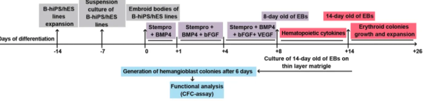
As shown in igure 1, seven-day-old aggregates were cultured in ultra-low attachment six-well plates
in the presence of aggregation media that consisted of Stem Pro-34 (Gibco,10639-011,USA) supplemented with 100 IU/ml penicillin, 100 mg/ml streptomycin (Invitrogen, USA), 10 ng/mL bone morphogenetic protein 4 (BMP-4) (R&D Systems, 314-BP, USA), 2 mM glutamine, 4×10⁻⁴ mM on othioglycerol (MTG)
(Sigma-Aldrich, 018K08122, USA), and 50 μg/mL
ascorbic acid (Sigma-Aldrich, A4403, USA). The ag-gregates were incubated at 37˚C in 5% CO2. After 24 hours, half of the media was carefully removed and
replaced with fresh aggregation media supplemented with 5 ng/mL hbFGF (induction media 1) and cells were incubated for 72 hours. After incubation, cells were harvested and re-suspended in induction me-dia1, which also contained 10 ng/mL vascular en-dothelial growth factor (VEGF) (R&D Systems, 293-VE, USA) for an additional four days. To gen-erate hemangioblast colonies, 14-day-old aggregates
were plated in Iscoveʼs Modiied Dulbeccoʼs Me -dium (IMDM) (Biowest, Lo192-500, France) with 1% methylcellulose (Sigma-Aldrich, 274429, USA) supplemented with 10% FBS, 2 mM L-glutamine,
50 μg/mL ascorbic acid, 4×10⁻⁴ M MTG, 150 μg/
mL holo-transferrin (Sigma-Aldrich, T0665, USA), 1 ng/mL hbFGF, 10 ng/mL hVEGF, 100 ng/mL hu-man stem cell factor (hSCF) (R&D Systems, 255-SC, USA), 20 ng/mL human interleukin-6 (hIL-6) (R&D Systems, 206-IL, USA), 2 U/mL human Erythropoietin (hEPO) (R&D Systems, 287-TC, USA), 40 ng/mL hIL-3 (R&D Systems, 203-IL, USA), and 25 g/mL human Insulin-like Growth Factors I (hIGF-I, R&D Systems, 291-G1,USA). Plated aggregates were maintained at 37˚C in a 5% CO2 incubator for six days. For direct differentia -tion of erythroid cells, 14-day-old aggregates were plated in IMDM with 1% methylcellulose, 10% FBS, 2 mM L-glutamine, 100 ng/mL hSCF, 2 U/ mL hEPO, 5 ng/mL hIL-6, 40 ng/mL hIL-3, 40 ng/ mL recombinant human thrombopoietin (rhTPO) (R&D Systems, 288-TP, USA), 25 ng/mL hIGF-1, 10 ng/mL hVEGF, and 1 ng/mL recombinant human granulocyte macrophage colony-stimulat-ing factor (hGM-CSF) (R&D Systems, 215-GM, USA) (12). Plated aggregates were maintained at 37˚C in a 5% CO2 incubator for 12 days.
Phenotypic analysis of ES and iPS cell-derived blast cells
We evaluated the cellsʼ differentiation stages with
monoclonal PE-conjugated antibodies against hu-man KDR, CD34, GPA (Glycophorin A), fetal hemo-globin, and FITC-conjugated antibodies against CD31and CD71. All antibodies were obtained from BD Pharmingenand used for immunophenotyping. The results were determined by BD FACS Calibur and analyzed with WinMDI version 2.9 software.
Clonogenicity potential of B-hiPSC and HSC-derived cells
For evaluation of colony formation capability, we plated 14-day-old aggregates onto a thin layer of matrigel in 96-well plates and cultured them in IMDM medium supplemented with 10% FBS, 10% horse serum, 2 mM L-glutamine, 4×10⁻⁴M
MTG, 150 μg/ml holo-transferrin, 5 ng/ml bFGF,
10 ng/mL hVEGF, 100 ng/mL hSCF, 20 ng/mL hIL-6, 2 U/mL hEPO, and 25 ng/mL hIGF-1 for
six days. Subsequently, six-day-old grape-like
blast cells were isolated and platedon methylcellu-lose base media for 14 days up to 28 days at 37˚C in a 5% CO2 incubator.
Immunocytochemistry
Differentiated cells and colonies in
methylcel-lulose were washed with PBS and ixed in 4%
paraformaldehyde (PFA) for 15 minutes,
permea-bilized with 0.2% triton X-100 for 30 minutes for
primary antibody, anti CD31 (1:100, BD, 555849) for endothelial cells was performed for 1 hour at 37˚C. Cells were then washed and incubated with FITC-conjugated secondary antibody, anti-mouse IgG (1:100, BD, 04611) as appropriate, for 1 hour at 37˚C, then washed. PE-conjugated human anti fetal hemoglobin (1:500.BD, 560041) was used for staining erythroid cells. Incubation of cells was performed for 30 minutes at 37˚C. Followo -ing the development of distinct adherent popula-tions, Low Density Lipoprotein from Human
Plas-ma, Acetylated, DiI complex (DiI-AcLDL, 5 µg/
mL) (Biomedical Technologies, Stoughton, MA) was added to the media for 2 hours. The cultures
were then washed in PBS and the cells were ixed
with 4% PFA, 3% sucrose in PBS for 20 minutes
at room temperature. Nuclei were stained with
4, 6 Diamidino-2-phenylindole (DAPI) (Sigma, D8417, USA). Finally, cells were analyzed under
a luorescent microscope (Olympus, Japan).
Gene expression analysis
RNA was extracted from the different samples
of iPSC and ES cell aggregates, at days 8 and 14 of differentiation using TRIzol Reagent
(Invitro-gen, USA). Total RNA was treated with DNase I to remove genomic DNA contamination. Two mi
-crograms of total RNA was used for the reverse transcription reaction with the irst strand cDNA synthesis kit (fermentas, UK) and random hex -amer primer, following manufacture’s instruction. Quantitative polymerase chain reactions (PCR) were set up in three biological replications with the
Power SYBR GreenMaster Mix (Applied Biosys -tems, USA) and analyzed with the 7500 real-time (RT) PCR system (Applied Biosystems, USA).
Expression values were normalized to the average expression of the housekeeping gene
Glyceralde-hyde 3-phosphate dehydrogenase (GAPDH). The
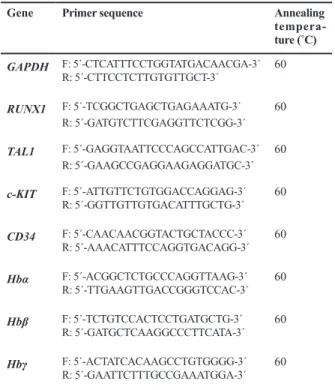
primer sequences are presented in table 1.
Table 1: Real-time polymerase chain reaction (RT-PCR) primers Annealing tempera-ture (˚C) Primer sequence Gene 60 F: 5´-CTCATTTCCTGGTATGACAACGA-3´ R: 5´-CTTCCTCTTGTGTTGCT-3´ GAPDH 60 F: 5´-TCGGCTGAGCTGAGAAATG-3´ R: 5´-GATGTCTTCGAGGTTCTCGG-3´ RUNX1 60 F: 5´-GAGGTAATTCCCAGCCATTGAC-3´ R: 5´-GAAGCCGAGGAAGAGGATGC-3´ TAL1 60 F: 5´-ATTGTTCTGTGGACCAGGAG-3´ R: 5´-GGTTGTTGTGACATTTGCTG-3´ c-KIT 60 F: 5´-CAACAACGGTACTGCTACCC-3´ R: 5´-AAACATTTCCAGGTGACAGG-3´ CD34 60 F: 5´-ACGGCTCTGCCCAGGTTAAG-3´ R: 5´-TTGAAGTTGACCGGGTCCAC-3´ Hbα 60 F: 5´-TCTGTCCACTCCTGATGCTG-3´ R: 5´-GATGCTCAAGGCCCTTCATA-3´ Hbβ 60 F: 5´-ACTATCACAAGCCTGTGGGG-3´ R: 5´-GAATTCTTTGCCGAAATGGA-3´ Hbγ
Statistical analysis
Data are presented as mean ± standard deviation. Multiple comparisons were performed with the re-peated measure test. Differences were considered
statistically signiicant at p≤0.01 or p≤0.05.
Results
Differentiation of B-hiPSCs and hESCs into he-mangioblast progenitors
The initial step in erythroid differentiation
is hemangioblast formation, for which we ex -panded B-hiPS and hES cells on matrigel-coat-ed plates. Cells in both lines formmatrigel-coat-ed compact colonies (Fig 2Ai, iii). Then, the suspension culture was used for cell proliferation and ag-gregate formation (Fig 2Aii, iv). We used a two-step method for hemangioblast differen-tiation. The first step lasted for eight days and
the combination of BMP-4, b-FGF, and VEGF was added between days 1 and 8 in serum-free medium. In the second step, the culture was al-lowed to continue until day 14 in the presence of VEGF, SCF, IL-6, EPO, IL-3, and IGF-1.
Co-expression of kinase insert domain receptor
(KDR or FLK-1), clusters of differentiation 34 (CD34) and CD31 has been shown to determine early stage hematopoietic development (13-15),
therefore we tested the cells for expressions of
those markers. According to our results,
undif-ferentiated ES cells and iPSCs expressed KDR
at low levels, while culture condition changed
to erythrocyte differentiation media, expression
of KDR significantly increased on day 14 in all
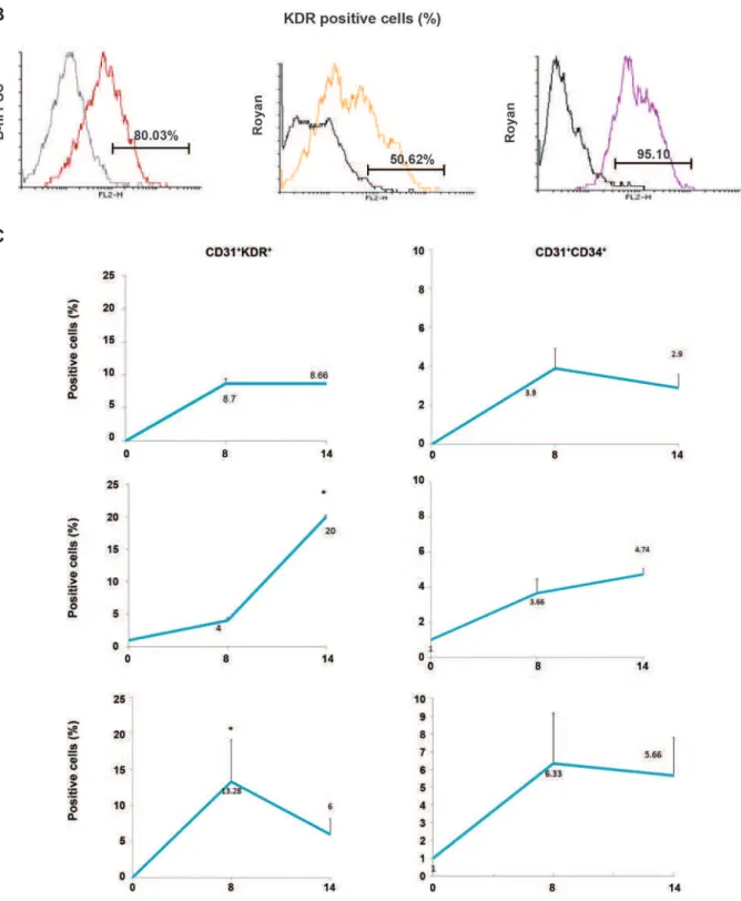
lines (Fig 2B, p≤0.01). However this expression
was prominent in differentiated iPSCs (80%) and RH5SCs (95%), when compared with RH-6SCs (50%).
Fig 2: Morphology, phenotype and mRNA analysis of B-hiPSC-derived cells before and after differentiation. A. Photograph of B-hiPSC and ESC colonies on a thin layer of matrigel (i, iii). Aggregate formation from B-hiPSC and ESC colonies under suspension conditions (ii, iv). B. Flow cytometry analysis was used to investigate KDR expression differences between B-hiPSC and ESC lines on day 14 of differentiation. C. Flow cytometry analysis showing the expression of early hematopoietic surface antigens in aggregates at days 8 and 14 of differentiation in iPSC and ES cell lines. Undifferentiated iPSC and ES cells (day 0) were used as a negative control (n=3). B-hiPSC; Bombay human-induced pluripotent stem cells, ESC; Embryonic stem cell, CD; Clusters of differentiation, KDR; Kinase insert domain receptor and *; P≤0.05.
B
As shown in figure 2B, co-expression of KDR
and CD3l was not detected in undifferentiated cells. During eight days of culture, the percent of CD31+KDR+ (8.7%) and CD31+CD34+ (3.9%)
cells significantly increased in the iPSC cell lines in addition to the significant increase that was also noted for CD31+KDR+ (13.28%) and
CD31+CD34+ (6.33%) cells in the RH5 ES cell
lines (p≤0.05). Expression of these cells per -sisted through 14 days of culture in iPSC lines and RH5 ES cells. In contrast, CD31+KDR+ and
CD31+CD34+ cells increased after 14 days of
culture in RH6 ES cells (Fig 2B). However, the CD31+KDR+ cells showed a more prominent
increase than CD31+CD34+ cells. We observed
that all CD31+KDR+ cells expressed CD34+
sur-face markers which might have been related to their hemangioblast origin (data not shown). A
comparison of all cell lines for expression of
hemangioblast markers showed that the
pat-tern of expression was similar in both the ES cell and iPSC lines, with the exception of RH6 that had a significant increase in expression of
CD31+ KDR+ up to day 14 (p≤0.05).
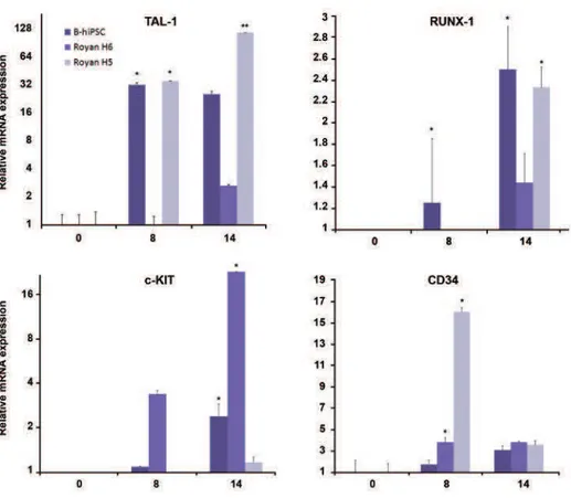
To conirm the above data, the expressions of
major hematopoietic genes, CD34, RUNX-1, c-KIT, and SCL (TAL-1) (16-20) were assessed in ES cells and iPSCs on days 8 and 14 of differentiation by quantitative RT-PCR. Our results determined
that the expression of TAL-1, RUNX-1, c-KIT, and
CD34 up-regulated at day eight and continued or increased up to day14 in both ES cells as well
as iPSCs. Expression of CD34 decreased only in RH5 signiicantly until day 14 (Fig 3). Therefore,
we proposed that the ES and iPS cells in the two-step protocol differentiated into hemangioblasts.
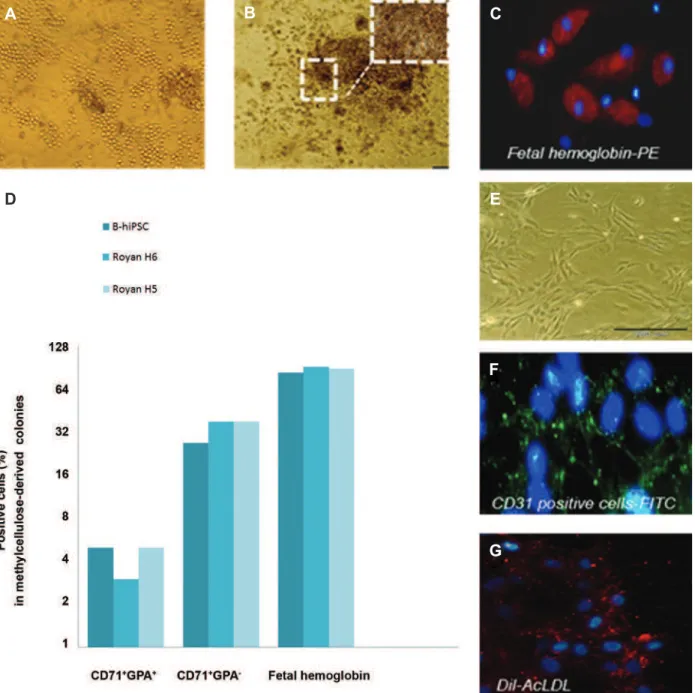
Identiication of hemangioblast functionality As previously mentioned, cells from early-stage aggregates (14-day) were cultured in con-ditions known to support the growth of blast colonies. As shown in figure 4A, colonies with grape-like morphology of hemangioblast colo-nies were detected in ES and iPSC lines after
seeding on a thin layer of matrigel for six days.
Cells isolated from these colonies at days 3, 4, 5, and 6 were sub-cultured on methylcellulose to form hematopoietic progenitor cells. As shown
in figure 4B, six-day-old colonies formed two
types of cells on methylcellulose, adhesive and non-adhesive (or loosely adhesive). Interest-ingly, non-adhesive cells formed small color-ed colonies, their color changcolor-ed to rcolor-ed pale
and more than 80% of them expressed fetal
hemoglobin (Fig 4B, C). It seems the culture
includes mixed cells. For further evaluation of
the erythroid cells, we chose colonies cultured on methylcellulose to be pooled and analyzed
for CD71 and GPA expressions by flow cytom -etry. According to our findings, about 5-8%
of cells from all lines expressed CD71+ GPA+
(p≤0.05). There was a similar pattern of CD71+
GPA+ and fetal hemoglobin expression seen in
iPSCs and RH5SCs. However, there was a
dif-ference in expression of CD71+GPA- in the ES
cell group (38%) compared to the iPSC group (27%) (Fig 4D). As during erythroid
develop-ment, the expression of CD71 happens earlier, followed by co-expression with GPA. In ma
-ture erythrocytes, expression of GPA increased
(21), therefore we have proposed that to pro-mote erythrocyte maturation in vitro, conditions should be conducive to support more erythroid maturation.
We sought to determine the characteristics of
the adhesive cells which developed from
six-day-old hemangioblast-like cells. There were endothelial-like cells in our culture (Fig 4E),
therefore we evaluated CD31 expression and
uptake of Dil-AcLDL, both of which are spe-cific for endothelial cells. Immunostaining of the blast colony-derived adhesive population revealed that over 30% were positive for CD31 surface antigen (Fig 4F) and had the potential for uptake of Dil-AcLDL (Fig 4G) in all lines. However, the condition of culture was sup-ported hematopoietic coloniesfurther, hence,
the adhesive cells could not proliferate more. These results have demonstrated the capability of hiPS and ES cells to differentiate into he-mangioblasts, erythroid, and endothelial line-ages under our differentiation system.
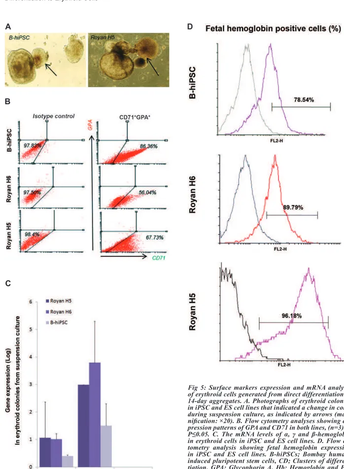
Direct differentiation of 14-day aggregates into fetal-like erythroid cells
Our previous results determined that 14-day
aggregates expressed KDR, CD31 and CD34;
therefore, these aggregates were chosen for di-rect erythroid differentiation without passage through the hemangioblast formation step. Ag-gregates cultured in the suspension media that included SCF, IL-6, VEGF, IL-3, EPO, GM-CSF, and IGF-1. At 12 days post-culture, colors of colonies changed which indicated with
in-creased expression of hemoglobin in some parts
of the colonies (Fig 5A). For obtaining more accurate results, we continued the culture of these aggregates up to day 16, then character-ized their erythroid surface antigens, intracel-lular proteins, and globin genes. According to
phenotype analysis, co-expression of CD71/
GPA, which is related to erythroid maturation (16) was not detected. In contrast, we observed
that both ES cells expressed approximately 40%
CD71+GPA- (Fig 5B) compared to more than
80% of erythroid cells that were derived from
iPSCs which expressed CD71+GPA.
Up-regula-tion of α- and γ-globin mRNA was detected in both ES cells related to iPSCs. However, ex
-pression of β-globin was not detected in both lines (Fig 5C). Also, there was approximately an 80% expression of fetal hemoglobin in RH6
line and iPSCs compared with 60% observed in the RH5 line (Fig 5D). These results confirmed that the fetal like erythroid cells produced from
both lines in this experiment and also suggested
these conditions are more supportive for fe-tal not adult characteristic. It appears that the combination of the aforementioned cytokines induced early erythroid differentiation, main-ly EPO, which played an essential role in the emergence of erythroid cells. The synchronous presence of two specific cytokines, SCF and EPO, produced a signal that markedly affected
Fig 4: Clonogenicity of blast colonies. A. Photograph of grape-like blast colonies generated from day14
aggre-gates on a thin layer of matrigel. B. Photograph showing erythroid and non-erythroid colonies (mixed-colonies) which differentiated from six-day-old blast colony in methylcellulose base media in B-hiPSC line. C. Immu -nostaining of erythroid cells by anti-human fetal hemoglobin confirmed the expression of fetal hemoglobin that
had been shown by flow cytometry. Nuclei were stained with DAPI (magnification: ×40). D. Mixed colonies picked
from CFU-culture. Expression of erythroid-specific marker CD71 (transferring receptor), CD235 (GPA) and fetal
hemoglobin as analyzed by flow cytometry. E-G.Endothelial cells appeared from B-hiPSCs; expression of CD31 markers shown by green fluorescence and LDL uptake by red fluorescence. Nuclei were stained with DAPI (mag
-nification: ×40). B-hiPSCs; Bombay human-induced pluripotent stem cells, CD; Clusters of differentiation, GPA;
Glycophorin A and DAPI; 4,6 Diamidino-2-phenylindole.
A B C
E
F
Fig 5: Surface markers expression and mRNA analysis of erythroid cells generated from direct differentiation of 14-day aggregates. A. Photographs of erythroid colonies in iPSC and ES cell lines that indicated a change in color during suspension culture, as indicated by arrows (mag -niication: ×20). B. Flow cytometry analyses showing ex -pression patterns of GPA and CD71 in both lines, (n=3).*; P≤0.05. C. The mRNA levels of α, γ and β-hemoglobin in erythroid cells in iPSC and ES cell lines. D. Flow cy -tometry analysis showing fetal hemoglobin expression in iPSC and ES cell lines. B-hiPSCs; Bombay human-induced pluripotent stem cells, CD; Clusters of differen-tiation, GPA; Glycophorin A, Hb; Hemoglobin and ES; Embryonic stem.
A
B
C
Discussion
Although transfusion of RBCs is a
well-es-tablished cellular therapy; the lack of healthy
donors, possibility of human viral infection and increasing the requirement for immunological matching limit its use. However, new discov-eries in stem cell research and introduction of pluripotent stem cells (PSCs), such as ES cells (22, 23) and iPS cells (8, 24, 25) with their po-tential to form any cell type in vitro, have been sought as possible sources to candidate for the production of unlimited numbers of erythroid cells. Most studies have shown hES cells and hiPS cells cultured in the presence of animal serum and OP9 or MS-5 mouse fibroblasts as feeder layers (26-30) in order to produce
eryth-rocytes. Few data exists for hiPSC differentia -tion into erythrocytes in feeder-free and serum-free medium.
Thus, this study employed the use of feeder-free culture for erythrocyte differentiation in
vitro with the intent to propose new, unlimited
cell sources that can be an appropriate source for those who need cell therapy in future. For first time, we used iPSCs which have been de-rived from adult cells that carry the Bombay
phenotype which fails to express ABH antigens
on RBCs (31, 32). These cells have been used to generate histocompatible erythroid cells and introduce a universal red blood source that is not patient-specific and compatible with all pa-tients’ immune systems. We have attempted to
examine the potential for erythroid differentia -tion of B-hiPSCs derived from adult cells that carry the Bombay phenotype, and then we com-pared their capability with ES cells.
Previous research in our lab has shown that ES cells and iPSCs could be maintained and
expanded as aggregate suspensions over an extended period and then induced for specific
differentiation into cardiac and hepatic cells (11). In this study, we used a feeder-free sus-pension culture and have produced aggregates that underwent induction of differentiation to-ward erythroid cells in the presence of several cytokines which are necessary for erythroid dif-ferentiation in a suspension culture.
Our results determined that B-hiPS, hRH5SC
and hRH6SC have expressed the crucial genes
TAL-1, RUNX-1, c-KIT and CD34 which are
essential during early development of heman-gioblasts in humans (16, 18, 33, 34) and can differentiate to hemangioblastsat the beginning of differentiation which is concomitant with up-regulation of TAL-1, RUNX-1 and c-KIT genes that correlated with their mesodermal-hemat-opoietic properties.
According to our analyses, KDR was ex -pressed on undifferentiated iPSCs and ES cells, and then it increased between days 8 and 14 of differentiation. KDR as a tyrosine kinase-receptor binds to its ligand, VEGF and KDR/
VEGF activates expression of genes which are
crucial in erythroid development. In primitive
streak–stage embryos, KDR expression is first detectable in cells within and exiting the primi
-tive streak as well as in the extra-embryonic
mesoderm that is crucial for development of cardiac, endothelial and hematopoietic pro-genitor cells (35, 36). It seems that the KDR+
population involves hematopoietic along with cardiac and endothelial progenitors. Addition-ally the combination of BMP4 and VEGF in-creased the numbers of KDR-positive cells in 14-day embryoid bodies (EBs) (37). Our current study also showed that iPSC- and ES cell-derived
hemangioblasts expressed KDR. Thus expres -sion of KDR increased progressively up to day
8 of differentiation, rather than expressions of
CD31 and CD34. In contrast to the other lines,
co-expression of CD31KDR and CD31CD34
markers increased significantly in RH6 line on day 8 then persisted up to day 14, although
co-expression of these surface antigens did not
changed significantly at day 14. A number of previous studies have shown the development of hES cell-derived cell types with hemangio-blast properties. Some research has shown that the CD31+VE-cadherin+KDR+CD45- population
in day-ten aggregates displayed the potential to generate both hematopoietic and
endothe-lial progeny (38). Although we did not exam
-ine the expression of CD45, however there was expression of hemangioblast-specific markers.
Subsequently, we demonstrated that iPSCs were similar to ES cell lines by their ability to dif-ferentiate into erythroid cells, the type of
glo-bin expression, surface antigen expression, and the ability to form mixed colonies. However the
A new finding in our results was the forma-tion of endothelial-like cells at the time heman-gioblasts were formed. The earliest stage of hematopoietic development in the human and mouse embryo begins in the yolk sac, within blood islands that consist of emerging primi-tive erythroblasts surrounded by endothelial cells. Consequently, researchers hypothesize that these lineages share a common origin, a progenitor known as hemangioblasts (39-41). Interestingly, when mouse-derived hemangio-blasts are cultured in methylcellulose media, these progenitors generate immature blast colo-nies that display both hematopoietic and vascu-lar potentials (42). The cell that produces these colonies, the blast colony-forming cell
(BL-CFC) or hemangioblast, expresses the receptor
tyrosine kinase Flk-1 and the mesodermal gene
T (brachyury), which demonstrates that it repre-sents a population undergoing mesoderm speci-fication to hematopoietic and vascular lineages (13, 43, 44).
Thus we have suggested that our culture meth-od induced mesmeth-odermal-hematopoietic progeni-tors (36) which easily gave rise to endothelial and hematopoietic progeny, which is similar
to the mixed colonies obtained from CD133 or
CD34 and mononuclear cells present in bone marrow, peripheral or cord blood progenitors.
Although co-expression of CD71/GPA was shown in erythroid cells, expression of fetal
hemoglobin was significantly high.
Interest-ingly, most cells in the mixed colonies were erythroid and had high expression of hemo -globin. The duration of the culture was greater
than previous and the number of cells that ex -pressed hemoglobin increased (data not shown). It seemed that the long-term presence of VEGF was effective in increasing the erythroid
popu-lation (45). We did not examine the expression of megakaryocytic lineage markers in mixed
colonies, although studies have indicated that erythroid and megakaryocytic lineage commit-ment take place together and potentially arise from a common precursor population (38).
Giv-en that adherGiv-ent cells express CD31 surface an -tigen, possibly they arose from bipotential cells as nominated hemangioblast that differentiated
from iPS and ES cells in our experiment. How -ever, they need to be cultured as single-cells for
more detailed characterization.
We have attempted to differentiate iPSCs and hES cells directly into erythroid cells in sus-pension culture over a short period of time. Our results revealed that iPS and hES cells could produce erythroid cells in this system. Although the majority of cells were hemoglobinized,
there was a low-level co-expression of CD71/
GPA. Possibly the presence of FBS promoted the generation of CD71+GPA- cells and reduced
the number of cells that expressed GPA. Ac -cording to Chang et al. (3), when non-adherent
cells were expanded in serum-free medium in
the absence of FBS they gave rise to a higher frequency of GPA cells.
According to our results, erythroid cells ex
-pressed high levels of fetal hemoglobin. mRNA expression analyses also confirmed that they expressed α- and γ-globin, where remarkably, the expression level of γ-globin was more. This
was possibly related to the presence of several cytokines as EPO, SCF. It has been shown that the combination of EPO, SCF and transforming
growth factor-beta (TGF-β) signal transduction produce a marked increase in γ-globin transcript and protein expression (46). Erythroblasts cul -tured in the presence of these cytokines reveal a significant enhancement of fetal hemoglobin (HbF) without significant effect on erythroblast maturation (47, 48). SCF also has anti-apop-totic effects on cultured erythroblasts (49, 50). Due to the lack of mature erythroid cells, we have proposed that the cytokine signals used in
our test most likely have changed the expres -sion levels of transcription factors essential for erythroid commitment and have not affected terminal differentiation or maturation of eryth-roblasts. Accordingly, our culture conditions have supported increase in fetal hemoglobin
expression.
Conclusion
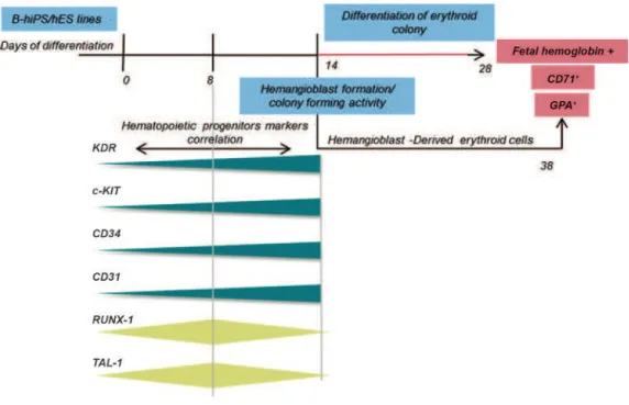
Finally, our study provided evidence that B-hiPS differentiated to erythroid cells similar to ES cells and produced a population with KDR+CD31+CD34+
characteristics. In addition, they were able to pro-duce colonies with hemangioblast properties (Fig
6) and in the inal step, they differentiated to
Fig 6: Summary of cellular and molecular events during differentiation of iPSCs and hESCs. Developmental progression of hemangioblast and erythroid gene expression during differentiation and correlation of key genes and markers. B-hiPS; Bom-bay human-induced pluripotent stem, hES; Human embryonic stem, CD; Clusters of differentiation and GPA; Glycophorin A.
Our ability to produce erythroid cells with a fetal phenotype from iPSCs and ES cells might assist with studies on the development of early erythropoiesis in
humans and be of practical use for examining thera -pies for different blood disorders, particularly
hemo-globinopathies characterized by insuficient produc
-tion of β-globin chains due to muta-tions that affect the β-globin gene complex. However, our inability
to produce adult RBCs from iPSCs or ES cells will
affect possibility of these experiments that exposure beneicial in the near future. It seems that production
of RBCs with an adult phenotype from pluripotent cells is a critical step and an issue that remains unre-solved, thus necessitating the need to develop and use more advanced techniques.
Acknowledgments
This study was funded by a grant provided from Royan Institute, Tehran, Iran.
The authors express their appreciation to Mr. Ehsan Janzamin and Mr. Sahraneshine-Samani for their
technical support in the Flow Cytometry Laboratory.
F.G. researched the data, contributed to the dis-cussion, wrote and edited the manuscript. S.A.
con-tributed to the discussion, wrote and reviewed the manuscript. M.E. contributed to the discussion and
reviewed the manuscript. N.A. contributed to the
discussion. H.B. contributed to the discussion and
re-viewed the manuscript. The authors have no conlict
of interest in this article.
References
1. Liu QP, Yuan H, Bennett EP, Levery SB, Nudelman E, Spence J, et al. Identiication of a GH110 subfamily of alpha 1,3-galactosi -dases: novel enzymes for removal of the alpha 3Gal xenotrans -plantation antigen. J Biol Chem. 2008; 283(13): 8545-8554. 2. Douay L, Andreu G. Ex vivo production of human red blood cells
from hematopoietic stem cells: what is the future in transfusion?. Transfus Med Rev. 2007; 21(2): 91-100.
3. Chang KH, Nelson AM, Cao H, Wang L, Nakamoto B, Ware CB, et al. Deinitive-like erythroid cells derived from human embryonic stem cells coexpress high levels of embryonic and fetal globins with little or no adult globin. Blood. 2006; 108(5): 1515-1523. 4. Park IH, Zhao R, West JA, Yabuuchi A, Huo H, Ince TA, et al. Re
-programming of human somatic cells to pluripotency with deined factors. Nature. 2008; 451(7175): 141-146.
5. Boland MJ, Hazen JL, Nazor KL, Rodriguez AR, Gifford W, Martin G, et al. Adult mice generated from induced pluripotent stem cells. Nature. 2009; 461(7260): 91-94.
6. Lengner CJ. iPS cell technology in regenerative medicine. Ann N Y Acad Sci. 2010; 1192: 38-44.
7. Choi KD, Yu J, Smuga-Otto K, Salvagiotto G, Rehrauer W, Vody -anik M, et al. Hematopoietic and endothelial differentiation of human induced pluripotent stem cells. Stem Cells. 2009; 27(3): 559-567.
9. Seiinejad A, Taei A, Totonchi M, Vazirinasab H, Hassani SN, Aghdami N, et al. Generation of human induced pluripotent stem cells from a Bombay individual: moving towards "universal-donor" red blood cells. Biochem Biophys Res Commun. 2010; 391(1): 329-334.
10. Baharvand H, Ashtiani SK, Taee A, Massumi M, Valojerdi MR, Yazdi PE, et al. Generation of new human embryonic stem cell lines with diploid and triploid karyotypes. Dev Growth Differ. 2006; 48(2): 117-128.
11. Larijani MR, Seiinejad A, Pournasr B, Hajihoseini V, Hassani SN, Totonchi M, et al. Long-term maintenance of undifferentiated hu -man embryonic and induced pluripotent stem cells in suspension. Stem Cells Dev. 2011; 20(11): 1911-1923.
12. Kennedy M, DSouza SL, Lynch-Kattman M, Schwantz S, Kel -ler G. Development of the hemangioblast deines the onset of hematopoiesis in human ES cell differentiation cultures. Blood. 2007; 109(7): 2679-2687.
13. Fehling HJ, Lacaud G, Kubo A, Kennedy M, Robertson S, Keller G, et al.Tracking mesoderm induction and its speciication to the hemangioblast during embryonic stem cell differentiation. Devel -opment. 2003; 130(17): 4217-4227.
14. Kabrun N, Buhring HJ, Choi K, Ullrich A, Risau W, Keller G. Flk-1 expression deines a population of early embryonic hematopoi -etic precursors. Development. 1997; 124(10): 2039-2048. 15. Mikkola HK, Fujiwara Y, Schlaeger TM, Traver D, Orkin SH. Ex
-pression of CD41 marks the initiation of deinitive hematopoiesis in the mouse embryo. Blood. 2003; 101(2): 508-516.
16. Yokochi T, Brice M, Rabinovitch PS, Papayannopoulou T, Stama -toyannopoulos G. Monoclonal antibodies detecting antigenic de -terminants with restricted expression on erythroid cells: from the erythroid committed progenitor level to the mature erythroblast. Blood. 1984; 63(6): 1376-1384.
17. Krause DS, Fackler MJ, Civin CI, May WS. CD34: structure, biol -ogy, and clinical utility. Blood. 1996; 87(1): 1-13.
18. Vodyanik MA, Bork JA, Thomson JA, Slukvin II. Human embryon -ic stem cell-derived CD34+ cells: ef-icient production in the cocul -ture with OP9 stromal cells and analysis of lymphohematopoietic potential. Blood. 2005; 105(2): 617-626.
19. Park C, Afrikanova I, Chung YS, Zhang WJ, Arentson E, Fong Gh Gh, et al. A hierarchical order of factors in the generation of FLK1- and SCL-expressing hematopoietic and endothelial pro -genitors from embryonic stem cells. Development. 2004; 131(11): 2749-2762.
20. Yamaguchi TP, Dumont DJ, Conlon RA, Breitman ML, Rossant J. lk-1, an lt-related receptor tyrosine kinase is an early marker for endothelial cell precursors. Development. 1993; 118(2): 489-498.
21. Scicchitano MS, McFarland DC, Tierney LA, Narayanan PK, Schwartz LW. In vitro expansion of human cord blood CD36+ erythroid progenitors: temporal changes in gene and protein ex -pression. Exp Hematol. 2003; 31(9): 760-769.
22. Evans MJ, Kaufman MH. Establishment in culture of pluripotential cells from mouse embryos. Nature. 1981; 292(5819): 154-156. 23. Thomson JA, Itskovitz-Eldor J, Shapiro SS, Waknitz MA, Swier
-giel JJ, Marshall VS, et al. Embryonic stem cell lines derived from human blastocysts. Science. 1998; 282(5391): 1145-1147. 24. Takahashi K, Tanabe K, Ohnuki M, Narita M, Ichisaka T, Tomoda
K, et al. Induction of pluripotent stem cells from adult human ibro -blasts by deined factors. Cell. 2007; 131(5): 861-872.
25. Park IH, Arora N, Huo H, Maherali N, Ahfeldt T, Shimamura A, et al. Disease-speciic induced pluripotent stem cells. Cell. 2008; 134(5): 877-886.
26. Ma F, Ebihara Y, Umeda K, Sakai H, Hanada S, Zhang H, et al. Generation of functional erythrocytes from human embryonic stem cell-derived deinitive hematopoiesis. Proc Natl Acad Sci USA. 2008; 105(35): 13087-13092.
27. Lapillonne H, Kobari L, Mazurier C, Tropel P, Giarratana MC, Zanella-Cleon I, et al. Red blood cell generation from human in -duced pluripotent stem cells: perspectives for transfusion medi -cine. Haematologica. 2010; 95(10): 1651-1659.
28. Lu SJ, Feng Q, Park JS, Vida L, Lee BS, Strausbauch M, et al. Biologic properties and enucleation of red blood cells from human embryonic stem cells. Blood. 2008; 112(12): 4475-4484. 29. Honig GR, Lu SJ, Feng Q, Vida LN, Lee BS, Lanza R.
alpha-Thalassemia-like globin gene expression by primitive erythro -cytes derived from human embryonic stem cells. Hemoglobin. 2010; 34(2): 145-150.
30. Chang CJ, Mitra K, Koya M, Velho M, Desprat R, Lenz J, et al. Production of embryonic and fetal-like red blood cells from hu -man induced pluripotent stem cells. PLoS One. 2011; 6(10): e25761.
31. Koda Y, Soejima M, Johnson PH, Smart E, Kimura H. Missense mutation of FUT1 and deletion of FUT2 are responsible for Indian Bombay phenotype of ABO blood group system. Biochem Bio -phys Res Commun. 1997; 238(1): 21-25.
32. Koda Y, Soejima M, Wang B, Kimura H. Structure and expression of the gene encoding secretor-type galactoside 2-alpha-L-fuco -syltransferase (FUT2). Eur J Biochem. 1997; 246(3): 750-755. 33. Tian X, Morris JK, Linehan JL, Kaufman DS. Cytokine require
-ments differ for stroma and embryoid body-mediated hemat -opoiesis from human embryonic stem cells. Exp Hematol. 2004; 32(10): 1000-1009.
34. Okuda Y, Sakoda S, Fujimura H, Yanagihara T. Pentoxifylline de -lays the onset of experimental allergic encephalomyelitis in mice by modulating cytokine production in peripheral blood mononu -clear cells. Immunopharmacology. 1996; 35(2): 141-148. 35. Tavian M, Peault B. Embryonic development of the human he
-matopoietic system. Int J Dev Biol. 2005; 49(2-3): 243-250. 36. Zambidis ET, Peault B, Park TS, Bunz F, Civin CI. Hematopoi
-etic differentiation of human embryonic stem cells progresses through sequential hematoendothelial, primitive, and deinitive stages resembling human yolk sac development. Blood. 2005; 106(3): 860-870.
37. Pearson S, Sroczynska P, Lacaud G, Kouskoff V. The stepwise speciication of embryonic stem cells to hematopoietic fate is driv -en by sequ-ential exposure to Bmp4, activin A, bFGF and VEGF. Development. 2008; 135(8): 1525-1535.
38. Wang L, Li L, Shojaei F, Levac K, Cerdan C, Menendez P, et al. Endothelial and hematopoietic cell fate of human embryonic stem cells originates from primitive endothelium with hemangioblastic properties. Immunity. 2004; 21(1): 31-41.
39. Haar JL, Ackerman GA. A phase and electron microscopic study of vasculogenesis and erythropoiesis in the yolk sac of the mouse. Anat Rec. 1971; 170(2): 199-223.
40. Palis J, McGrath KE, Kingsley PD. Initiation of hematopoiesis and vasculogenesis in murine yolk sac explants. Blood. 1995; 86(1): 156-163.
41. Keller G. Embryonic stem cell differentiation: emergence of a new era in biology and medicine. Genes Dev. 2005; 19(10): 1129-1155.
42. Choi K, Kennedy M, Kazarov A, Papadimitriou JC, Keller G. A common precursor for hematopoietic and endothelial cells. De -velopment. 1998; 125(4): 725-732.
43. Park TS, Zimmerlin L, Zambidis ET. Eficient and simultaneous generation of hematopoietic and vascular progenitors from hu -man induced pluripotent stem cells. Cytometry A. 2013; 83(1): 114-126
44. Peters A, Burridge PW, Pryzhkova MV, Levine MA, Park TS, Rox -bury C, et al. Challenges and strategies for generating therapeu -tic patient-speciic hemangioblasts and hematopoie-tic stem cells from human pluripotent stem cells. Int J Dev Biol. 2010; 54(6-7): 965-990.
45. Palis J, Robertson S, Kennedy M, Wall C, Keller G. Development of erythroid and myeloid progenitors in the yolk sac and embryo proper of the mouse. Development. 1999; 126(22): 5073-5084. 46. Sripichai O, Kiefer CM, Bhanu NV, Tanno T, Noh SJ, Goh SH, et
al. Cytokine-mediated increases in fetal hemoglobin are associ -ated with globin gene histone modiication and transcription factor reprogramming. Blood. 2009; 114(11): 2299-2306.
47. Wojda U, Leigh KR, Njoroge JM, Jackson KA, Natarajan B, Stitely M, et al. Fetal hemoglobin modulation during human erythropoie -sis: stem cell factor has "late" effects related to the expression pattern of CD117. Blood. 2003; 101(2): 492-497.
48. Gabbianelli M, Testa U, Massa A, Morsilli O, Saulle E, Sposi NM, et al. HbF reactivation in sibling BFU-E colonies: synergistic inter -action of kit ligand with low-dose dexamethasone. Blood. 2003; 101(7): 2826-2832.
49. Endo T, Odb A, Satoh I, Haseyama Y, Nishio M, Koizumi K, et al. Stem cell factor protects c-kit+ human primary erythroid cells from apoptosis. Exp Hematol. 2001; 29(7): 833-841.