Mesenchymal stem cell therapy promotes the improvement and recovery of
renal function in a preclinical model
Antônio Urt-Filho
1,2,*, Rodrigo Juliano Oliveira
1,2,3,*, Larissa Correa Hermeto
4, João Renato Pesarini
1,2,
Natan de David
1,3, Wilson de Barros Cantero
1,2, Gustavo Falcão
5, Guido Marks
2and
Andréia Conceição Milan Brochado Antoniolli-Silva
1,2.
1Centro de Estudos em Células Tronco, Terapia Celular e Genética Toxicológica, Hospital Universitário
“Maria Aparecida Pedrossian”, Empresa Brasileira de Serviços Hospitalares, Campo Grande, MS, Brazil.
2
Programa de Pós-Graduação em Saúde e Desenvolvimento na Região Centro-Oeste, Faculdade de
Medicina “Dr. Hélio Mandetta”, Universidade Federal de Mato Grosso do Sul, Campo Grande, MS, Brazil.
3
Programa de Mestrado em Farmácia, Centro de Ciências Biológicas e da Saúde,
Universidade Federal de Mato Grosso do Sul, Campo Grande, MS, Brazil.
4
Programa de Pós-Graduação em Clínica Veterinária, Faculdade de Ciências Agrária e Veterinária,
Universidade Estadual Paulista “Júlio de Mesquita Filho”, Jaboticabal, SP, Brazil.
5
Faculdade de Medicina “Dr. Hélio Mandetta”, Universidade Federal de Mato Grosso do Sul,
Campo Grande, MS, Brazil.
Abstract
Acute renal failure (ARF) is an extremely important public health issue in need of novel therapies. The present study aimed to evaluate the capacity of mesenchymal stem cell (MSC) therapy to promote the improvement and recovery of renal function in a preclinical model. Wistar rats were used as the experimental model, and our results show that cisplatin (5mg/kg) can efficiently induce ARF, as measured by changes in biochemical (urea and creatinine) and histological parameters. MSC therapy performed 24h after the administration of chemotherapy resulted in normal-ized plasma urea and creatinine levels 30 and 45d after the onset of kidney disease. Furthermore, MSC therapy sig-nificantly reduced histological changes (intratubular cast formation in protein overload nephropathy and tubular hydropic degeneration) in this ARF model. Thus, considering that current therapies for ARF are merely palliative and that MSC therapy can promote the improvement and recovery of renal function in this model system, we suggest that innovative/alternative therapies involving MSCs should be considered for clinical studies in humans to treat ARF.
Keywords: bone marrow, chemotherapy, nephrotoxicity, kidney disease, tissue regeneration. Received: July 09, 2015; Accepted: December 29, 2015.
Introduction
Acute renal failure (ARF) is characterized by the ac-cumulation of nitrogen-containing compounds (urea and creatinine) in the blood (with or without oliguria) followed by the loss of renal function, ultimately blocking the excre-tion of waste nitrogen and maintenance of homeostasis (Thadhaniet al., 1996).
ARF is associated with high mortality and morbidity rates and is reported in approximately 4-5% of all hospital-ization cases. This disease can develop into more severe forms, resulting in death in approximately 50-70% of
pa-tients, making ARF an extremely important public health issue (Hoste and Schurgers, 2008; Wenet al., 2010).
ARF can be caused by a number of factors, including decreased renal perfusion without cellular injury, an is-chemic, toxic or obstructive insult to the renal tubules, a tu-bule-interstitial process with inflammation and edema, or a primary reduction of glomerular filtering capacity, among others (Thadhaniet al., 1996). Chemo- and radiotherapy
are also important causes of ARF (Humphreyset al., 2005).
Among chemotherapeutic drugs, cisplatin, which is used to treat the majority of solid and hematological tumors (Ser-peloniet al., 2013), is most associated with nephrotoxicity
due to its high concentrations in the kidneys, even at non-toxic plasma levels, as well as its adverse impact on the re-nal transport system (Gordon and Gattone 1986; Renet al.,
2002; Serpeloniet al., 2013). DOI: http://dx.doi.org/10.1590/1678-4685-GMB-2015-0178
Send correspondence to Rodrigo Juliano Oliveira, Federal Univer-sity of Mato Grosso do Sul, Medical School (FAMED), Cidade Universitária, S/N. Campo Grande, MS, Brazil. E-mail: [email protected]
*These authors contributed equally to this work
Generally, ARF is incurable, and current therapeutic strategies involve simply removing or treating its cause (Fry and Farrington, 2006). As ARF is prone to complica-tions and chronicity, innovative/alternative therapies for the quick, safe and efficient recovery of renal function are increasingly required.
Currently, models of ARF involving cell therapy have been effective and promising (Yokoo et al., 2003;
Brodie and Humes, 2005; Zerbiniet al., 2006 Choiet al.,
2010; Papazovaet al., 2015). In the present study, we
in-vestigated the capacity of mesenchymal stem cell (MSC) therapy to promote the improvement and recovery of renal function in a pre-clinical model.
Material and Methods
Experiment 1
Chemical agents
ARF was induced by means of a single-dose adminis-tration of the chemotherapeutic agent cisplatin (Fauldcispla LIBBS®, Brazil, Lot: 12GO702) at 5mg/kg body weight (bw) via intraperitoneal (ip) injection.
Animals
Fifty-nine sexually mature male Wistar rats were ob-tained from the Central Animal House of the Center of Bio-logical and Health Sciences of the Federal University of Mato Grosso do Sul (Centro de Ciências Biológicas e da Saúde da Universidade Federal de Mato Grosso do Sul – CCBS/UFMS). The animals were housed in groups of three in polypropylene cages containing wood shavings as bed-ding and were fed a commercial diet (Nuvital®, Brazil) and filtered waterad libitum. The cages were kept in a
venti-lated cabinet (Alesco®, Brazil) under standard climate-controlled conditions with a 12-h photoperiod (12:12 h LD), a temperature of 222 °C, and a relative humidity of 5510%. The experiment was conducted in accordance with the guidelines of the Universal Declaration of Animal Rights and was approved by the Animal Research Ethics Committee of the UFMS (Protocol #499/2012).
Experimental design
Initially, the animals were divided into two lots. Lot I consisted of 10 animals, which were used as bone mar-row-derived MSC donors. Lot II consisted of 49 animals, which were further subdivided into three experimental groups. Control Group (CG;n= 14) animals were treated
with a single dose of 1 mL/100g (bw; ip) phosphate-buff-ered saline (PBS), followed by an intravenous (iv) dose of 1 mL PBS via tail vein injection after 24 h. Control animals were evaluated at 30 days (CG-30d;n = 7) and 45 days
(CG-45d; n = 7) after the onset of treatment. The ARF
Group (ARFG;n= 21) animals were treated with a single
dose of cisplatin at 5 mg/kg (bw; ip), followed by 1 mL PBS (iv) 24-h later. ARF animals were then evaluated at 48 h
(ARFG-48h;n = 7), 30 days (ARFG-30d;n= 7), and 45
days (ARFG-45d; n = 7) after cisplatin treatment. The
ARFG + MSC Group (ARFG+MSC;n= 14) animals were
treated with a single dose of cisplatin at 5mg/kg (bw; ip), followed by treatment with MSCs in 1 mL PBS (iv) 24h later. Animals were then evaluated at 30days (ARF+MSC-30d;n = 7) and 45days (ARFG+MSC-45d; n= 7) after cisplatin treatment.
Assessment of renal function and confirmation of acute renal failure
ARF was confirmed indirectly by measuring serum urea and creatinine levels. Peripheral blood was collected under anesthesia (ketamine – 50 mg/kg bw, ip; xylazine – 10mg/kg bw, ip) from the retro-orbital plexus, either at 48h in the ARFG group (used as the standard for the establish-ment of ARF), or at 30d and 45d after cisplatin administra-tion in the other groups. The first blood sample was drawn from animals in the ARFG-48h group 7d prior to the ad-ministration of cisplatin, which was used as an internal con-trol for the experiment. After collection, the blood was allowed to sediment at 4 °C, and the serum was then stored at -20 °C until the time of analysis. Analyses were per-formed using an automated COBAS 6000 analyzer (Roche Diagnostics ®, Brazil), according to the manufacturer’s recommendations.
Isolation and expansion of bone marrow-derived mesenchymal stem cells
Donor animals from Lot I were euthanized by anes-thetic overdose (Thiopentax® Cristalia Laboratories, Bra-zil, Lot: 12075226). Femurs were then collected under ster-ile conditions, and the bone marrow was flushed with 5mL PBS supplemented with 1% antibiotics (penicil-lin/streptomycin, LGB Biotecnologia®, Brazil). Cell sus-pensions were centrifuged for 5 min at 670 x g, and the pel-let was resuspended with 5 mL sterile PBS; resuspension was performed three times. The pellet was then resuspend-ed with 4 mL Dulbecco’s Low Glucose Modifiresuspend-ed Eagle’s Medium (DMEM-low glucose; LGC Biotecnologia®, Bra-zil), transferred to a conical tube containing 4 mL SeptCell (LGC Biotecnologia®, Brazil), and then centrifuged for 30 min at 241 x g. The mononuclear cell fraction was collected and resuspended with 5 mL PBS, centrifuged for 5 min at 670 x g, and then resuspended with PBS. This last step was repeated five times before the pellet from each donor ani-mal was seeded into two 25-cm2culture flasks containing
10 mL of DMEM-low glucose supplemented with 10% Fe-tal Calf Serum (LGC Biotecnologia®, Brazil) and 1% anti-biotics (penicillin/streptomycin, LGB Biotecnologia, Brazil). Cell viability was assessed by adding an equal vol-ume of trypan blue (LGC Biotecnologia®, Brazil) to 10
mL samples from each donor.
Cells were cultured in a CO2incubator (Thermo
Sci-entific®, USA) at 37 °C under 5% CO2. During expansion,
During each passage (cultures at 70-80% confluence), the cells were washed three times with 5 mL PBS and then trypsinized by adding 1 or 2 mL of 0.25% trypsin (LGC Biotecnologia®, Brazil) to the 25- or 75-cm2flasks,
respec-tively. The trypsin was inactivated by adding 5 volumes of supplemented culture medium. The cell suspension was then centrifuged for 5 min at 670 x g, and the pellet was re-suspended with 1 mL of supplemented culture medium. For the first passage, each 25-cm2flask was divided into two
25-cm2flasks. For the second and third passages, cells were
seeded into 75-cm2flasks at 1:1 and 1:2 ratios, respectively.
During each passage, 10mL samples were collected for trypan blue analysis of cell viability.
Osteogenic, adipogenic and chondrogenic differentiation Flasks intended for differentiation experiments were trypsinized as described above after reaching 80% conflu-ence; four 25-cm2flasks were seeded with 2 105cells each,
and two 15-mL conical tubes were seeded with 1 106cells
each. Two 25-cm2flasks and one conical tube were used as
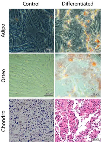
controls and cultured with supplemented culture medium. Cells used for osteogenic and adipogenic differentiation were preincubated with supplemented culture medium for 24 h and then cultured for 14 d with culture medium from the STEMPRO Osteogenesis or Adipogenesis Differentia-tion Kit (Invitrogen®, USA), respectively. For chondro-genic differentiation, cells were pre-incubated in supple-mented medium for 2 h and then cultured for 21 d with culture medium from the STEMPRO Chondrogenesis Dif-ferentiation Kit (Invitrogen®, USA). Culture media from all of the above differentiation cultures were changed twice weekly. At the end of the respective culture periods, media were removed and cells were fixed in paraformaldehyde for 1 h. Adipogenic and osteogenic differentiation were as-sessed by staining with Oil Red O and Alizarin Red S, re-spectively. Chondrogenic differentiation was assessed by submitting the pellet to conventional histological process-ing and stainprocess-ing with toluidine blue (Hermetoet al., 2015).
Mesenchymal stem cell transplantation
After reaching confluence during the third passage, 49 culture flasks originated from the first unique 10 donors were trypsinized and randomly transplanted to the 49 re-ceptor animals from the different experimental groups. Suspended cells were centrifuged for 5 min at 670 x g, and the pellet was resuspended with PBS to obtain 1.0 x 10⁶ cells for use in cell therapy. Animals from the ARFG+MSC received MSC transplant (iv) 24 h after cisplatin adminis-tration, whereas animals from the remaining groups were treated with 1mL of MSC suspension vehicle (sterile PBS; iv). For transplantation, the animals were anesthetized as described above.
Histopathological analysis
At the end of each experimental period (48 h, 30 d and 45 d), the animals were euthanized by an anesthetic
over-dose, as described above. Kidneys were collected, sec-tioned, fixed in 10% buffered formaldehyde and prepared according to routine histopathological practices. Briefly, tissue fragments were dehydrated after fixation, cleaned, and then embedded in paraffin. Samples were prepared us-ing a microtome (4-mm thick slices) and stained with hema-toxylin/eosin (HE) for histopathological analysis by bright-field microscopy (1000x magnification).
Slides were submitted to double-blind histopatho-logical analysis according to the Banff 97 Classification Criteria: 1) tubulitis: intratubular lymphocyte infiltration (necrosis and the degree of fibrosis were not evaluated); 2) tubular hydropic degeneration: cytoplasmic ballooning of cells in the proximal convoluted tubule (1+ < 25%; 2+ 25–75%; 3+ > 75%); 3) intratubular cast formation: eosino-philic deposits within tubules (proximal convoluted and distal tubule, Henle’s loop, failure of glomerular filtration, failure of tubule reabsorption) (1+ < 25%; 2+ 25–75%; 3+ > 75%); 4) glomerulitis: lymphocyte infiltration in the glomeruli; 5) arteritis: lymphocyte infiltration in the arteri-oles; 6) interstitial infiltration: lymphocytes in the intersti-tial space (1+ 0–25%; 2+ 25–75%; 3+ > 75%); 7) interstitial fibrosis: scar collagen deposition; 8) glomerulo-sclerosis: fibrosis in the glomeruli (nephron failure); 9) calcifications; 10) cortical retraction/atrophy: partial sub-stitution of kidney parenchyma by fibrosis and/or inflam-matory infiltration; 11) tubular apoptosis/necrosis: total or partial tubule degeneration (1+ < 25%; 2+ 25–75%; 3+ > 75%; and 12) global necrosis: involvement of the glomeruli and tubules (Racusenet al., 1999).
Statistical Analysis
Statistical analysis was performed using the SigmaPlot statistics software program (Version 12.5), with the significance level set at 5%. The applied non-para-metric tests included the Mann-WhitneyUtest for
compar-isons between two experimental groups, and the Kruskal-Wallis test, followed by Dunn’s test, for comparisons be-tween more than two experimental groups.
Experiment 2: Mesenchymal stem cell localization and migration
Experimental animals, design and conditions
Six sexually mature male Wistar rats were divided into two experimental groups (n = 4): ARFG and
ARFG+MSC. Animals were evaluated 48 h after the trans-plantation of MSCs that were previously stained with the nuclear marker 4’,6-diamidino-2-phenylindole (DAPI, Life Technologies®). Animal housing conditions, determi-nation of renal function, confirmation of ARF, anesthesia, and euthanasia, as well as isolation, expansion and trans-plantation of MSCs were performed as described for Ex-periment 1. However, prior to transplantation, MSCs were incubated with 10mg/mL of DAPI in the dark for 30 min at
Histological fluorescence analysis
After euthanasia, animal kidneys were collected and immediately frozen in liquid nitrogen. Organs were then mounted on a holder with cryostat embedding medium (EasyPath), and 5-mm thick slices were cut at -10 °C in the
dark. Sections were placed on slides and analyzed by fluo-rescence microscopy (BA410 FL) with a DAPI filter (EX D350/50x; DM 400DCLP; BA D460/50m) at 400x magni-fication.
Results
Expansion of mesenchymal stem cells
After the isolation procedure, MSCs were subjected to an expansion protocol with consecutive passages. For all passages, only cultures with > 95% viability were main-tained.
Mesenchymal stem cells differentiate into
osteogenic, adipogenic and chondrogenic cells in vitro
Bone-marrow-derived MSCs obtained from donor rats were induced to differentiate, and characteristic mor-phological changes were observed in these cell types. Fig-ure 1 shows representative images of undifferentiated con-trol cells cultured on an adherent surface (Concon-trol), as well as adipogenic and osteogenic differentiation (Differentia-tion). Osteogenesis was assessed by means of Alizarin Red S staining, which labels calcium deposits in differentiated cells, and adipogenesis was assessed by means of Oil Red O staining, which labels fat vesicles and vacuoles in adipo-genic cells. The lower row shows undifferentiated cells from pellet cultures (Control), and chondrogenic differenti-ated MSCs, the latter showing toluidine blue staining, indi-cating increased extracellular matrix deposition by differ-entiated chondrocytes.
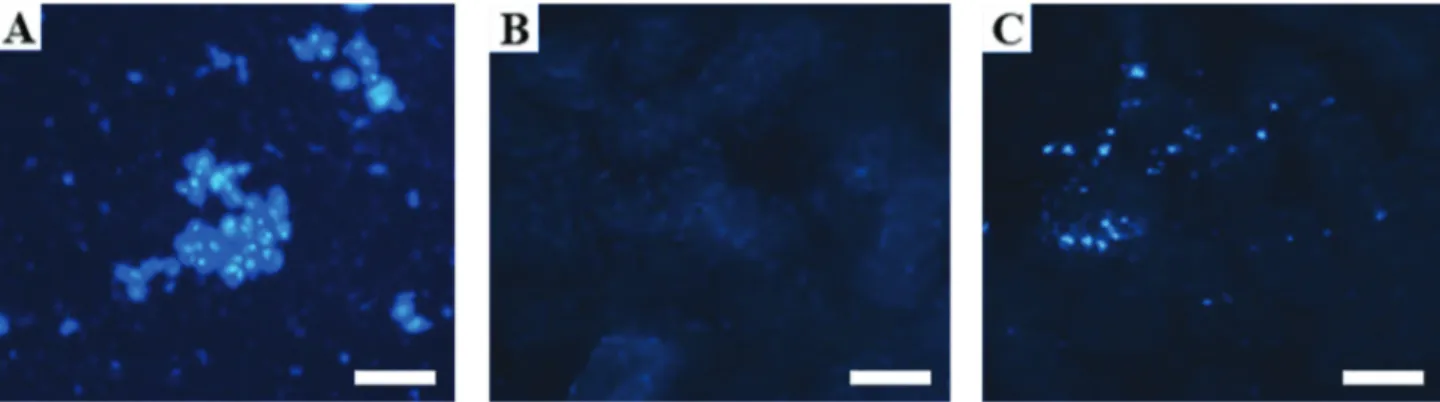
Mesenchymal stem cell localization and migration
DAPI-stained MSCs migrated from the bloodstream to the kidneys, and their localization was confirmed by flu-orescence microscopy. Figure 2 shows a stained MSC sus-pension (A), a renal cortex with no evidence of MSC infiltration (B), and a renal cortex exhibiting MSCs stained with DAPI (C).
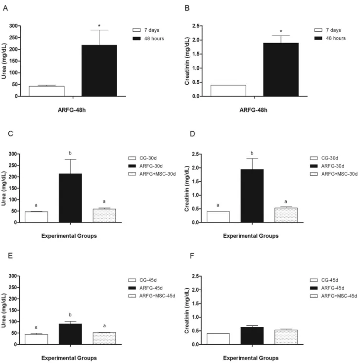
Validation of the cisplatin-induced acute renal failure model
Plasma creatinine and urea levels were measured in the ARFG-48h group 7 d before and 48 h after the adminis-tration of cisplatin, and statistically significant increases in the levels of these biomarkers confirmed the onset of ARF (Figure 3A,B). Mean urea and creatinine levels were 43.43 ± 4.07 and 0.40 ± 0.00, respectively; after cisplatin
admin-istration, levels increased (p£0.05) to 218.00 ± 64.42 and
1.89 ± 0.26, respectively.
Based on urea and creatinine levels 30 d after the ad-ministration of chemotherapy in the ARFG group, we con-cluded that cisplatin treatment successfully induced ARF, which persisted throughout the experimental period, exhib-iting mean values of 213.29 ± 63.23 and 1.94 ± 0.40, respectively. By contrast, in the ARFG+MSC, transplanta-tion of MSCs 24h after chemotherapy led to improved renal function, with mean urea and creatinine values of 59.14 ± 4.53 and 0.53 ± 0.05, respectively. Importantly, these val-ues were statistically similar (p > 0.05) to those from the CG group (47.43 ± 1.54 and 0.40 ± 0.00 for urea and creatinine, respectively).
At 45 d, creatinine levels were no longer signifi-cantly different (p > 0.05) between experimental groups, whereas urea levels remained high (p£0.05) in the ARFG
group (90.5 ± 10.56) compared with the CG (44.14 ± 4.36) and ARFG+MSC (52.57 ± 1.19) groups; the latter two groups were not significantly different from one another (p > 0.05).
Histopathological analysis
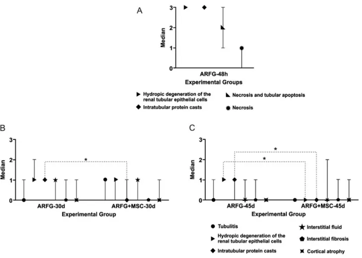
Histopathological analysis of the ARFG-48h group revealed tubular hydropic degeneration, intratubular cast formation, tubular necrosis/apoptosis, and global necrosis, with medians of 3, 3, 2 and 1, respectively.
In the ARFG and ARFG+MSC groups, the observed changes were tubulitis, tubular hydropic degeneration, intratubular cast formation, interstitial infiltration, intersti-tial fibrosis, and cortical atrophy – irrespective of the period of analysis (30 d or 45 d) (Figure 4A, Figure 5).
Comparisons between the ARFG-30d and the ARFG+MSC-30d groups revealed a statistically significant reduction in intratubular cast formation, with medians of 1 and 0, respectively. Conversely, there was no statistically significant difference between the ARFG-30d and the ARFG+MSC-30d groups with respect to tubulitis, tubular hydropic degeneration, interstitial infiltration, interstitial fibrosis, or cortical atrophy (medians of 0, 1, 1, 0 and 0vs.
1, 1, 1, 0 and 1, respectively) (Figure 4B).
When comparing the ARFG-45d and the ARFG+MSC-45d groups, there was a statistically signifi-cant reduction in tubular hydropic degeneration (medians of 1 and 0, respectively) and intratubular cast formation (medians of 1 and 0, respectively). No significant differ-ences were observed between the ARFG-45d and ARFG+MSC-45d groups with respect to tubulitis, intersti-tial infiltration, interstiintersti-tial fibrosis, or cortical atrophy (me-dians of 0, 1, 1, 0vs.0, 0, 0, 0, respectively) (Figure 4C).
Discussion
Preclinical models are currently being used to assess the effects of MSCs on various nephropathies. Of the sev-eral types of kidney disease that affect humans, renal failure is the primary target of modern bioengineering approaches, as cell therapy can replace specific cells types that have been lost in renal patients (Brodie and Humes, 2005). Such studies have addressed how cell therapy can help regener-ate convoluted tubules and promote the normalization of biochemical parameters after an ischemic or toxic insult to
the kidney (Lin et al., 2003; Lin, 2006). However, the
mechanisms involved in these processes remain poorly un-derstood, and further studies are needed to extend these findings.
The MSCs used in the present study were character-ized by adipogenic, osteogenic and chondrogenic differen-tiation. These results are consistent with previous studies (Bagnaninchiet al., 2011; Pendletonet al., 2013; Vieiraet al., 2014; Hermetoet al., 2015), as was the efficiency of the
selection and expansion methods used here.
The migration of MSCs to the kidney and the perma-nence of these cells remain controversial topics. However, in the present study, MSCs were stained with DAPI prior to intravenous transplantation, and they were identified in the kidney 48h after cell therapy. This result is consistent with other studies showing the presence of MSCs in the kidney (Rosset al., 2012; Tanget al., 2012; Panet al., 2014). In
ad-dition to nuclear staining with DAPI, other protocols have also been used to detect MSCs in the kidneys, including tagging with green fluorescent protein (Qiuet al., 2014),
detection ofb-galactosidase (b-Gal) (Klinkhammeret al.,
2014), and transplantation of cells derived from different sexes (e.g., male donor and female recipient) (Grimmet al.,
2001). Despite these studies, some authors have reported an absence of MSCs in the kidney in experiments performed with MSCs and ARF (Duffieldet al., 2005).
These discrepancies aside, studies involving MSC transplants have yielded good results (Altunet al., 2012;
Liu et al., 2012; Xinaris et al., 2013; Erpicum et al.,
2014), and in general, exogenous MSC donor sources are used (Morigiet al., 2004), as was the case in the present
study.
Our results demonstrate that ARF is efficiently in-duced by treatment with cisplatin – a widely used cancer therapy drug whose major complication is nephrotoxicity (Nishihara et al., 2013; Hagar et al., 2015). Urea and
creatinine levels were used as biomarkers to confirm ARF induction, and these were increased 48h after chemother-apy administration. ARF was also confirmed histologically by detecting tubular hydropic degeneration, intratubular
cast formation, tubular necrosis and apoptosis, and global necrosis. We also observed that these biochemical and histological changes persisted 30d after cisplatin adminis-tration. At 45 d, only increased urea levels and histological damage could still be observed, whereas creatinine levels were back to normal (i.e., not significantly different from
the control group). These observations suggest that a single dose of 5 mg/kg (bw; ip) cisplatin in Wistar rats is sufficient
to efficiently induce ARF by toxic insult in this experiment model. Thus, this model can be used in studies to evaluate the events associated with patients under chemotherapy during the occurrence of ARF.
With respect to MSC transplantation, this method proved to be effective at improving the overall health of the animals undergoing cell therapy. Specifically, plasma urea and creatinine levels were efficiently reduced to levels
ilar to those found in the control group 30 d after the admin-istration of chemotherapy. Additionally, histological analysis revealed significantly reduced intratubular cast formation over the same time period.
Forty-five days after cisplatin administration, the transplanted MSCs were still able to reduce plasma urea levels, yielding values similar to those in the control group, and histological analysis revealed statistically significant reductions in both tubular hydropic degeneration and intra-tubular cast formation.
The above results are consistent with other studies showing reductions in urea and creatinine levels in animals with ARF following cell therapy (Shihet al., 2013; Genget al., 2014; Jianget al., 2015). Furthermore, in the studies by
Imbertiet al.(2007) and Qiuet al.(2014), improvements in
histological parameters were also observed in animals with ARF who received MSC transplants.
Considering these results, we conclude that cell ther-apy with MSCs has therapeutic potential for ARF. Unlike some other organs, such as the heart or brain, the kidneys possess a high regenerative capacity following ischemic or
toxic insult, and tissue repair is thought to involve three phases: (I) inflammation – recruitment of immune cells; (II) regeneration – substitution of injured cells with new cells of the same type; and (III) repair – healing of intersti-tial tissues (Bonventre, 2003; Linet al., 2008). More
spe-cifically, tubules regenerate to recover renal function through elongation, mitosis and differentiation of the re-maining uninjured cells, which ultimately line the denuded basement membrane (Thadhaniet al., 1996; Sheridan and
Bonventre, 2000; Devarajan, 2006; Humphreys et al.,
2008).
With respect to the capacity of MSCs to promote re-nal tissue repair, these cells are thought to act by: (I) modu-lating the inflammatory response, which can induce phago-cytosis in the spleen or liver to remove the MSCs from the bloodstream, where they are present for a short time (Duf-field and Bonventre, 2005; Duf(Duf-fieldet al., 2005); (II)
se-creting growth factors; (III) merging with injured cells; or (IV) differentiating into new resident renal cells (Duffield and Bonventre, 2005; Tögelet al., 2005; Humphreys and
Bonventre, 2008).
Our results support the notion that the effects of cell therapy are not based on repopulation and/or fusion of MSCs with the renal tubule (Duffield and Bonventre, 2005; Tögel et al., 2005; Humphreys and Bonventre, 2008).
However, in the present study, MSCs were found in the kid-ney 48h after cell therapy, and according to Morigiet al.
(2006), Humphreys and Bonventre (2007, 2008), this time period is insufficient for the repopulation, fusion and/or transdifferentiation of MSCs towards reconstituting the re-nal epithelium. Also, the absence of MSCs on the rere-nal tis-sue, observed by DAPI staining on days 5, 15, 30 and 45 (data not shown), corroborate these facts. An alternative explanation for the observed results is modulation at the paracrine level, considering that MSC therapy is related to increases in vascular endothelial growth factor (VEGF), hepatocyte growth factor (HGF) and insulin-like growth factor (IGF) 1 (Langeet al., 2005; Tögelet al., 2005), as
well as with reduced inflammation, through the induction of anti-inflammatory cytokines (Krampera et al., 2006).
Another possibility is that MSCs trigger renal stem cells to undergo mitosis, differentiate, and migrate, thus promoting renal tissue regeneration. Indeed, the latter hypothesis ap-pears to be the most widely accepted mode of action (Oli-ver, 2004; Oliveret al., 2004; Linet al., 2005).
In view of the above, the present study is original be-cause it demonstrated that the therapy with MSCs could be an alternative for patients under chemotherapy treatment who developed ARF. Also, considering the fact that cancer is a growing public health concern and the majority of treat-ments involve aggressive chemical components, this kind of therapy could be an important alternative to improve the health quality of patients. Thus, considering that current therapies for ARF are merely palliative and that MSC ther-apy promotes the recovery of renal function in this model, we suggest that innovative/alternative therapies involving MSCs should be considered for clinical studies in humans to treat ARF.
Acknowledgments
We would like to thank Roseana Silveira Leite for helping in laboratory procedures and Jean Gleydson Cor-reia da Silva for helping in histo-pathological procedures. This study was supported by the Mato Grosso do Sul Foun-dation for the Developmental of Education, Science and Tecnology (Fundação de Apoio ao Desenvolvimento do Ensino, Ciência e Tecnologia do Estado de Mato Grosso do Sul FUNDECT).
References
Angeli JP, Ribeiro LR, Gonzaga ML, Soares S de A, Ricardo MP, Tsuboy MS, Stidl R, Knasmueller S, Linhares RS and Man-tovani MS (2006) Protective effects of beta-glucan extracted fromAgaricus blazeiagainst chemically induced DNA dam-age in human lymphocytes. Cell Biol Toxicol 22:285-291. Angeli JP, Ribeiro LR, Angeli JL and Mantovani MS (2009a)
Pro-tective effects of beta-glucan from barley against ben-zo[a]pyrene-induced DNA damage in hepatic cell HepG2. Exp Toxicol Pathol 61:83-89.
Angeli JP, Ribeiro LR, Bellini MF and Mantovani MS (2009b) Beta-glucan extracted from the medicinal mushroom Agaricus blazei prevents the genotoxics effects of ben-zo[a]pyrene in the human hepatoma cel line HepG2. Arch Toxicol 83:81-86.
Best TM, Fiebig R, Corr DT, Brickson S and Ji L (1999) Free radi-cal activity, antioxidant enzyme, and glutathione changes with muscle stretch injury in rabbits. J Appl Physiol 87:74-82.
Brickson S, Hollander J, Corr DT, Ji LL and Best TM (2001) Oxi-dant production and immune response after stretch injury in skeletal muscle. Med Sci Sports Exerc 33:2010-2015. Brodie JC and Humes HD (2005) Stem cell approaches for the
treatment of renal failure. Pharmacol Rev 57:299-313. Butterfield TA, Best TM and Merrick MA (2006) The dual roles
of neutrophils and macrophages in inflammation: A critical balance between tissue damage and repair. J Athl Train 41:457-465.
Camargo JLV, Oliveira MLCS, Rocha NS and Ito N (1994) A detecção de substâncias cancerígenas em estudos experi-mentais. Rev Bras Cancerol 40:21-30.
Figure 5- Histological sections of the kidneys from cisplatin treated and MSC transplanted animals. A) Hydropic degeneration (>) and interstitial infil-tration (*); scale bar: 100mm. B) Intratubular cast formation (>>); scale bar: 25mm. C) interstitial fibrosis (>) and interstitial infiltration (*); scale bar:
Choi SJ, Kim JK and Hwang SD (2010) Mesenchymal stem cell therapy for chronic renal failure. Expert Opin Biol Ther 10:1217:1226.
Chorvatovicová D (1991) Suppressing effects of glucan on micro-muceli induced by Co60 in mice. Strahlenther Onkol 167:612-614.
Chorvatovicová D, Machová E and Sandula J (1996) Effect of ultrasonicated carboxymethilglucan on cyclophosphamide induced mutagenicity. Mutat Res 371:115-120.
Chorvatovicová D, Machová E and Sandula J (1998) Ultrasoni-cation: The way to achieve antimutagenic effect of carboxy-methyl-chitin-glucan by oral administration. Mutat Res 412:83-89.
Cisneros RL, Gibson FC and Tzianabos AO (1996) Passive trans-fer of poly-(1-6)-b- glucotriosyl-(1-3)-b-glucopyranose glu-cana protection against lethal infection in an animal model of intra-abdominal sepsis. Infect Immun 64:2201-2205. Cline SD and Hanawalt PC (2003) Who’s on first in the cellular
response to DNA damage? Nat Rev Mol Cell Biol 4:361-372.
De Flora S (1998) Mechanisms of inhibitors of mutagenesis and carcinogenesis. Mutat Res 402:151-158.
Demir G, Klein HO, Mandel-Molinas N and Tuzuner N (2007) b-glucan induces proliferation and activation of monocytes in peripheral blood of patients with advanced breast cancer. Int Immunopharmacol 7:113-116.
Di Luzio NR, Williams DL, Mcnamee RB, Edwards BF and Kitahama A (1979) Comparative tumor-inhibitory and anti-bacterial activity of soluble and particulate glucan. Int J Can-cer 24:773-779.
Dragsted LO, Strube M and Larsen JC (1993) Cancer-protective factors in fruits and vegetables: Biochemical and biological background. Pharmacol Toxicol 72:116-135.
Dusse LMS, Vieira LM and Carvalho MG (2003) Nitric oxide re-vision. J Bras Patol Med Lab 39:343-350.
Erpicum P, Detry O, Weekers L, Bonvoisin C, Lechanteur C, Briquet A, Beguin Y, Krzesinski JM and Jouret F (2014) Mesenchymal stromal cell therapy in conditions of renal ischaemia/reperfusion. Nephrol Dial Transplant 29:1487-1493.
Fearon ER and Jones PA (1992) Progressing toward a molecular description of colorectal cancer development. FASEB J 6:2783-2790.
Fry AC and Farrington K (2006) Management of acute renal fail-ure. Postgrad Med J 82:106-116.
Gordon JA and Gattone 2nd VH (1986) Mitochondrial alterations in cisplatin-induced acute renal failure. Am J Physiol 250:F991-F998.
Grimm PC, Nickerson P, Jeffery J, Savani RC, Gough J, McKenna RM, Stern E and Rush DN (2001) Neointimal and tubulointerstitial infiltration by recipient mesenchymal cells in chronic renal-allograft rejection. N Engl J Med 345:93-97.
Hayashi M, Morita T, Kodama Y, Sofuni T and Ishidate Jr M (1990) The micronucleus assay with mouse peripheral blood reticulocytes using acridine orange-coated slides. Mutat Res 245:245-249.
Hermeto LC, Oliveira RJ, Matuo R, Jardim PH, DeRossi R, Antoniolli AC, Deffune E, Evaristo TC and Santana ÁE (2015) Evaluation of pH effects on genomic integrity in
adi-pose-derived mesenchymal stem cells using the comet as-say. Genet Mol Res 14:339-348.
Hoste EA and Schurgers M (2008) Epidemiology of acute kidney injury: How big is the problem? Crit Care Med 36:S146-S151.
Humphreys BD, Soiffer RJ and Magee CC (2005) Renal failure associated with cancer and its treatment: An update. J Am Soc Nephrol 16:151-161.
Kada T, Inoue T and Namiki N (1982) Environmental desmu-tagens and antidesmudesmu-tagens. In: Klekowski EJ (ed) Environ-mental Mutagenesis and Plant Biology. Praeger, New York, pp 137-151.
Kada T and Shimoi K (1987) Desmutagens and bio-antimutagens: Their modes of action. BioEssays 7:113-115.
Kaneno Y, Chihara G and Taguchi T (1989) Activity of lentinan against cancer and AIDS. Int J Immunother 4:203-213. Kobayashi H, Sugiyama C, Morikawa Y, Hayashi M and Sofuni T
(1995) A comparison between manual microscopic analysis and computerized image analysis in the single cell gel elec-trophoresis assay. MMS Communications 2:103-115. Krampera M, Cosmi L, Angeli R, Pasini A, Liotta F, Andreini A,
Santarlasci V, Mazzinghi B, Pizzolo G, Vinante F,et al. (2006) Role for interferon-gamma in the immunomodu-latory activity of human bone marrow mesenchymal stem cells. Stem Cells 24:386-398.
Kumar V, Sinha AK, Makkar HP, de Boeck G and Becker K (2012) Dietary roles of non-starch polysachharides in hu-man nutrition: A review. Crit Rev Food Sci Nutr 52:899-935.
Lange C, Tögel F, Ittrich H, Clayton F, Nolte-Ernsting C, Zander AR and Westenfelder C (2005) Administered mesenchymal stem cells enhance recovery from ischemia/reperfusion-induced acute renal failure in rats. Kidney Int 68:1613-1617. Lazarová M, Lábaj J, Kogan G and Slamenová D (2006) Carbo-xymethyl chitin-glucan enriched diet exhibits protective ef-fects against oxidative DNA damage induced in freshly iso-lated rat cells. Neoplasma 53:434-439.
Lin H, She Y, Cassileth B, Sirotnak F and Rundles SC (2004) Maitake beta-glucan MD-fraction enhances bone marrow colony formation and reduces doxorubicin toxicity in vitro. Int. Immunopharmacology 4:91-99.
Luiking YC, Poeze M, Ramsay G and Deutz NE (2005) The role of arginine in infection and sepsis. J Parenter Enteral Nutr 29:70-74.
Magnani M, Castro-Gomez RJH, Mori MP, Kyasne H, Gregório EP, Libos-Jr F and Cólus IMS (2011) Protective effect of carboxymethyl-glucan (CM-G) against DNA damage in pa-tients with advanced prostate cancer. Genet Mol Biol 34:131-135.
Manoharan K and Banerjee MR (1985) beta-Carotene reduces sis-ter chromatid exchanges induced by chemical carcinogens in mouse mammary cells in organ culture. Cell Biol Int Rep 9:783-789.
Mantovani MS, Bellini MF, Angelia JPF, Oliveira RJ, Silva AF and Ribeiro LR (2008)b-Glucans in promoting health: Pre-vention against mutation and cancer. Mutat Res 658:154-161.
Masihi KN (2000) Immunomodulators in infectious diseases pan-oply of possibilities. Int J Immunopharmacol 22:1083-1091. McCord JM (1993) Human disease, free radicals, and the
Oliveira RJ, Ribeiro LR, Silva AF, Matuo R and Mantovani MS (2006) Evaluation of antimutagenic activity and mecha-nisms o action of beta-glucan from barley, in CHO-k1 and HTC cell lines using the micronucleus test. Tox in Vitro 20:1225-1233.
Oliveira RJ, Matuo R, Silva AF, Matiazi HJ, Mantovani M and Ribeiro LR (2007) Protective effect of beta-glucan extracted fromSaccharomyces cerevisiae, against DNA damage and cytotoxicity in wild-type (k1) and repair-deficient (xrs5) CHO cells. Tox in Vitro 21:41-52.
Oliveira RJ, Salles MJ, da Silva AF, Kanno TYN, Lourenço ACS, Freiria GA, Matiazi HJ, Ribeiro LR and Mantovani MS (2009a) Effects of the polysaccharideb-glucan on clasto-genicity and teratoclasto-genicity caused by acute exposure to cyclophosphamide in mice. Regul Toxicol Pharmacol 53:164-173.
Oliveira RJ, Baise E, Mauro MO, Pesarini JR, Matuo R, Silva AF, Ribeiro LR and Mantovani MS (2009b) Evaluation of chemopreventive activity of glutamine by the comet and the micronucleus assay in mice’s peripheral blood. Environ Toxicol Pharmacol 28:120-124.
Papazova DA, Oosterhuis NR, Gremmels H, van Koppen A, Joles JA and Verhaar MC (2015) Cell-based therapies for experi-mental chronic kidney disease: A systematic review and meta-analysis. Dis Model Mech 8:281-293.
Patchen ML, D’Alesandro MM, Brook I, Blakely WF and Mac Vittie TJ (1987) Glucan: Mechanisms involved in its radio-protective effect. J Leukocyte Biol 42:95-105.
Pinkerton pH and Dubé ID (1991) Chronic myeloid leukemia as a paradigm for oncogenesis. Diagnon Oncol 1:288-297. Pitot HC (1993) The molecular biology of carcinogenesis. Cancer
72:962-970.
Pitot HC and Dragan Y (1991) Facts and theories concerning the mechanisms of carcinogenesis. FASEB J 5:2280-2286. Racusen LC, Solez K, Colvin RB, Bonsib SM, Castro MC,
Caval-lo T, Croker BP, Demetris AJ, Drachenberg CB, Fogo AB, et al.(1999) The Banff 97 working classification of renal allograft pathology. Kidney Int 55:713-723.
Ren L, Zhou Q, Hou M, Qiu M, He J and Chen H (2002) Moni-toring of the renal function changes during chemotherapy based on high-dose cisplatin in patients with lung cancer. Zhongguo Fei Ai Za Zhi 5:363-365.
Salvadori DMF, Ribeiro LR and Fenech M (2003) Teste do micronúcleo em células humanasin vitro. In: Ribeiro LR, Salvadori DMF and Marques EK (eds) Mutagênese Am-biental. ULBRA, Canoas, pp 201-219.
Serpeloni JM, Batista BL, Angeli JP, Barcelos GR, Bianchi M de L, Barbosa Jr F and Antunes LM (2013) Antigenotoxic properties of chlorophyll b against cisplatin-induced DNA damage and its relationship with distribution of platinum and magnesium in vivo. J Toxicol Environ Health A 76:345-353.
Silva AF, Oliveira RJ, Niwa AM, D’Epiro GFR, Ribeiro LR and Mantovani MS (2012) Anticlastogenic effect ofb-glucan,
extracted fromSaccharomyces cerevisiae, on cultured cells exposed to ultraviolet radiation. Cytotechnology 65:41-48. Singh NP, McCoy MT, Tice RR and Schneider EL (1988) A
sim-ple technique for quantification of low levels of DNA dam-age in individual cells. Exp Cell Res 175:184-191. Slamenová D, Lábaj J, Krizková L, Kogan G, Sandula J, Bresgen
N and Eckl P (2003) Protective effects of fungal (1® 3)-b-D-glucan derivatives against oxidative DNA lesions in V79 hamster lung cells. Cancer Lett 198:153-160.
Sugimura T (1992) Multistep carcinogenesis: A 1992 perspective. Science 258:603-607.
Tögel F, Isaac J, Hu Z, Weiss K and Westenfelder C (2005) Ad-ministered mesenchymal stem cells protect against ischemic acute renal failure through differentiation-independent mechanisms. Am J Physiol Renal Physiol 289:F31-F42. Tohamy AA, El-Ghor AA, El-Nahas SM and Noshy MM (2003)
b-Glucan inhibits the genotoxicity of cyclophosphamide, adramycin and cisplatin. Mutat Res 541:45-53.
Torrinhas RSMM, Campos LDN and Mazza RPJ (2006) Me-todologia na pesquisa de dieta, nutrição e câncer: Estudos experimentais em modelos animais e culturas de células. In: Waitzberg DL (ed) Dieta, Nutrição e Câncer. Ateneu, São Paulo, pp 665-674.
Turnbull JL, Patchen ML and Scadden DT (1999) The poly-saccharide, PGG-glucan, enhances human myelopoiesis by direct action independent of and additive to early-acting cytokines. Acta Haematol 102:66-71.
Vieira MH, Oliveira RJ, Eça LP, Pereira IS, Hermeto LC, Matuo R, Fernandes WS, Silva RA and Antoniolli AC (2014) Ther-apeutic potential of mesenchymal stem cells to treat Achilles tendon injuries. Genet Mol Res 13:10434-10449.
Xiao Z, Trincado CA and Murtaugh MP (2004)b-Glucan en-hancement of T cell IFN-gamma response in swine. Vet Immunol Immunopathol 102:315-320.
Waters MD, Brady AL, Stack HF and Brockman HE (1990) Antimutagenicity profiles for some model compounds. Mutat Res 238:57-85.
Wen X, Murugan R, Peng Z and Kellum JA (2010) Pathophysio-logy of acute kidney injury: A new perspective. Contrib Nephrol 165:39-45.
Yokoo T, Sakurai K, Ohashi T and Kawamura T (2003) Stem cell gene therapy for chronic renal failure. Curr Gene Ther 3:387-394.
Zerbini G, Piemonti L, Maestroni A, Dell’Antonio G and Bianchi G (2006) Stem cells and the kidney: A new therapeutic tool? J Am Soc Nephrol 17:S123-S126.
Zimmerman JW, Lindermuth J, Fish PA, Palace GP, Stevenson TT and DeMong DE (1998) A novel carbohydrate-glyco-sphingolipid interaction between ab-(1-3)-glucan immuno-modulator, PGG-glucan, and lactosylceramide of human leukocytes. J Biol Chem 273:22014-22020.
Associate Editor: Maria Rita Passos-Bueno