Osteoprogenitor cells can enhance early bone formation in
critical bone defects in dogs
Células osteoprogenitoras podem aumentar a formação óssea precoce em defeitos ósseos críticos de cães
Endrigo Gabellini Leonel Alves1, 2* Rogéria Serakides2 Isabel Rodrigues Rosado1, 2
Omar Leonardo Aristizabal Paez2 Jéssica Alejandra Castro Varon2 Felipe Nemer Machado2 Fabíola Bono Fukushima2 Alfredo Miranda Góes2 Cleuza Maria de Faria Rezende2
ISSNe 1678-4596
INTRODUCTION
One of the greatest challenges of human and veterinary orthopedics is the treatment of critical bone defects, characterized by large bony losses and the inability to regenerate naturally without additional treatment. Generally, critical bone defects are treated with bone grafts, preferably autogenous, that favor regeneration of the bone tissue by means of osteoconduction, osteoinduction, and osteogenesis (ALVES et al., 2015b). However, some drawbacks
such as increased surgical time, complications at the collection site, local embrittlement, and a limited amount of bone graft collected, are associated with the use of autogenous bone grafts, despite this method being considered the best to date (KRAUS & BAYER, 2012). Because of this, studies have been
devoted to the search for efficient alternatives to
replace this form of grafting, and stem cell therapy is one of the most promising alternatives (KRAUS & KIRKER-HEAD, 2006; KANG et al., 2012). Mesenchymal stem cells (MSCs) are multipotent
1Universidade de Uberaba (UNIUBE), Uberaba, MG, Brasil. E-mail:[email protected]. *Corresponding author. 2Universidade Federal de Minas Gerais (UFMG), Belo Horizonte, MG, Brasil.
ABSTRACT: The aim of this study was to evaluate the effect of osteoprogenitor cells derived from mesenchymal stem cells from adipose tissue (OC-AD-MSCs), and differentiated into osteoblasts, in the treatment of critical bone defects in dogs. Adipose tissue derived mesenchymal stem cells (AD-MSCs) were subjected to osteogenic differentiation for 21 days and used in the treatment of bone defects in dogs radius. Either
three experimental groups were bone defects treated with OC-AD-MSCs (OC), defects filled with autogenous bone (Control- C +), or empty
defects (Control- C -). Bone regeneration was assessed by radiology, densitometry, and histomorphometry. The area of new bone formation was higher in the OC group compared to the control group (C-) on postoperative day 15. Defects treated with OC-AD-MSCs showed greater neovascularization than the other two groups at 90 days. We concluded that treatment with OC-AD-MSCs increased the area of new bone formation 15 days after surgery; however, it didn’t complete the bone union in critical bone defects in the radius of dogs at 90 days.
Key words: bone regeneration, orthopedics, cellular therapy, fracture non-union.
RESUMO: O objetivo deste estudo foi avaliar o efeito das células osteoprogenitoras derivadas de células tronco mesenquimais do tecido adiposo (CO-CTM-AD) no tratamento de defeitos ósseos críticos de cães. As células tronco mesenquimais do tecido adiposo (CTM-AD) foram submetidas à diferenciação osteogênica por 21 dias e usadas no tratamento de defeitos ósseos em rádios de cães. Foram constituídos
três grupos experimentais: defeitos ósseos tratados com CO-CTM-AD (OC), defeitos preenchidos com osso autógeno (C+) e defeitos não preenchidos (C-). A regeneração óssea foi avaliada por meio de exames radiográficos, densitométricos e histomorfométricos. A área de
neoformação óssea foi maior no grupo OC em relação ao grupo C- no 15o dia de pós-operatório. Os defeitos tratados com
CO-CTM-AD mostraram maior neovascularização que os demais grupos aos 90 dias de avaliação. Conclui-se que o tratamento com CO-CTM-CO-CTM-AD
aumentou a área de osso neoformado no 15o dia de pós-operatório, mas não foi suficiente para que houvesse a completa união óssea em
defeitos ósseos críticos no rádio de cães aos 90 dias.
Palavras-chave: regeneração óssea, ortopedia, terapia celular, não união de fraturas.
cells, with self-renewal and differentiation functions, found among differentiated cells in tissue. These cells contribute to the different cell types of the tissue in which they reside (STRIOGA et al., 2012).
To improve osteogenic differentiation of adipose-derived mesenchymal stem cells (AD-MSCs), this step can be performed in vitro before clinical use (LIU et al., 2013). Thus, the aim of this study was to evaluate osteoprogenitor cells derived from mesenchymal stem cells from adipose tissue (OC-AD-MSCs) in the treatment of critical bone defects in dogs radio.
MATERIALS AND METHODS
Adipose MSCs collection and cuture
Three healthy male mongrel dogs (aproximatively 2 years-old, body mass of 18±1,6kg) were used as AD-MSC donors. Adipose tissue collection was performed on dogs under general anesthesia (Propofol). For analgesia, meloxicam (Maxicam, Ouro Fino, Brazil) (0.2mg kg-1 IM)
was used every 24 hours for three days. Adipose tissue (1cm3 of each animal) was collected from the
subcutaneous gluteal region and processed according
to ALVES et al. (2014). Briefly, the samples were
washed with 0.15M PBS and digested in 20mL of 0.1% w/v collagenase B solution (Roche, Germany), in a 5% CO2 incubator at 37°C for 45 minutes. Next, the samples were centrifuged at 1,400rpm for 10 minutes; the supernatant was discarded, and the pellet
was seeded and cultured in T75 flasks containing basal low-glucose Dulbecco’s modified Eagle’s medium
(DMEM; Gibco, USA) with gentamicin (60μg L-1),
penicillin (100U mL-1), streptomycin (100μg mL-1),
and amphotericin (25μg mL-1) (PSA, Sigma-Aldrich,
USA)], and supplemented with 10% fetal bovine serum (Soralis, Brazil). Culture medium was changed twice a week, and cells were cultured until 80-90 %
confluence. Cells were then trypinized and passage.
Osteogenic differenciation
The third passage cells were induced by
lineage-specific induction factor to osteoprogenitor
cells (OC-AD-MSCs) according to ALVES et al.
(2015a). The AD-MSCs were seeded in T75 flasks at
density of 1x10⁴ cells cm-1 in DMEM with ascorbic
acid (50mg mL-1), ß-glycerophosphate (10mM)
(Sigma, USA), dexamethasone (0.1mM) (Aché, Brazil), and 10% fetal bovine serum (Soralis, Brazil).
To verify osteogenic differentiation of AD-MSCs after 21 days of cultivation in osteogenic
medium, we performed relative quantification of
gene transcripts of osterix (OSX), bone sialoprotein (BSP), and osteocalcin (OC) by real time RT-PCR using the 2-delta delta CT method. The RT-PCR reactions
were performed using 2µg of cDNA, 5pM of each primer, and 12.5µL of the SYBR Green reagent (SuperScriptTM III Platinum® Two-Step qRT-PCR Kit
with SYBR Green, Invitrogen, CA, USA) in a final
volume of 25µL per well, and using the SmartCycler Systemapparatus. Parameters used for amplification were as follows: 50°C for 120 seconds, 95°C for 150 seconds, and 45 cycles of 95°C for 15 seconds and 60°C for 30 seconds. We used the primers F-5´-ACGACACTGGGCAAAGCAG-3´ and R-5´-CATGTCCAGGGAGGTGTAGAC-3´ for OSX, F-5´-TTGCTCAGCATTTTGGGAAT-3´ and R-5´-AACGTGGCCGATACTTAAAGAC-3´ for BSP, and F-5´-GAGGGCAGCGAGGTGGTGAG and R-5´-TCAGCCAGCTCGTCACAGTTGG-3´ for OC. The glyceraldehyde 3-phosphate dehydrogenase gene (GAPDH) was used as housekeeping control, with the primers F- 5´-CCATCTTCCAGGAGCGAGGAT-3´ and R- 5´-TTCTCCATGGTGGTGAAGAC-3´ (ALVES et al., 2015a). Canine osteoblasts were used as the expression standard of the transcripts.
Confirmation of osteogenesis was also
done by means of Von Kossa highlight ECM
calcification.
Creation of bone defects and OC-AD-MSCs implantation
For the evaluation of cell therapy, we have used 10 healthy male mongrel dogs (approximatively 2 two-year-old, body weight of 25±5,6kg). Dogs were randomly divided into two experimental groups:
the negative control group (C-), animals with unfilled
critical bone defects (n=5) or the group treated with OC-AD-MSCs (OC), animals with critical bone defects treated with a single local injection of OC-AD-MSCs at the end of the surgery (n=5). The positive control (C+) was performed in all animals using the contralateral radius (n=10), with the critical
defect filled with the bone fragment removed from
the ostectomy.
Animals were operated under inhalation
general anesthesia (isoflurane). To control pain and
inflammation we administered 3mg kg-1 SC TID
tramadol (Tramadol Hydrochloride, Teuto, Brazil) and 0.2mg kg-1 IM SID meloxicam (0.2% injectable
Maxicam, Ouro Fino, Brazil) for five consecutive
days, as well as antibiotic therapy with cephalexin (30mg kg-1 VO, BID) for seven consecutive days.
a bone fragment from the middle third of the radius with an extension corresponding to 1.5 times the diameter of the bone. Bone stabilization was performed with an F318 steel bridge plate positioned
on the cranial surface of the radius and fixed with six bicortical screws. After bone fixation, the defect was
treated according to the experimental group. For the OC group, 3x10⁶ cells cm-3 of defect were implanted
according to ARINZEH et al. (2003).
Radiographic evaluations were performed in the mid-lateral and craniocaudal radiographic projections of both radii immediately before and after the operation, and at 15, 30, 45, 60, 75, and 90 days after surgery. Craniocaudal projection was used for monitoring the bone alignment and the mid-lateral projection, to measure areas of bone growth in the lines of osteotomy. Measurements were performed using Digimizer Image Analysissoftware. The areas corresponding to bone growth were expressed as a percentage relative to the total area of the bone defect for each animal (ALVES et al., 2015b).
Animals were euthanized using propofol to induce cardiac arrest, followed by administration of 40ml of potassium chloride. The radio and ulnae
were collected, dissected, and fixed in 10% buffered
formalin for 30 days. Densitometric evaluation was performed using a dual energy X-ray densitometer (DXA Densitometer, DPX-Alpha Lunar model). Bone mineral density (g cm-2) was measured in
the region corresponding to bone defects with an incidence on mid-lateral projection. Density values of each defect were converted to percentage of region corresponding to the defects treated with autograft, considered 100%. (ALVES et al., 2015b).
For histological and histomorphometric evaluation, radii and ulnae were sectioned transversely at the screws adjacent to the failure. The central bone segment containing the defect was subjected to a demineralization protocol with 50% v/v aqueous formic acid and 9% w/v sodium citrate for 90 days, changed every four days. After this period, a longitudinal section of bone segment corresponding to the defect was removed, and each
half was processed by routine paraffin embedding. Histological sections (5μm) were obtained from each
block and stained with hematoxylin-eosin. Slides were assessed qualitatively by optical microscopy.
The area of new bone formation was quantified with
the aid of an ocular micrometer, having a graticule with 121 points and an objective of four, covering the entire length of the defect. The area of new bone formation was expressed relatively to the total defect
area of each animal. We also quantified the number of
blood vessels per field in 12 fields taken in the defect
area by microscopy using a 20 x objective.
We used a completely randomized design with a paired positive control. Data were subjected to analysis of variance (ANOVA), and the means were compared by the Student Newman Keuls (SNK) test using Graphpad Instat 3 software. Differences were
significant if P<0.05.
RESULTS
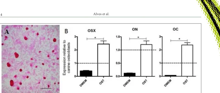
After 21 days of culture in osteogenic medium, AD-MSCs formed mineralized nodules (Figure 1) and an increase in the relative expression of gene transcripts for osterix, bone sialoprotein, and osteocalcin were observed (Figure 1).
The negative control (C-) and the group treated with OC-AD-MSCs (OC) showed partial
bone filling of the defect (a maximum of 56.68 %
and 63.72 %, respectively), without the formation of a bone bridge between the ends; the positive control (C+) showed complete bone healing in all animals at
90 days (Figure 2). The areas of bone filling in the C- and OC groups were significantly lower than those
of the positive control (C+) at all time points tested.
The OC group showed greater bone filling than the
C- group, at 15 days post-operation; however, no difference was observed at the subsequent evaluation times (Figure 3).
Histological evaluation of C- and OC groups showed new bone formation in the defect ends toward the center, without a union between them. The neoformed bone in all defects of C- and OC groups was spongy (Figure 2), with thick trabeculae predominantly covered with bulky osteoblasts. Predominance of active osteocytes was also found in the neoformed bone tissue in extended gaps. Histomorphometric and densitometric evaluations revealed similar areas of new bone formation and bone mineral mass between groups C- and OC at 90 days post operatively (Figure 3).
We have observed that the center of the defects in the C- and OC groups were formed by
disorganized fibrous connective tissue; however, in
the defects of the MSC group, connective tissue was less dense, with multiple cell clusters arranged in small caliber vascular channels lined with endothelial cells.
Neovascularization was significantly greater in the
OC group compared to the C- group, corresponding
to an average value of 6.75±2.39 vessels per field in
the C- and OC groups. There was complete bone healing in the defects treated with autogenous bone (C+) at 90 days post-operation. The presence of
inflammatory infiltrate was not observed in any of
the defects of all groups.
DISCUSSION
For the clinical use of a new therapy for bone regeneration, prior experimental study in subjects, preferably of the same species, is required. As the biological response may vary according to species, extrapolation from one species to another is not allowed; for this reason, we selected the dog as an experimental model. Dogs are widely used in bone regeneration studies and may be used as a model for preclinical studies when proposing an application for use in human patients.
The partial filling of bone defects observed
at 90 days post-operation in the negative control group (C-) (Figures 2, 3) demonstrated that the bone
defect was sufficient to cause a critical defect that
required support for regeneration, and thus validated the methodology used. A bone defect segment 1.5-fold greater than the diaphyseal diameter exceeds the bone regenerative capacity in dogs with skeletal maturity, resulting in non-union (KANG et al., 2012; ALVES et al., 2015b).
Complete bone regeneration was observed at 90 days post-operation in all defects treated by autograft (C+ group) (Figures 2, 3); this showed that all animals had normal bone regeneration capacity and that the bone stabilization technique was adequate.
Improper bone fixing is a major cause of problems
in bone consolidation and the most frequent cause of non-union fractures in small animals (KRAUS & BAYER, 2012). Therefore, it is necessary to ensure adequate bone stabilization when studying new therapies to promote bone regeneration.
A larger neoformed bone area was observed radiographically on group OC, at 15 days post-operation compared to that of the C- group (Figure 3). This showed that OC-AD-MSCs were able to enhance bone healing in this evaluation period. The presence of osteoprogenitor cells and mesenchymal stem cells (MSCs), with the ability to differentiate into bone cells, is probably the predominant factor contributing to the increase in osteogenic activity and consequently, repair of bone injury. When MSCs are added to a bone injury site, they can differentiate into osteoblasts and increase bone matrix synthesis (KANG et al., 2012; ARINZEH et al., 2003). MSCs have the capacity to differentiate into any mesenchymal cell such as tenocytes, myoblasts, adipocytes, and chondrocytes (STRIOGA et al., 2012). To maximize osteogenic differentiation, in vitro
differentiation is recommended before clinical use (LIU et al., 2013), as was performed in the present study (Figure 1). The formation of mineralized nodules and increases in expression of OSX, BSP, and OC (Figure 1) are considered reliable markers of osteogenic differentiation (ALVES et al., 2014; ALVES et al., 2015a).
Similarity in new bone formation area and bone mineral mass between C- and OC Figure 1 - Micrograph of mineralized nodules in AD-MSC cultures undergoing osteogenic differentiation after 21 days Bar = 350µm Von
groups, obtained by the histomorphometric and densitometric evaluations (Figure 3), showed that a single application of OC-AD-MSCs at the defect focus did not increase osteogenesis at 90 days post-operation. Number of cells used is a factor that may have contributed to this low bone regeneration
efficiency. In theory, the use of more cells would
result in greater matrix production and better bone regeneration; however, more cells require longer time for expansion in vitro before therapeutic use, thus resulting in a longer time to perform the
treatment. For this reason, there is a need to find the
minimum number of cells per volume of bone defect able to promote effective bone regeneration. Most studies have been successful using 106
OC-AD-MSC cm-3 of defect (ZHU et al., 2010), similar to
the number of cells used in this study. Some studies have suggested the use of fewer cells (105MSC cm-3
of defect) (BELOTI et al., 2012); however, other studies recommend higher cell numbers of 107MSC
cm-3 (LIU et al., 2013) and 108MSC cm-3 for effective
bone regeneration (WU et al., 2006).
Another factor that may be responsible
for the low efficiency of OC-AD-MSCs in the
regeneration of critical bone defects is the absence of a scaffold for cell adhesion and conduction. Studies showed that the presence of a scaffold for osteoconduction favors the regeneration of critical bone defects (ARINZEH et al., 2003; KANG et al., 2012; LIU et al., 2013).
Allogeneic MSCs, differentiated or not, have immunomodulatory capacity (NIERMEYER Figure 2 - Critical bone defects in radii of untreated dogs (A and D), dogs treated with OC-AD-MSC (B and E) and dogs treated with
autogenous bone (C and F), obtained at 90 days post-operation. Radiographic (A, B and C) and histological (D, E and F)
evaluation using hematoxylin-eosin. Bar = 93µm. In image A and B the bone defect was partially filled with cancellous bone
et al., 2007) and are not rejected when implanted in immunologically compatible individuals (ARINZEH et al., 2003). However, it is known that there is a gradual reduction in the number of MSCs in the focus of the injury over time (NANDOE TEWARIE et al.,
2009), which may explain the transient beneficial
effect of OC-AD-MSCs observed in this study. It is believed that therapy using multiple applications of
OC-AD-MSCs may result in an effective treatment with complete bone regeneration, but to prove this hypothesis another study is required. The therapeutic use of allogeneic cells, as performed in this study, showed advantages over the use of autogenous cells; when working with allogeneic cells it is possible to create cryopreserved cell banks, which enables a readily available reserve for clinical use Figure 3 - Mean percentage and standard deviation of the bone-filled area (A and B) and bone mineral mass (C) in critical defects in
in emergency cases (KRAUS & KIRKER-HEAD, 2006). In addition, young donors in perfect health can be selected, which resulted in the isolation of MSCs with high regenerative potential, thus allowing therapeutic use in geriatric patients and those with other conditions that may affect the regenerative potential of autogenous MSCs, such as the presence of genetic diseases.
The increased neovascularization observed in the OC group at 90 days post-operation (Figure 4) is another factor that may favor bone healing. In theory, increased tissue vascularization results in greater perfusion and enhanced delivery of nutrients and other
cells, providing a microenvironment more favorable for bone regeneration.
As the major limitation of the present study, we had only one application of osteoprogenitor
cells, which seem insufficient for complete boner
regeneration. Phenotypic characterization of osteogenic cells would bring interesting information about these cells; however, it was not performed. As a positive point of the study, the application of osteoprogenitor cells improve bone regeneration temporarily, suggesting that multiple application of
these cells could be an efficient therapy for critical
bone defects in dogs.
Figure 4 - Micrograph of the center of critical bone defects in dog radio, untreated (A), treated with OC-AD-MSC (B) or autogenous bone
(C+), 90 days after surgery. The presence of fibrous connective tissue in groups C- and OC and increased vascularization in the
OC group is shown by hematoxylin-eosin staining. Bar = 186µm. Image (D) shows the mean percentage and standard deviation
of the number of vessels p er field in the center of critical defects in dog radio in the C-, OC and C+ groups at 90 days
CONCLUSION
Based on the conditions of this study, we can say that treatment with OC-AD-MSCs increased newly formed bone areas at 15 days post-operation, but this was not enough for complete bone union in critical bone defects in dog radii at 90 days.
BIOETHICS AND BIOSSECURITY COMMITTEE APPROVAL
This study was conducted in accordance with the international standards of animal welfare after approval by the Ethics Committee in Animal Experimentation of UFMG (protocol no. 157/2009).
ACKNOWLEDGEMENTS
The authors thank the Fundação de Amparo à Pesquisa do Estado de Minas Gerais (FAPEMIG) for funding the
study, the Conselho Nacional de Desenvolvimento Científico e
Tecnológico (CNPq) for the doctoral grant, the Veterinary School of the Universidade Federal de Minas Gerais (UFMG) and The Veterinary School of the Universidade de Uberaba (UNIUBE) for the support needed to conclude the present study.
REFERENCES
ALVES, E.G.L. et al. Comparison of the osteogenic potential of mesenchymal stem cells from the bone marrow and adipose tissue of young dogs. BMC Veterinary Research, v.10, p.1-10, 2014. Available from: <http://www.ncbi.nlm.nih.gov/ pubmed/25178540>. Accessed: Aug. 03, 2015. doi: 10.1186/ s12917-014-0190-y.
ALVES, E.G.L. et al. Effect of ionic product of bioglass 60S in osteogenic differentiation of mesenchymal stem cells from the adipose tissue do dogs. Arquivo Brasileiro de Medicina Veterinária e Zootecnia, v.67, p.969-978, 2015a. Available from: <http://cpro4576.publiccloud.com.br:8080/editora// upload/trabalho/10086-21355.pdf>. Acessed: Aug. 03, 2015. doi: 10.1590/1678-4162-7539.
ALVES, E.G.L. et al. Porous matrix of BG60S in the treatment of critical bone defects in the radius of dogs. Arquivo Brasileiro de Medicina Veterinária e Zootecnia, v.67, p.993-1002, 2015b. Available from: <http://cpro4576.publiccloud.com.br:8080/ editora//upload/trabalho/10301-21358.pdf>. Accessed: Aug. 03, 2015. doi: 10.1590/1678-4162-7744.
ARINZEH, T.L. et al. Allogeneic mesenchymal stem cells regenerate bone in a critical-sized canine segmental defect.
Journal of Bone & Joint Surgery, v.85, p.1927-1935, 2003. Available from: <http://jbjs.org/content/85/10/1927>. Accessed: Aug. 03, 2015.
BELOTI, M.M. et al. The influence of osteoblast differentiation
stage on bone formation in autogenously implanted cell-based
poly(lactide-co-glycolide) and calcium phosphate constructs.
Tissue Engineering, v.18, p.999-1005, 2012. Available from: <http://www.ncbi.nlm.nih.gov/pubmed/22150110>. Accessed: Aug. 03, 2015. doi: 10.1089/ten.TEA.2011.0405.
KANG, B.J. et al. Comparing the osteogenic potential of canine mesenchymal stem cells derived from adipose tissues, bone marrow, umbilical cord blood, and Wharton’s jelly for treating bone defects. Journal Veterinary Science, v.13, p.299-310, 2012. Available from: <http://www.ncbi.nlm.nih.gov/pmc/ articles/PMC3467406/>. Accessed: Mar. 23, 2016. doi: 10.4142/ jvs.2012.13.3.299.
KRAUS, K.H.; KIRKER-HEAD, C. Mesenchymal stem cells and bone regeneration. Veterinary Surgery,v.35, p.232-242, 2006. Available from: <http://onlinelibrary.wiley.com/doi/10.1111/ j.1532-950X.2006.00142.x/abstract>. Accessed: Aug. 03, 2015. doi: 10.1111/j.1532-950X.2006.00142.x.
KRAUS, K.H.; BAYER, B.J. Delayed unions, nonunions, and malunions. In: TOBIAS, K.M.; JOHNSTON, S.A. Veterinary surgery small animal. St. Louis:Saunders, 2012. Cap.46, p.647-656.
NIERMEYER, P. et al. Comparison of immunological properties of bone marrow stromal cells and adipose tissue-derived stem cells before and after osteogenic differentiation in vitro. Tissue Engineering,v.13, p.111-121, 2007. Available from: <http://www. ncbi.nlm.nih.gov/pubmed/17518585>. Accessed: Aug. 03, 2015. doi: 10.1089/ten.2006.0114.
LIU, G. et al. Bone regeneration in a canine model using allogeneic adipose derived stem cells and coral scaffold. Biomaterials,v.34, p.2655-2664, 2013. Available from: <http://www.ncbi.nlm.nih. gov/pubmed/23343633>. Accessed: Aug. 03, 2015. doi: 10.1016/j. biomaterials.2013.01.004.
NANDOE TEWARIE, R.D. et al. Bone marrow stromal cells elicit tissue sparing after acute but not delayed transplantation into the contused adult rat thoracic spinal cord. Journal Neurotrauma, v.26, p.2313-2322, 2009. Available from: <http://online.liebertpub. com/doi/abs/10.1089/neu.2009.0987>. Accessed: Mar. 28, 2016. doi: 10.1089/neu.2009.0987.
STRIOGA, M. et al. Same or not the same? Comparison of adipose tissue-derived versus bone marrow-derived mesenchymal stem and stromal cells. Stem Cells and Development, v.21, p.2724-2752, 2012. Available from: <http://www.ncbi.nlm.nih. gov/pubmed/22468918>. Accessed: Aug. 03, 2015. doi: 10.1089/ scd.2011.0722.
WU, W. et al. Bone marrow-derived osteoblast seeded into porous beta-tricalcium phosphate to repair segmental defect in canine´s mandibular. Turkish Journal of Trauma & Emergency Surgery,v.12, p.268-276, 2006. Available from: <http://www. journalagent.com/travma/pdfs/UTD_12_4_268_276.pdf>. Acessed: Aug. 03, 2015.
ZHU, L. et al. Enhanced healing of goat femur-defect using
BMP7 gene-modified BMSCs and load-bearing