Vol.61: e18160308, 2018
http://dx.doi.org/10.1590/1678-4324-2018160308 ISSN 1678-4324 Online Edition
BRAZILIAN ARCHIVES OF BIOLOGY AND TECHNOLOGY
A N I N T E R N A T I O N A L J O U R N A L
Decreased mRNA Expression of Cystathionine Gamma lyase
and H2S Concentration in Gastric Mucosal Tissue in
Diabetic Rats
Seyyed Ali Mard
1*, Kobra Alinejad Dehkohneh
1, Akram Ahangarpour
1.
1Physiology Research Center (PRC), Dept. of Physiology, School of Medicine, Ahvaz Jundishapur University of
Medical Sciences Ahvaz, Iran.
ABSTRACT
Background and aim: It is well established that the rate of gastric lesions increases in diabetic rats. Recently, the protective effect of hydrogen sulfide (H2S) in gastric mucosa has been proven. This study aimed to determine the
release of H2S and mRNA expression of cystathionine gamma lyase (CSE) in gastric mucosa in alloxan-diabetic rats
in response to distention-induced gastric acid secretion.
Twenty-four rats were randomly assigned to 4 groups (6 in each). They were the normal-control, distention-control, diabetic-control, and distention-diabetic groups. Under anesthesia, animals underwent a tracheotomy and midline laparotomy. To washout the gastric contents, a catheter was inserted in the stomach through the duodenum. To determine the effect of distention-induced gastric acid secretion on H2S release and mRNA expression of CSE, the
stomachs were distended by normal saline. At the end of experiments, animals were sacrificed and the gastric mucosa was collected to determine H2S concentration and to quantify mRNA expression of CSE by quantitative
real-time PCR.
Mucosal release of H2S and mRNA expression of CSE significantly increased in response to stimulated gastric acid
secretion in normal rats (P<0.01), while the increases in diabetic rats were not significant. Basal release of H2S and
mRNA expression of CSE in gastric mucosa were significantly in diabetic rats lower than normal rats.
On the basis of the results, we conclude that the decreased release of H2S in response to basal and stimulated
gastric acid output in alloxan-diabetic rats compared to normal rats is largely due to downregulation of mRNA expression of CSE.
Key words: gastric acid secretion; cystathionine gamma lyase; hydrogen sulfide; alloxan-diabetic rats
*
Author for correspondence:[email protected]
INTRODUCTION
In mammals, hydrogen sulfide (H2S) is naturally produced by enzymatic and
non-enzymatic pathways 1. It has been shown that cystathionine gamma lyase (CSE) is
the main enzyme responsible for H2S synthesis in the gastric mucosa in rats 2. H2S
has been shown to inhibit gastric acid release 3, stimulate mucus and bicarbonate
secretion4, 5, and increase gastric mucosal blood flow (GMBF)6.
Recently, it has been shown that stimulated gastric acid secretion and mucosal
acidification increase mucosal release of H2S in the rat stomach 2, 7.
It has been shown that the increased susceptibility of gastric mucosal tissue to
irritants in diabetic animals8, 9 is due to different alterations in gastric mucosal tissue
such as impairment of GMBF, decreased release of calcitonin-gene related peptide
(CGRP) from CGRP neurons9, impairment of the antioxidant system 10, and
suppression of basic fibroblast growth factor 11.CGRP mediates the hyperemic
response following the back-diffusion of gastric acid, aimed at maintaining the
integrity of the gastric mucosal barrier 12, 13.
As far as we know, there is no report about the effect of stimulated gastric acid
secretion on gastric H2S release in diabetic rats. Therefore, the present study aimed
to determine the gastric release of H2S and mRNA expression of CSE in diabetic rats
in response to distention-induced gastric acid secretion.
MATERIALS AND METHODS
Animals
Male Wistar rats (150-200 g) were purchased from the animal house of Ahvaz Jundishapur University of Medical Sciences. The animals were fed conventional diet and had free access to tap water. They were maintained under standard conditions of humidity, temperature (22±2°C) and 12-h light/dark cycle. The animals were deprived of food but not water overnight before intervention. All experiments were carried out in accordance with the regulations set by Ethics Committee of Ahvaz Jundishapur University of Medical Sciences (APRC-94-16).
Animal Grouping and Experimental Procedures
Twenty-four rats were randomly assigned to 4 groups (6 in each). The animals were anesthetized with 60 mg/kg ketamine and 15 mg/kg xylazine, i.p., and subjected to midline laparotomy. The depth of anesthesia was checked throughout the experiment by the pedal withdrawal (toe pinch) reflex every 30 min. If the pedal withdrawal reflex was observed, a supplemental dose of ketamine+xylazine (1/3 of initial dose) was administered to maintain adequate anesthesia. Animal body temperature was measured with a rectal thermometer and maintained at 37°C using a homeothermic blanket control system (Harvard, UK). At the beginning of each experiment, the lumen of the stomach was gently rinsed with isotonic saline (37°C, pH7) until gastric washout was clear.
Thirty minutes after surgical operation, 1 ml of isotonic saline (pH 7 and 37°C) was instilled into the stomach in control groups (normal and diabetic) through a duodenal
catheter. To evaluate the effect of gastric distention on mucosal release of H2S and
Gastric H2S release decreases in diabetic rats
saline. For measuring the mRNA expression of CSE by quantitative real-time PCR
and determining H2S release in the gastric mucosa by an ELISA kit, two samples of
gastric mucosal tissue were quickly snap-frozen and stored in liquid nitrogen.
Induction of diabetes
To induce diabetes, overnight fasted rats were given a single intraperitoneal injection of 175 mg/kgalloxan monohydrate. After 72 h, blood glucose was checked with a glucometer (Elegance, Germany). Rats with fasting blood glucose higher than 250 mg/dl were considered diabetic. Two weeks later, animals with fasting blood sugar
higher than 250 mg/dl were considered gastroparetic4, 14.
Quantitative real-time PCR
The mRNA levels of CSE, and the housekeeping gene glyceraldehyde-3-phosphate dehydrogenase (GAPDH) were measured by quantitative real-time PCR (qRT-PCR) using a LightCycler® 480 System (Roche Diagnostics). The specific primers (Bioneer, South Korea) for measurement of CSE and GAPDH were used, and the lengths for amplified products were as follows:
GAPDH (forward primer:5´-TGCTGGTGCTGAGTATGTCGTG-3 and reverse primer:5´- CGGAGATGATGACCCTTTTGG-3´, 101 bp) and CSE (forward
primer:5´- TGTTGTCATGGGCTTAGTG-3´and reverse primer:5´-
CCATCCCATTCCTGAAGTG -3, 167 bp). All PCR amplifications were performed
in duplicate and in final volume of 20 μl containing 2 μlof cDNA, 0.8 μl of specific
primers, 10 μl of SYBR green master mix [TAKARA SYBR® Premix Ex Taq ™ II
(TliRNaseH Plus), Bulk] and 6.4 μl of ddH2O, using the following protocol:
pre-incubation at 95°C for 2 min to activate DNA Taq polymerase and 40 two-step cycles with denaturation at 95°C for 20 s, and annealing/extension at 60°C for 1 min. In addition, the no-template negative control (H2O) was routinely run in every PCR. The melting curve was examined at the end of amplification process to ensure the specificity of PCR products. Expression level of CSE was normalized against GAPDH expression (internal calibrator for equal RNA template loading and normalization). To determine the relative quantification of gene expression, the
comparative threshold cycle (Ct) method with arithmetic formulae (2−QQCt) was
used.
Determination of mucosal H2S levels
To determine the effect of distention-induced gastric acid secretion on H2S release, a commercial ELISA (enzyme-linked immunosorbent assay) Kit (ABIN771902, antibodies-online, USA) was used.
Statistical analysis
Data are shown as mean ± SEM. Statistical analysis was performed by two-way
ANOVA followed by Tukey’s post hoc test. Significance was set at P<0.05.
RESULTS
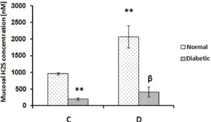
Effect of Distention-Induced Gastric Acid Secretion on Mucosal Release of Hydrogen Sulfide in Normal and Alloxan-Induced Diabetic Rats.
Figure1- Effect of distention-induced gastric acid secretion on mucosal release of H2S in the rat stomach. C: control groups (normal and diabetic rats) and D: distention groups [the acid output was stimulated by gastric distention (normal saline, 1.5 ml/100 g body weight, pH7 and 37°C)]. Analysis of ELISA results showed that the increased mucosal release of H2S in response to distention-induced gastric acid secretion in normal rats was significantly higher than in diabetic rats. **P<0.01 versus normal control group; βP<0.01 versus normal distention group. Data are expressed as mean±SEM.
Effect of Distention-Induced Gastric Acid Secretion on mRna Expression of Cse in Normal and Alloxan-Induced Diabetic Rats
As illustrated in Figure 2, mucosal mRNA expression of CSE significantly increased in response to stimulated gastric acid secretion in normal rats (P<0.01), while the increase in diabetic rats was not significant. Analysis of qRT-PCR results also showed that basal level and acid-induced expression of CSE mRNA in the gastric mucosa was significantly lower in diabetic rats than normal rats (P<0.01 in both cases).
Figure 2- Effect of distention-induced gastric acid secretion on mucosal expression of CSE in the rat stomach.
Analysis of qRT-PCR results showed that the increased expression of CSE in response to distention-induced gastric acid secretion in normal rats was significantly higher than in diabetic rats. **P<0.01 versus normal control group; βP<0.01 versus normal distention group. Data are expressed as mean±SEM.
Effect of Gastric Distention on pH of Gastric Contents in Normal and Alloxan-Induced Diabetic Rats
Gastric H2S release decreases in diabetic rats
the pH of gastric contents in states of control and distention was significantly lower in normal rats than diabetic rats (P<0.01 in both cases). As illustrated in Figure 4, there is a direct correlation between the gastric acid output and mucosal H2S release. This correlation in normal rats was more direct than in diabetic rats. As demonstrated in Figure 3, mucosal acid response to gastric distention and basal acid output were significantly lower in diabetic rats than normal rats as evidenced by the pH of gastric contents.
Figure 3- Effect of gastric distention on pH of gastric effluents in normal and alloxan-induced diabetic rats.
Gastric distention significantly decreased pH of gastric contents in normal and diabetic rats. pH of gastric contents in states of control and distention in normal rats was significantly lower than in diabetic rats. *P<0.05 and **P<0.01 versus normal control group; βP<0.01 versus normal distention group. αP<0.01 versus diabetic control group. Data are expressed as mean±SEM.
Figure 4- The correlation between gastric acid output and the release of H2S in normal and diabetic rats.
DISCUSSION
The present results showed that mucosal H2S release in the rat stomach increased in response to distention-induced gastric acid secretion in normal rats. Recently, mucosal release of H2S was shown to increase in response to gastric acid and mucosal acidification in normal Wistar rats 2, 7.
Therefore, these findings together demonstrate that any increase in gastric acid output is accompanied by increased mucosal H2S production in the rat stomach. Consistent with previous findings 2, 7, the present study showed that CSE mRNA expression increased in response to distention-induced gastric distention in normal rats but not significantly in diabetic rats. Therefore, it can be concluded that decreased mucosal release of H2S in response to gastric acid in diabetic rats compared to normal rats is largely due to decreased expression of CSE.
As shown in Figure 3, mucosal acid response to gastric distention and basal acid output were significantly lower in diabetic rats than normal rats as evidenced by the pH of gastric contents.
Reports have shown that gastric acid response to secretagogues changed in diabetes, although some studies showed an increase while others a decrease. One study showed that the rate of basal gastric secretion was similar between normal and diabetic rats, while it was significantly higher in response to pentagastrin and histamine in streptozocin-diabetic rats15. On the other hand, it has been shown that basal and stimulated gastric acid secretion decreases in alloxan- and streptozocin-induced diabetes rats16, 17. Accordingly, our findings showed that basal and stimulated gastric acid output was significantly reduced in alloxan-diabetic rats compared to normal rats. As shown in the current study and previous investigations 2, 7, there is a direct correlation between gastric acid output and H2S release in normal rats, but this correlation was not observed in alloxan-diabetic rats. Increased acid output without enough expected increase in mucosal release of H2S in diabetic rats maybe related to the chemical ablation or decreased activity of CGRP neurons or a decrease in mucosal NO release.
CGRP neurons have been shown to be activated in response to back-diffused gastric acid, mediating the hyperemic response by activating the release of H2S 12, 13. This study did not evaluate the activity of CGRP neurons, but another study showed that the activity of CGRP neurons decreases in the gastric mucosa of diabetic rats 9.Therefore, these results suggest that the decreased level of H2S in diabetic rats is due to a decrease in the activity of CGRP neurons.
Another factor involved in acid-induced H2S release is nitric oxide (NO). Recently, NO was shown to increase the activity and protein expression of CSE in gastric mucosa in response to distention and pentagastrin-induced gastric acid secretion 2. Additionally, NO has been shown to mediate gastric hyperemic response to capsaicin18.One study showed that the luminal release of NO decreased in streptozocin-diabetic rats 19. Therefore, it could be concluded that decreased production of NO leads to a decrease in H2S release in diabetic rats.
Gastric H2S release decreases in diabetic rats
CONCLUSION
The present findings showed that basal and acid-induced release of H2S decreases in alloxandiabetic rats. Therefore, we conclude that decreased release of H2S in diabetic rats is largely due to downregulation of CSE mRNA expression.
ACKNOWLEDGMENT
The source of data used in this paper was from the M.Sc thesis of Miss Kobra Alinejad Dehkohneh a student of the Ahvaz Jundishapur University of Medical Sciences. The authors gratefully acknowledge the help and financial support of the Physiology Research Center of Ahvaz Jundishapur University of Medical Sciences (grant No. APRC-94-16), Ahvaz, Iran.
REFERENCES
1. Pozsgai G, Hajna Z, Bagoly T, Boros M, Kemeny A, Materazzi S, et al. The role of transient receptor potential ankyrin 1 (TRPA1) receptor activation in hydrogen-sulphide-induced CGRP-release and vasodilation. Eur J Pharmacol. 2012; 689(1-3):56-64.
2. Mard SA, Veisi A, Ahangarpour A, Gharib-Naseri MK. Gastric acid induces mucosal H2S release in rats by upregulating mRNA and protein expression of cystathionine gamma lyase. J Physiol Sci. 2015; 65(6):545-554.
3. Mard SA, Askari H, Neisi N, Veisi A. Antisecretory Effect of Hydrogen Sulfide on Gastric Acid Secretion and the Involvement of Nitric Oxide. BioMed research international. 2014; 2014.
4. Qiu WC, Wang ZG, Lv R, Wang WG, Han XD, Yan J, et al. Ghrelin improves delayed gastrointestinal transit in alloxan-induced diabetic mice. World J Gastroenterol. 2008; 14 (16):2572- 2577.
5. Takeuchi K, Aihara E, Kimura M, Dogishi K, Hara T, Hayashi S. Gas Mediators Involved in Modulating Duodenal HCO3-Secretion. Current Medicinal Chemistry. 2012; 19(1):43-54.
6. Fiorucci S, Antonelli E, Distrutti E, Rizzo G, Mencarelli A, Orlandi S, et al. Inhibition of hydrogen sulfide generation contributes to gastric injury caused by anti-inflammatory nonsteroidal drugs. Gastroenterology. 2005; 129(4):1210-1224.
7. Mard SA, Veisi A, Ahangarpour A, Gharib-Naseri MK. Mucosal acidification increases hydrogen sulfide release through up-regulating gene and protein expressions of cystathionine gamma-lyase in the rat gastric mucosa. Iranian Journal of Basic Medical Sciences. 2016; 19(2):172- 177.
8. Takeuchi K, Ueshima K, Ohuchi T, Okabe S. Induction of gastric lesions and hypoglycemic response by food deprivation in streptozotocin-diabetic rats. Digestive Diseases and Sciences. 1994; 39(3):626-634.
9. Tashima K, Korolkiewicz R, Kubomi M, Takeuchi K. Increased susceptibility of gastric mucosa to ulcerogenic stimulation in diabetic rats–role of capsaicin-sensitive sensory neurons. British journal of pharmacology. 1998; 124(7):1395-1402.
10. Goldin E, Ardite E, Elizalde JI, Odriozola A, Panes J, Pique JM, et al. Gastric mucosal damage in experimental diabetes in rats: role of endogenous glutathione. Gastroenterology. 1997; 112(3):855-863.
11. Takeuchi K, Takehara K, Tajima K, Kato S, Hirata T. Impaired healing of gastric lesions in streptozotocin-induced diabetic rats: effect of basic fibroblast growth factor. J. Pharmacol. Exp. Ther. 1997; 281(1):200-2007.
13. Li D-S, Raybould HE, Quintero E, Guth PH. Calcitonin gene-related peptide mediates the gastric hyperemic response to acid back-diffusion. Gastroenterology. 1992; 102(4):1124-1118.
14. Mard SA AI, Ahangarpour A, Gharib-Naseri MK, Badavi M. Delayed gastric emptying in diabetic rats caused by decreased expression of cystathionine gamma lyase and H2S synthesis: in vitro and in vivo studies. Neurogastroenterol Motil. 2016. Doi: 10.1111/nmo.12867
15. Lin CY, Yeh GH, Hsu FC, Tsai SC, Lau CP, Pu HF, et al. Gastric acid secretion in streptozotocindiabetic female rats. Chin J Physiol. 1991; 34 (2):179-186.
16. Ozcelikay AT, Altan VM, Yildizoglu-Ari N, Altinkurt O, Onur F, Ozturk Y. Basal and histamineinduced gastric acid secretion in alloxan diabetic rats. Gen Pharmacol. 1993;24 (1):121-126.
17. Owu DU, Obembe AO, Nwokocha CR, Edoho IE, Osim EE. Gastric ulceration in diabetes mellitus: protective role of vitamin C. ISRN Gastroenterol. 2012; 2012:362805. 18. Raimura M, Tashima K, Matsumoto K, Tobe S, Chino A, Namiki T, et al. Neuronal nitric
oxide synthase-derived nitric oxide is involved in gastric mucosal hyperemic response to capsaicin in rats. Pharmacology. 2013; 92(1-2): 60-70.
19. Brzozowska I, Targosz A, Sliwowski Z, Kwiecien S, Drozdowicz D, Pajdo R, et al. Healing of chronic gastric ulcers in diabetic rats treated with native aspirin, nitric oxide (NO)-derivative of aspirin and cyclooxygenase (COX)-2 inhibitor. J Physiol Pharmacol. 2004; 55(4): 773-790.