O R I G I N A L A R T I C L E
Protective Effects of Simvastatin Against Alendronate-Induced
Gastric Mucosal Injury in Rats
Nathalia S. Carvalho1•Moˆnica M. Silva2•Renan O. Silva3•Lucas A. D. Nicolau3• Thiago S. L. Arau´jo2• Douglas S. Costa1•Nayara A. Sousa2•Luan K. M. Souza2• Pedro M. G. Soares3•Jand Venes R. Medeiros1,2,4
Received: 13 July 2015 / Accepted: 17 September 2015 / Published online: 24 September 2015
ÓSpringer Science+Business Media New York 2015
Abstract
Background It has been reported that simvastatin, a statin commonly prescribed for its anti-inflammatory and antioxidant effects, has gastroprotective effects in indo-methacin and ethanol-induced gastric ulcers. However, the effects of simvastatin on alendronate-induced gastric mucosal injury remain unexplored.
Aim This study investigated the use of simvastatin for the treatment of alendronate-induced gastric ulcers in rats. Methods Female rats were pretreated with vehicle or simvastatin (20 and 60 mg/kg p.o.). After 1 h, the rats received alendronate (50 mg/kg p.o.). Simvastatin was administered once daily for 7 days, and from the fourth day of simvastatin treatment, alendronate was administered once daily for 4 days. On the final day of treatment, 4 h after alendronate administration, animals were euthanized, their stomachs were removed, and gastric damage was measured. Samples of the stomach were fixed in 10 % formalin immediately after their removal for subsequent histopathological assessment. Unfixed samples were weighed, frozen at -80°C until assayed for glutathione
(GSH), malondialdehyde (MDA), and cytokine levels and myeloperoxidase (MPO) activity. A third group was used to measure mucus and gastric secretion.
Results Pretreatment with simvastatin prevented alen-dronate-induced macroscopic gastric damage and reduced the levels of MDA and GSH, TNF-a and IL-1b, MPO activity, and mucus levels, in the stomach.
Conclusions This study demonstrates the protective effects of simvastatin against alendronate-induced gastric ulceration. Maintenance of mucosal integrity, inhibition of neutrophil activity, and reduced oxidative stress associated with decreased gastric acidity may explain the gastropro-tective effects of simvastatin.
Keywords SimvastatinBiphosphonateGastric damageAntioxidantAnti-inflammatory
Abbreviations ALD Alendronate DMSO Dimethyl sulfoxide
ELISA Enzyme-linked immunosorbent assay GSH Glutathione
IL Interleukin MDA Malondialdehyde MPO Myeloperoxidase NO Nitric oxide SIM Simvastatin
TNF Tumor necrosis factor
Background
Osteoporosis is the most widespread bone disease, char-acterized by low bone mineral density and deterioration of bone microarchitecture leading to bone fragility fractures
& Jand Venes R. Medeiros
jandvenes@ufpi.edu.br
1 Post Graduation Program in Pharmacology, Medicinal Plant
Research Center (NPPM), Federal University of Piauı´, Teresina, PI, Brazil
2 Post Graduation Program in Biotechnology, Biotechnology
and Biodiversity Center Research (BIOTEC), Federal University of Piauı´, Parnaı´ba, PI, Brazil
3 Laboratory of Pharmacology of Inflammation and Cancer
(LAFICA), Department of Physiology and Pharmacology, Federal University of Ceara´, Fortaleza, CE, Brazil
4 BIOTEC/LAFFEX/UFPI, Av. Sa˜o Sebastia˜o, no. 2819,
[1]. Currently, biphosphonates, including alendronate (ALD), clodronate, etidronate, pamidronate, and tilu-dronate, are the most commonly prescribed drugs for the treatment of various bones diseases, including Paget’s disease, osteolytic bone metastases, and osteoporosis, and also for enhanced bone resorption [2]. However, these drugs are associated with several gastrointestinal distur-bances, such as abdominal discomfort, pain, diarrhea, and ulcers of the esophagus, stomach, or small intestine [3,4]. Recently, statins, a class of drugs used worldwide to lower plasma concentrations of cholesterol-carrying lipoproteins [5], were found to be associated with various pleiotropic effects including improvement of bone health owing to their interference with bone metabolism, reduc-tion in signal proteins regulating osteoclast activity [6], and upregulation of the expression of bone morphogenetic protein-2 gene as demonstrated in rat bone tissue [7]. Furthermore, studies have found that the risk of fractures markedly reduces in people using statins [8,9].
Simvastatin (SIM) is a commonly prescribed statin with anti-inflammatory [10, 11] and antioxidant effects [12]. The literature has also established that SIM has gastro-protective effects in indomethacin and ethanol-induced gastric ulcers [13, 14]. However, chronic use of ALD, a frequently used biphosphonate, also is associated with gastric ulcers via mechanisms differing from those of NSAIDs and ethanol. Adverse effects of ALD in the upper gastrointestinal tract have been attributed, in many cases, to adherence of the drug to the mucosal surface, causing lesions in the antrum that subsequently develop into ulcers (with white cap) [15, 16]. However, the effect of SIM on gastric mucosal injury induced by ALD has remained unexplored.
Owing to the lack of an effective therapy for gastropathy caused by ALD, few studies on the mechanisms involved in its toxicity, influence of interaction between ALD and SIM on the outcome, particularly in patients taking both agents simultaneously, the evidence presented in this work may provide a rationale for use of statins in the treatment of gastric ulcers induced by ALD.
Methods
Reagents
SIM was purchased from Sigma-Aldrich (St. Louis, MO, USA). ALD sodium was purchased from Farmafo´rmulaÒ, Fagron (Sa˜o Paulo, Brazil). ALD was dissolved in 2 % DMSO prior to use. All other chemicals were of analytical grade and obtained from standard commercial suppliers and dissolved in saline before use.
Animals
Female rats (Wistar strain, 100–150 g) were obtained from the Department of Physiology and Pharmacology, Federal University of Ceara´. The animals were housed in cages in a temperature-controlled environment under a 12 h light/ 12 h dark cycle and had constant access to drinking water and standard pellet diet. The animals were deprived of food for 18–24 h before the experiment, but had free access to water. All animal treatments and surgical procedures were performed in accordance with the Guide for Care and Use of Laboratory Animals (National Institutes of Health, Bethesda, MD, USA) and were approved by the local ethics committee (Protocol No. 0067/10).
Effect of Simvastatin on Alendronate-Induced Gastric Damage
The animals were initially pretreated with vehicle (0.9 % saline) or SIM (20 and 60 mg/kg) orally. After 1 h, ALD (50 mg/kg) was administered by gavage. The control group received only saline. SIM was administered once daily for 7 days, and, beginning on the fourth day, ALD was administered once daily for 4 days. On the last day of treatment, 4 h after ALD administration, pylorus ligation was performed to determine the gastric acidity. In other groups, the animals were euthanized and their stomachs removed. Gastric damage was measured using a computer planimetry program (ImageJ software, National Institutes of Health, Bethesda, MD, USA). Samples of the stomach were fixed in 10 % formalin immediately after their removal for subsequent histopathological assessment. Unfixed samples were then weighed, frozen, and stored at -80°C until assayed for glutathione (GSH) levels [17],
malondialdehyde (MDA) concentration [18], myeloperox-idase (MPO) activity [19], and cytokine levels [20].
Histological Evaluation of Gastric Damage
cells (a score of 0–3), yielding a maximum possible score of 14.
Glutathione Assay
The GSH levels in the segmented stomach tissue were measured using the method previously described [17]. The results were expressed as micrograms of GSH per gram of tissue (lg/g).
Malondialdehyde Assay
Lipid peroxidation was estimated by measuring the con-centration of MDA in the homogenate from each gastric sample according to the method previously described [18], which is based on a reaction with thiobarbituric acid. MDA concentrations are expressed as nanomoles per gram of tissue (nmol/g).
Myeloperoxidase Activity
MPO activity, an index of polymorphonuclear cell accu-mulation into various tissues including the gastrointestinal tract, was measured according to the method previously described [19]. The results were reported as the MPO units per mg (UMPO/mg) of tissue. A unit of MPO activity was defined as that sufficient for converting 1lmol of H2O2to
water in 1 min at 22°C.
Cytokine Measurements
The levels of tumor necrosis factor (TNF)-a and inter-leukin (IL)-1bwere determined by using an enzyme-linked immunosorbent assay (ELISA), as previously described [20]. ELISA results were expressed as pictograms of each cytokine per milliliter reaction mixture (pg/ml).
Determination of Gastric Mucus
The gastric tissues were collected and transferred to 1 % Alcian blue solution (0.16 M sucrose in 0.05 M sodium acetate, pH 5.8). Excess dye was removed by washing the tissue with 0.25 M sucrose solution. The mucus–dye com-plex was extracted by placing the tissue in 0.5 M MgCl2for
2 h. This extract was mixed with diethyl ether, centrifuged at 1400gfor 10 min, and the absorbance was measured at 598 nm [22]. The quantity of extracted Alcian blue (lg/g of tissue) was then calculated using a standard curve.
Anti-secretory Study
To evaluate secretory activity, the animals were treated according to the method described above. On the last day
of treatment, 4 h after ALD administration, pylorus liga-tion was performed. After 4 h of continuous observaliga-tion and monitoring, the animals were killed using a lethal dose of a combination of anesthetics, ketamine, and xylazine. Their stomachs were immediately opened, and the gastric content was collected. The final volume and pH were determined immediately after washing the stomach mucosa with 2 ml of distilled water. The total acidity of gastric juice was titrated with 0.01 N NaOH, using 2 % phe-nolphthalein as an indicator [23].
Statistical Analysis
All values are expressed as mean±SEM ANOVA, and a Student–Newman–Keuls test was used to determine sta-tistical significance of differences between groups. For histological assessment, the Kruskal–Wallis nonparametric test was used, followed by Dunn’s test for multiple com-parisons. Differences were considered to be significant when P\0.05.
Results
Effect of Simvastatin on Alendronate-Induced Gastric Damage
In this study, our results showed that pretreatment with SIM (60 mg/kg) administered orally prevented ALD-in-duced macroscopic gastric damage (Fig.1). Furthermore, we observed that ALD damaged both the corpus and the antral mucosa of the stomach, and this damage was mostly surface epithelial injury. The chronic exposure of the rat gastric mucosa to ALD for 4 days produced widespread damage to the epithelium, and the luminal surface was covered with cellular debris (Fig. 2c). The results of microscopic analysis indicated that ALD administration induced alterations in the gastric region characterized by intense hemorrhage, edema, epithelial cell loss, and inflammatory cells (Fig.2d; Table1). These changes were observed to be significantly reduced in rats treated with SIM (Fig.2e, f; Table1).
GSH and MDA Levels
effects caused by ALD (#P\0.05). The administration of SIM (72.2±10.73 mmol/g tissue) also significantly reduced both the concentration of gastric mucosal MDA and the levels of lipid peroxidation caused by ALD (#P\0.05). Pretreatment with SIM alone has no signifi-cant effects about the levels of GSH and MDA when compared to saline group.
Myeloperoxidase Activity During Gastric Injury Alendronate
From our data, we observe that the MPO activity in the saline control group is 1.2±0.35 U/mg tissue. Upon receipt of the ALD dose of 50 mg/kg, MPO levels are around 6.3±0.52 U/mg tissue, a significantly different value compared to the saline group (*P\0.05). However, the group receiving treatment with SIM exhibited decreased MPO activity levels (2.1±0.28 U/mg tissue), a significant difference compared to the group receiving the harmful agent (#P\0.05). This shows that SIM may reduce neu-trophil infiltration in gastric lesions caused by ALD (Fig.4).
Cytokine (TNF-aand IL-1b) Levels During Gastric Injury Alendronate
Quantification data for the levels of pro-inflammatory cytokines at the ALD-induced lesion show that both cytokines exhibited low levels upon use of SIM. In the saline group, TNF-aand IL-1blevels were 1313.0±154.61 and
586.6±28.08 pg/ml, respectively. Measured levels of TNF-a and IL-1bB in the group that received ALD were 2405.0±264.02 and 901.1±86.73 pg/ml, respectively, which is significantly increased compared to the saline group (*P\0.05). The groups treated with SIM clearly showed a significant reduction (#P\0.05) in the levels of the two cytokines, with TNF-a levels of 1535.0±192.61 and IL-1blevels of 525.7±56.82. This results shows that SIM is capable of reducing the levels of IL-1band TNF-ain the gastric damage caused by ALD (Fig.5).
Determination of Gastric Mucus and Gastric Acid Secretion
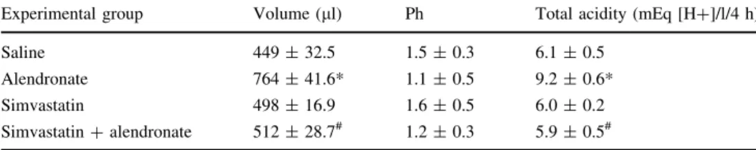
The injury caused by ALD significantly decreased the amount of gastric mucus adherent to gastric mucosa when compared to the saline treatment. The pretreatment with SIM (60 mg/kg) reversed the effect of ALD (*P\0.05). Thus, SIM significantly affects the level of adherent mucus in the stomach as compared to the control group (Fig.6). Table2 shows the measurements taken from the gastric contents after pylorus ligation test. ALD significantly increased the volume of gastric secretion compared to the control group (764.0±41.63, 449.1±32.55, respec-tively) (*P\0.05). In the group that was administered SIM (60 mg/kg) with ALD simultaneously, the gastric secretion levels decreased considerably when compared to the group that was administered ALD alone (512.2±28.71) (#P\0.05), thus demonstrating the anti-secretory capability of SIM. The pH of gastric secretions was also measured, but no difference between groups was significant and all remained acidic. Meanwhile, the total acidity levels of the groups that were administered only ALD were significantly increased compared to the control group (9.2±0.60, 6.1±0.50, respectively). In the group that was administered SIM and ALD, the total gastric acidity was significantly reduced compared to the group receiving only ALD (5.9±0.51,#P\0.05).
Discussion
Previous studies have established the beneficial effects of statins on bone [24], and statins currently represent a near ideal candidate family of anti-osteoporotic drugs, due to their potential dual anabolic and anti-resorptive effects on bone [25]. However, the protective effects of statins on gastrointestinal toxicity associated with biphosphonates have not yet been elucidated.
In the present study, the effect of SIM was studied on ALD-induced gastric injury in rats. Our finding that SIM-decreased ALD-induced gastropathy is consistent with those of other studies that reported that SIM-attenuated Fig. 1 Effect of simvastatin pretreatment on alendronate-induced
gastric damage. After 4 days of alendronate administration, the gastric mucosa of the control rats was intensely damaged. Treatment with simvastatin (60 mg/kg, p.o.) for 7 days significantly (P\0.05)
improved macroscopic gastric damage. Macroscopic gastric lesions were determined 4 h after alendronate administration on the fourth day. Data are expressed as the mean±SEM of five–six rats per group. *P\0.05 versus saline group;#P\0.05 versus alendronate
NSAID and ethanol-induced gastropathies [13,14]. Toge-ther, these findings highlight an important role of biphos-phonates in gastric protection.
Our results confirmed that ALD administration at high concentrations caused severe macroscopic gastric damage in both the corpus and antral mucosa of the stomach, and this damage was mostly localized to the epithelial surface. This injury is characterized by widespread damage to the epithelium, with the luminal surface covered in cellular debris subsequently developing into ulcers with a white cap.
Chronic oral administration of ALD also caused hemor-rhagic lesions in the mucosa of the glandular stomach, indicating true ulcer formation, and this was also supported by the histopathological findings of edema, increased inflammatory cells, and epithelial cell loss [15,16,26].
The mechanism through which ALD and other biphos-phonates cause mucosal injury has not been clearly iden-tified. However, ALD-induced neutrophil accumulation and subepithelial edema in the gastric mucosa have been shown to play major roles in the development of ulcers Fig. 2 Macroscopic and histopathological changes in the gastric
mucosa (920 magnification). a, b Control (saline) group showing gastric mucosal integrity. c Alendronate (50 mg/kg, p.o.) group showing widespread damage to the epithelium and cellular debris covering the luminal surface (indicated by arrows). d The micro-scopic analysis indicated that alendronate administration induced
[27]. In fact, the white cap that covers the damaged mucosa is composed mainly of inflammatory cells and fibrin-like substances [28]. Other studies have reported that toxic effects of ALD in the stomach have been linked to a direct effect of this agent on the mucosal surface [28, 29]. Additionally, when ALD is applied to the gastric mucosa, it decreases transmucosal potential difference of the stomach, which is suggestive of a direct disruption of surface epithelial cells [30].
In our study, we demonstrated that SIM inhibited the ALD-induced elevation of MPO activity and the increased TNF-a and IL-1b levels, suggesting that the gastroprotec-tive effect of SIM may be dependent on its inhibitory effect on neutrophil infiltration and the neutrophil-associated TNF-a and IL-1b response. In fact, several studies have shown that biphosphonates interfere with cell migration at the site of inflammation, and this protective effect is associated with decreased production of pro-inflammatory cytokines, TNF-a, IL-1b, and IL-6 [15,31,32]. Similarly, studies have reported the anti-inflammatory effects of sta-tins that have inhibitory effects on inflammatory mediators, Table 1 Protective effect of simvastatin in alendronate-induced microscopic gastric damage
Experimental group Hemorrhagic damage (score 0–4)
Edema (score 0–4)
Epithelial cell loss (score 0–3)
Inflammatory cells (score 0–3)
Total (score 0–14)
Saline 0 0 0 (0–1) 0 0 (0–1)
Alendronate 3 (2–4)# 2 (1–3)# 2 (1–3)# 2 (1–2)# 7 (2–12)
Simvastatin?alendronate 1 (0–2)* 0 (0–1)* 1 (0–2)* 0 (0–1)* 3 (0–6)*
Data shown are medians with minimal and maximal scores shown in parentheses. Kruskal–Wallis nonparametric test, followed by Dunn’s test, was used for multiple comparisons for histological assessment
*P\0.05 when compared with alendronate group
# P\0.05 when compared with control group
Fig. 3 Effect of simvastatin on glutathione (GSH) concentration and MDA levels in rats with alendronate-induced gastric damage. aAlendronate administration promoted a decrease in the GSH gastric concentration. This effect was reverted significantly when the animals were pretreated with simvastatin.bAlendronate promoted an increase in MDA gastric tissue levels; however, pretreatment with simvastatin
significantly prevented this effect. Results are expressed as the mean±SEM of at least five rats per group. *P\0.05 versus saline
group;#P\0.05 versus alendronate group. ANOVA and Newman–
Keuls test were used for evaluation.ANOVAanalysis of variance,SAL saline,SEMstandard error of the mean,SIMsimvastatin
Fig. 4 Effect of simvastatin on the activity of myeloperoxidase (MPO) in gastric injury induced by alendronate. The control group was treated with saline only. The administration of simvastatin reduced alendronate-induced MPO activity. Results are expressed as the mean±SEM of at least five rats per group. *P\0.05 versus
saline group; #P\0.05 versus alendronate group. ANOVA and
such as ILs, interferon-c, cytokines, and cyclooxygenase 2 [33,34]. Multiple lines of evidence also indicate that SIM influences the expression of vascular cell adhesion mole-cule-I (VCAM-I) and CD14 (an inducer of mononuclear cell infiltration), resulting in additional benefits, including beneficial effects on inflammation [35,36].
Reactive oxygen species (ROS) play a vital role in a variety of agents that induced gastric damage [37,38]. It seems that the important primary factor in indomethacin-induced gastric damage is ROS-mediated lipid peroxidation. It was demon-strated that indomethacin causes increase in lipid peroxidation
and decrease in GSH peroxidase activity. This effect can induce gastric erosions and apoptosis by DNA fragmentation [38,39]. Also, it has been suggested that oxygen-derived free radicals may contribute to ALD-induced gastric mucosal lesions [15,40,41]. However, the mechanism by which ALD generates ROS to cause mucosal damage has not yet been clarified. In fact, activated and migrating neutrophils induced by ALD are a potential source of oxygen metabolites that can contribute to gastric mucosal injury. ALD also increases the TNF-alevel that causes neutrophil infiltration and activation to generate ROS [42]. Other authors verified that severity of gastric lesions induced by ALD was reduced by treatment with allopurinol, suggesting the involvement of oxyradical production in this pathogenesis [43]. Thus, it seems that the ulcerogenic response in the antrum is related to the dysfunc-tion of the anti-oxidative mechanism.
These observations are consistent with our finding that ALD decreased GSH levels and increased MDA levels, and SIM reversed these damaging effects. This increase in lipid peroxidation may be due partly to the free radicals gener-ated by neutrophils and direct toxic effects of ALD on the mucosal surface. It is well established that GSH is important for gastric protection against aggressive agents [44]. Sub-stances containing sulfhydryl radicals protect the gastric mucosa in a manner similar to that of prostaglandins (PGs) and that sulfhydryl group blockers, such as diethylmaleate and iodoacetamide, reverse the gastroprotective effect of PGF2a. GSH administration also decreases ethanol-induced gastric damage by inactivating ROS and products of lipid peroxidation [44, 45]. Therefore, it is possible that SIM might decrease redox state in ALD-induced gastropathy. Thus, the mechanism through which SIM exerts its gas-troprotective effect appears to involve reduction in ALD-induced lipid peroxidation in the gastric mucosa.
Fig. 5 Effect of simvastatin on the cytokine levels of tumor necrosis factor (TNF)-a and interleukin (IL)-1b in rats with alendronate-induced gastric damage. Treatment with simvastatin (60 mg/kg) decreased the levels of TNF-aand IL-1b(a, b, respectively). The control group was treated with saline only. Results are expressed as
the mean±SEM of at least five rats per group.*P\0.05 versus
saline group; #P\0.05 versus alendronate group. ANOVA and
Newman–Keuls test were used for evaluation. ANOVA analysis of variance, SAL saline, SEM standard error of the mean, SIM simvastatin
Fig. 6 Gastroprotective effect of simvastatin by evaluating levels of mucus in alendronate-induced stomach lesions. The animals were pretreated by gavage with simvastatin (60 mg/kg) or with saline solution and were administered alendronate (50 mg/kg) after 30 min. The control group was administered only saline. All drugs were administered once daily during the 4 days. Results are expressed as the mean±SEM of least five–seven animals per group.*P\0.05
The biphosphonate-induced damage in the stomach also is accompanied by disruption of the mucosal barrier, leading to hydrogen proton back-diffusion into the mucosa [30]. The topical irritant property of ALD is, in part, attributable to the ability of biphosphonates to chemically associate with and destabilize the surface lining of phos-pholipids, resulting in a rapid disruption of the hydrophobic barrier of these tissues [46]. In this study, we confirmed that ALD significantly reduced gastric mucus production when compared with that observed for the negative control, whereas pretreatment with SIM significantly increased the amount of adherent mucus in the gastric mucosa wall. Similarly, Tariq et al. [13] demonstrated that anti-ulcer activity of SIM was associated with significant attenuation of adverse effects of ethanol on gastric wall mucus.
ALD is known to significantly increase gastric acidity when administrated orally, and this increased acidity may be owing to the local irritation caused by ALD [47]. At very low pH conditions (pH\2), ALD is in the acid form, which is known to be more irritating than the sodium salt [48]. Our results showed that SIM pretreatment reduced the total acidity and gastric pH, and these effects maybe contribute to their anti-ulcer activity. Therefore, by increasing the gastric pH, SIM may have prevented the ALD sodium from being converted to the acid form, thus decreasing the damaging effect of the free acid on gastric mucosa. Other studies indicate that SIM-induced gastroprotective effects may be attributed to its ability to reduce acidity [13], and this effect may be associated with an increase in endogenous nitric oxide production induced by SIM [49, 50]. In fact, the consensus seems to be that NO inhibits acid secretion under basal and stimulated conditions in several animal species [51–53], and recently, we demonstrated that eNOS- and iNOS-derived NO prevents ALD-induced gastric damage [15] and alterations in gastric emptying in rats [54].
Conclusions
In conclusion, the findings of this study clearly demonstrate the protective effects of SIM against ALD-induced gastric ulceration. Maintenance of mucosal integrity, inhibition of
neutrophil activity, and reduced oxidative stress associated with decreased gastric acidity explain the gastroprotective effects of SIM. These properties of SIM warrant its safe use as an anti-hyperlipidemic drug, for gastroprotective effects, or for the improvement of bone health in combination with biphosphonates.
Acknowledgments The authors gratefully acknowledge the finan-cial support from National Counsel of Technological and Scientific Development—CNPq (Brazil) and Research Foundation for the State of Piauı´—FAPEPI.
Compliance with ethical standards
Conflict of interest None.
Ethical standard All procedures performed in studies involving animals were in accordance with the ethical standards of the institu-tion or practice at which the studies were conducted.
References
1. Raisz LG. Pathogenesis of osteoporosis: concepts, conflicts, and prospects.J Clin Invest. 2005;15:3318–3325.
2. Fleisch H.Bisphosphonates in Bone Disease: From the Labora-tory to the Patient. New York: The Parthenon Group; 1997: 32–163.
3. Chestnut CH, McClung MR, Ensrud KE, et al. Alendronate treatment of the postmenopausal osteoporotic woman: effect of multiple dosages on bone mass and bone remodeling.Am J Med. 1995;29:144–152.
4. Groen PC, Lubbe DF, Hirsch LJ, et al. Esophagitis associated with the use of alendronate.N Engl J Med. 1996;335:1016–1021. 5. Stein EA. Management of dyslipidemia in the high-risk patient.
Am Heart J. 2002;144:43–50.
6. Beek E, Lowik C, Pluijm G, et al. The role of geranylgeranylation in bone resorption and its suppression by bisphosphonates in fetal bone explants in vitro: a clue to the mechanism of action of nitrogen-containing bisphosphonates. J Bone Miner Res. 1999; 14:722–729.
7. Mundy G, Garrett R, Harris S, et al. Stimulation of bone for-mation in vitro and in rodents by statins. Science. 1999;286: 1946–1949.
8. Meier CR, Schlienger RG, Kraenzlin ME, et al. HMG CoA reductase inhibitors and the risk of fractures. JAMA. 2000; 283:3205–3210.
9. Wang PS, Solomon DH, Mogun H, et al. HMG-CoA reductase inhibitors and the risk of hip fractures in elderly patients.JAMA. 2000;283:3211–3216.
Table 2 Effect of pretreatment with simvastatin on gastric acid secretion
Experimental group Volume (ll) Ph Total acidity (mEq [H?]/l/4 h)
Saline 449±32.5 1.5±0.3 6.1±0.5
Alendronate 764±41.6* 1.1±0.5 9.2±0.6*
Simvastatin 498±16.9 1.6±0.5 6.0±0.2
Simvastatin?alendronate 512±28.7# 1.2±0.3 5.9±0.5#
Data shown are mean±SEM of 5–7 rats
*P\0.05 when compared with saline group
# P
10. Scalia R, Gooszen ME, Jones SP, et al. Simvastatin exerts both anti-inflammatory and cardioprotective effects in apoliprotein E-deficient mice.Circulation. 2001;103:2598–2603.
11. Pruefer D, Scalia R, Lefer AM. Simvastatin inhibits leukocytes– endothelial cell interactions and protects against inflammatory processes in normocholesterolemic rats. Arterioscler Thromb Vasc Biol. 1999;19:2894–2900.
12. Ungureanu D, Filip C, Artenie A, Artenie R. Evaluation of simvastatin antioxidant effects. Rev Med Chir Soc Med Nat. 2003;107:66–71.
13. Tariq M, Khan HA, Elfaki I, et al. Gastric antisecretory and antiulcer effects of simvastatin in rats.J Gastroenterol Hepatol. 2007;22:2316–2323.
14. Heeba GH, Hassan MKA, Amin RS. Gastroprotective effect of simvastatin against indomethacin-induced gastric ulcer in rats: role of nitric oxide and prostaglandins. Eur J Pharmacol. 2009;607:188–193.
15. Silva RO, Lucetti LT, Wong DVT, et al. Alendronate induces gastric damage by reducing nitric oxide synthase expression and NO/ cGMP/KATP signaling pathway.Nitric Oxide. 2014;40:22–30. 16. Costa NRD, Silva RO, Nicolau LA, et al. Role of soluble
guanylate cyclase activation in the gastro protective effect of the HO-1/CO pathway against alendronate-induced gastric damage in rats.Eur J Pharmacol. 2013;700:51–59.
17. Sedlak J, Lindsay RH. Estimation of total, protein-bound, and nonprotein sulfhydryl groups in tissue with Ellman’s reagent. Anal Biochem. 1968;25:192–205.
18. Mihara M, Uchiyama M. Determination of malonaldehyde pre-cursor in tissues by thiobarbituric acid test. Anal Biochem. 1978;86:271–278.
19. Bradley PP, Christensen RD, Rothstein G. Cellular and extra-cellular myeloperoxidase in pyogenic inflammation. Blood. 1982;60:618–622.
20. Cunha FQ, Boukili MA, Motta JI, Vargaftig BB, Ferreira SH. Blockade by fenspiride of endotoxin-induced neutrophil migra-tion in the rat.Eur J Pharmacol. 1993;238:47–52.
21. Laine L, Weinstein WM. Histology of alcoholic hemorrhagic ‘‘gastritis’’: a prospective evaluation. Gastroenterology. 1988;94:1254–1262.
22. Corne SJ, Morrissey SM, Woods RJ. Proceedings: a method for the quantitative estimation of gastric barrier mucus. J Physiol. 1974;242:116–117.
23. Shay H. A simple method for the uniform production of gastric ulceration in the rat.Gastroenterology. 1945;5:43–61.
24. Jadhav SB, Jain GK. Statins and osteoporosis: new role for old drugs.J Pharm Pharmacol. 2006;58:3–18.
25. Uzzan B, Cohen R, Nicolas P, et al. Effects of statins on bone mineral density: a meta-analysis of clinical studies. Bone. 2007;40:1581–1587.
26. Nicolau LAD, Silva RO, Damasceno SRB, et al. The hydrogen sulfide donor, Lawesson’s reagent, prevents alendronate-induced gastric damage in rats.Braz J Med Biol Res. 2013;46:708–714. 27. Wallace JL, Dicay M, Mcknight W, et al. N-bisphosphonates
cause gastric epithelial injury independent of effects on the microcirculation.Aliment Pharmacol Ther. 1999;13:1675–1682. 28. Ohashi Y, Aihara E, Takasuka H, Takahashi K, Takeuchi K. Antral ulcers induced by alendronate, a nitrogen-containing bis-phosphonate, in rat stomachs—prophylactic effect of rebamipide. J Physiol Pharmacol. 2009;60:85–93.
29. Wallace JL, Ma L. Inflammatory mediators in gastrointestinal defense and injury.Exp Biol Med. 2001;226:1003–1015. 30. Kanatsu K, Aihara E, Okayama M, Kato S, Takeuchi K. Mucosal
irritative and healing impairment action of risedronate in rat stomachs: comparison with alendronate.J Gastroenterol Hepatol. 2004;19:512–520.
31. Thieabaud D, Sauty A, Burckhard A, et al. An in vitro and in vivo study of cytokines in the acute-phase response associated with bisphosphonates.Calcif Tissue Int. 1997;61:386–392.
32. Yamaguchi K, Motegi M, Iwakura Y, Endo Y. Involvement of interleukin-1 in the inflammatory actions of aminobisphospho-nates in mice.Br J Pharmacol. 2000;130:1646–1654.
33. Weitz-Schmidt G. Statins as anti-inflammatory agents. Trends Pharmacol Sci. 2002;23:482–487.
34. Scho¨nbeck U, Libby P. Inflammation, immunity, and HMG-CoA reductase inhibitors: statins as antiinflammatory agents? Circu-lation. 2004;109:II18–II26.
35. Sager PT, Melani L, Lipka J, et al. Effect of coadministration of ezetimibe and simvastatin on high-sensitivity C-reactive protein. Am J Cardiol. 2003;15:1414–1418.
36. Bracht L, Barbosa CP, Caparroz-Assef SM, et al. Effects of simvastatin, atorvastatin, ezetimibe, and ezetimibe?simvastatin combination on the inflammatory process and on the liver metabolic changes of arthritic rats. Basic Clin Pharmacol. 2012;26:722–734.
37. Vaananenn PM, Medding JB, Wallace JL. Rol of oxygen derived free radicals in indomethacin-induced gastric injury. Am J Physiol. 1991;261:G470–G475.
38. Yoshikawa T, Naito Y, Kishi A, et al. Role of active oxygen, lipid peroxidation and antioxidants in the pathogenesis of gastric mucosal injury induced by indomethacin in rats. Gut. 1993;34:732–737.
39. Chattopadhyay I, Bandyopadhyay U, Biswas K, Maity P, Ban-erjee RK. Indomethacin inactivates gastric peroxidase to induced reactive-oxygen-mediated gastric mucosal injury and curcumin protects it by preventing peroxidase inactivation and scavenging reactive oxygen.Free Radic Biol Med. 2006;40:1397–1408. 40. Sener G, Paskaloglu K, Kapucu C, Cetinel S, Contuk G,
Aya-noglu-Dulger G. Octreotide ameliorates alendronate-induced gastric injury.Peptides. 2004;25:115–121.
41. Sener G, Kapucu C, Cetinel S, Cikler E, Ayanoglu-Dulge RG. Gastroprotective effect of leukotriene receptor blocker mon-telukast in alendronate-induced lesions of the rat gastric mucosa. Prostaglandins Leukot Essent Fatty Acids. 2005;72:1–11. 42. Wallace JL, Keenan CM, Granger DN. Gastric ulceration induced
by nonsteroidal anti-inflammatory drugs is a neutrophil depen-dent process.Am J Physiol. 1990;259:G462–G467.
43. Ohashi Y, Aihara E, Amagase K, Takeuchi K. Induction of antral ulcers by alendronate, a nitrogen-containing bisphosphonate, in rat stomachs (abstract).Gastroenterology. 2008;134:A-237. 44. Szabo S, Trier JS, Frankel PW. Sulfhydryl compounds may
mediate gastric cytoprotection.Science. 1981;214:200–202. 45. Szabo S, Nagy L, Plebani M. Glutathione, protein sulfhydryls and
cysteine proteases in gastric mucosal injury and protection.Clin Chim Acta. 1992;206:95–105.
46. Lichtenberger LM, Romero JJ, Gibson GW, Blank MA. Effect of bisphosphonates on surface hydrophobicity and phosphatidyl-choline concentration of rodent gastric mucosa. Dig Dis Sci. 2000;45:1792–1801.
47. Sener G, Goren FO, Ulosoy NB, Ersoy Y, Arbak S, Dulger GA. Protective effect of melatonin and omeprazole against alen-dronat-induced gastric damage.Dig Dis Sci. 2005;50:1506–1512. 48. Peter CP, Handt LK, Smith SM. Esophageal irritation due to
alendronate sodium tablets.Dig Dis Sci. 2002;43:1998–2002. 49. Dobrucki LE, Kalinowski L, Dobrucki IT, Malinski T.
Statin-stimulated nitric oxide release from endothelium.Med Sci Monit. 2001;7:622–627.
51. Kim H, Kim KH. Effects of a nitric oxide donor and nitric oxide synthase inhibitors on acid secretion of isolated rabbit gastric glands.Pharmacology. 1996;53:331–339.
52. Kato S, Kitamura M, Korolkiewicz RP, Takeuchi K. Role of nitric oxide in regulation of gastric acid secretion in rats: effects of NO donors and NO synthase inhibitor. Br J Pharmacol. 1998;123:839–846.
53. Berg A, Redeen S, Ericson AC, Sjo¨strand SE. Nitric oxide—an endogenous inhibitor of gastric acid secretion in isolated human gastric glands.BMC Gastroenterol. 2004;4:1–9.