Universidade de Lisboa
Faculdade de Medicina Dentária
Effect of pre-etching on in vitro enamel bond
strength of two adhesive systems: Universal
Vs. Self-etch
Daniela Sofia de Gonçalves Rocha
Mestrado Integrado em Medicina Dentária
2014
Universidade de Lisboa
Faculdade de Medicina Dentária
Efeito do pré-condicionamento ácido nas
forças de adesão em esmalte in vitro de dois
sistemas adesivos: Universal Vs. Self-etch
Daniela Sofia de Gonçalves Rocha
Dissertação orientada pelo Doutor António Amorim Afonso Co-orientador: Professor Doutor Alexandre Cavalheiro
Mestrado Integrado em Medicina Dentária
2014
i
“Choose a job you love, and you will never have to work a day in your life”
ii
Agradecimentos
Ao Dr. Amorim Afonso pela confiança durante a orientação desta dissertação. Ao Professor Alexandre Cavalheiro pela disponibilidade e dedicação demonstradas durante a realização desta dissertação. A todo o departamento de Dentisteria Conservadora da FMDUL pela ajuda e disponibilidade durante os trabalhos experimentais e disponibilidade. Ao Professor Doutor Luís Pires Lopes pela disponibilização do laboratório de materiais Dentários da FMDUL. Aos meus pais por me terem proporcionado esta jornada, apoio e pelos valores que me transmitiram, que me tornou na pessoa que sou hoje. A toda a minha família que sempre me apoiou e esteve presente durante este processo. Ao Bruno, Kiko, Pepa, Gonçalo, Açores, Luís Miguel e Marcos, por estarem sempre ao meu lado, me apoiarem em tudo e por todos os momentos e vivências que partilhámos juntos. À Margarida, Inês, Rita, Sara e Ju por terem tornado este percurso mais fácil e toda a companhia e compreensão que me transmitiram. A todos os que de forma direta ou indireta contribuíram para a realização desta dissertação.
iii
Resumo
Objetivos: O presente trabalho tem como objetivo avaliar o efeito do
pré-condicionamento ácido nas forças de adesão em esmalte in vitro de dois sistemas adesivos: um adesivo universal e um self-etch de dois passos.
Materiais e Métodos: Neste estudo utilizaram-se 8 dentes molares humanos livres de
cárie. Foram realizados cortes de maneira a hemissecionar as coroas dos dentes mesiodistalmente e estas foram aleatoriamente divididas em 2 grupos (n=8). Às superfícies em esmalte foram aplicados 2 sistemas adesivos distintos: Scotchbond Universal (3M ESPE Seefeld, Germany) segundo instruções do fabricante pela técnica total-etch; Clearfil SE Bond (Kuraray, Okayama, Japan) pela ténica total-etch, tendo sido restauradas com o compósito ENAMEL plus HRi (Micerium S.p.A. Avegno (GE) Italy) fotopolimerizados em três incrementos, cada um com 2mm. Os espécimenes foram armazenados em água destilada a 37ºC durante 24h e depois seccionados em palitos de aproximadamente 1mm2 com um disco de diamante sob refrigeração com água, nas direções X e Y de maneira a obter palitos. Todas as amostras foram testadas até à fratura em testes de microtração, a uma velocidade de 1mm/minuto. A análise estatística foi feita utilizando o T-student. As fraturas foram observadas num estereomicroscópio e registadas.
Resultados: Os dois grupos estudados não apresentam diferenças estatisticamente
significativas (p<0,05).
Conclusão: Um adesivo universal parece proporcionar forças de adesão em esmalte
similares a adesivos self-etch de dois passos quando efectuado pré-condicionamento ácido.
iv
Abstract
Objectives: The purpose of this study was to evaluate the effect of pre-etching on in
vitro enamel bond strength of two adhesive systems: one universal adhesive and one two-step self-etch.
Methods: In this study were used 8 caries-free human molars. The specimens were
partially split in two halves and were assigned to two groups (n=8). On the enamel surfaces were applied two different adhesive systems: Scotchbond Universal (3M ESPE Seefeld, Germany) following manufacturer's instructions as a total-etch; Clearfil SE Bond (Kuraray, Okayama, Japan) applied as a total-etch. Build-ups were constructed with ENAMEL plus HRi (Micerium S.p.A. Avegno (GE) Italy) and cured in three increments of 2mm each. Specimens were kept in 37ºC destilated water for 24 hours and then sectioned with a slow-speed Diamond saw under water in X and Y directions to obtain bonded beams that were tested to failure in tension at a crosshead speed of 1 mm/minute. Statistical analyses were computed using T-student. The failures interfaces were observed under an optical microscope and registered.
Results: There were not statistically significant differences among the two groups. Conclusions: A new universal adhesive system has similar bond strength than a
two-step self-etch adhesive when pre-etching is performed.
v
Table of contents
Agradecimentos ... ii Resumo ... iii Abstract ... iv 1. Introduction ... 1 Adhesives classification ... 1Etch-and-rinse/ Total-etch approach ... 2
Self-Etch/ Etch-and-dry Approach ... 2
Bond Strength Testing ... 4
2. Materials and Methods ... 6
2.1 Type of study ... 6
2.2 Design of the study ... 6
2.3 Teeth preparation ... 6
2.4 Distribution and treatment of the crown segments ... 9
2.5. Specimens preparation for the microtensile tests ... 13
2.6. Microtensile tests ... 14 2.7. Statistical Analysis ... 15 3. Results ... 16 4. Discussion ... 18 5. Conclusions ... 22 6. References ... i 7. Appendix ... viii
I – Stereomicroscopy photographs of the different types of failures: ... viii
A) Adhesive failures ... viii
B) Cohesive failures ... viii
II – Tables ... xi
III - Graphics ... xii
vi
Figures index
Figure 1 - Diamond wafering blade……… 6
Figure 2 - Isomet 1000 Precision Saw, hard tissue microtome……….. 6
Figure 3 - Tooth attached to an acrylic holder with sticky wax………. 7
Figure 4 - Roots cutting 1-2 mm below cementoenamel junction………. 7
Figure 5, 6 and 7 – Removal of the pulp tissues………. 7
Figure 8 – The crown fixed to the acrylic holder………... 8
Figure 9 – Mesiodistal crown cutting………. 8
Figure 10 – Crowns glued with cyanoacrylate glue to acrylic holders……….. 8
Figure 11 – Scotchbond Universal Adhesive………. 10
Figure 12 – Clearfil SE Bond plus the Scotchbond Universal etchant……….. 11
Figure 13 – Resin composite ENAMEL Plus HRi………. 12
Figure 14 – Resin composite build-up……… 12
Figure 15 and 16 – The teeth after being sectioned in both ―x‖ and ―y‖ directions... 13
Figure 17 – The sticks obtained after the final cut………. 13
Figure 18 – Specimens attached to Geraldelli’s jig……… 14
Figure 19 – Instron 4502, universal testing machine………. 14
Figure 20 – The specimen attached to a Geraldelli’s jig……… 14
Tables Index
Table 1 – Manufacturer, components and lot code of the adhesives, acid and composite used……….. 12Table 2 – Characterization of sticks obtained, per group and per tooth………... 14
Table 3 – Mean and SD of µTBS (MPa) per group……….. 16
Table 4 – t-test……… 17
Table 5 – Characterization of the different types of failures per group……… 17
Graphic index
Graphic 1 – Distribution of the µTBS (MPa) values for the two experimental groups………. 16vii
Abreviations
E&R or TE – Etch-and-rinse or total-etch SE or E&D – Self-etch adhesives
SBU – Scotchbond Universal Adhesive CSE – Clearfil SE Bond Adhesive pH – potential of hydrogen
µTBS – Microtensile bond strength
MDP - 10- Methacryloyloxydecyl dihydrogen phosphate HEMA – 2-Hydroxyethyl methacrylate
Bis-GMA - Bisphenol A diglycidylmethacrylate SD - Standard deviation
PA – Phosphoric acid HA – Hidroxyapatite
Daniela Rocha 1
1. Introduction
Bonding procedures to enamel play an important role in restorative and preventive dental procedures, and for many years, the bond obtained using phosphoric acid (PA) etching of enamel was the mainstay of such procedures (Erickson et al., 2009).
The task of achieving an intimate adaptation between the restorative material and the dental substrate is different for enamel and dentin, since the bonding processes are not alike. That is, dentin is more humid and more organic than enamel, while enamel is composed of 96% hydroxyapatite (mineral) by weight, dentin contains more water and organic material, mainly type-I collagen (Asmussen and Uno, 1992). This makes bonding to dentin a challenge.
When tooth structure is cut with a bur or other instrument, the residual components form a ―smear layer‖ of debris on the surface, this debris forms a uniform coating on enamel and dentin, reducing their permeability (Bowen et al., 1984), making bonding even more challenging
Almost sixty years ago Buonocore (1955) reported the use of 85% phosphoric acid to improve the retention of an acrylic resin to enamel. The micromechanical nature of the interaction of dental adhesives with enamel is a result of the infiltration of resin monomers into the microporosities left by the acid dissolution of enamel and subsequent enveloping of the exposed hydroxyapatite crystals with the polymerized monomers within the pores on enamel surfaces (Swift et al., 1995).
Adhesives classification
In spite of different classifications of adhesive systems, the current adhesion strategies depend exclusively on how dental adhesives interact with smear layer. One strategy involves total-etch or etch-and-rinse adhesives (TE or E&R), which remove the smear layer and the superficial hydroxyapatite through etching with a phosphoric acid gel that is later rinsed away. The second strategy involves self-etching or etch-and-dry adhesives (SE or E&D), containing acidic monomers, which will not be rinsed and will make the smear layer permeable without completely removing it (Perdigao, 2007; Tay and Pashley, 2002; Van Meerbeek et al., 2003).
Daniela Rocha 2
priming and bonding that can be either separate or combined, depending on the adhesive system. So, the current classification of adhesives relies also on the number of the application steps (Van Meerbeek et al., 2003). E&R adhesive systems can be either three- or two-step depending on whether the primer and the adhesive are separated or combined on a single bottle. Similarly self-etch adhesives can be either two- or one-step systems depending on whether the etching/primer agent is separated from the adhesive or combined with it to allow a single application procedure.
Etch-and-rinse/ Total-etch approach
This E&R technique is still the most effective approach of achieving efficient and stable bonding to enamel and basically only requires two steps. Selective dissolution of hydroxyapatite crystals through etching (commonly with a 30-40% phosphoric-acid gel) is followed by in situ polymerization of resin that is readily absorbed by capillary attraction within the created etch pits, thereby, enveloping individually exposed hydroxyapatite crystals (Van Meerbeek et al., 2003). Most critical on the E&R approach is the priming step. When an acetone-based adhesive is used, the highly technique-sensitive “wet-bonding” technique is mandatory (Pashley et al., 2011; Tay et al., 1996). Otherwise, gentle post-conditioning air-drying of acid-etch dentin and enamel following a “dry-bonding” technique still guarantees effective bonding when a water/ethanol-based adhesive is used (Van Meerbeek et al., 1996; Van Meerbeek et al., 1998).
Self-Etch/ Etch-and-dry Approach
Regarding to SE adhesives, without the need of rinsing, the application time is shorter and the technique-sensivity lower (De Munck et al., 2005a). Another important advantage of the self-etch approach is that infiltration of resin occurs simultaneously with the self-etching process, by which the risk of discrepancy between both processes is low or non-existent (Van Meerbeek et al., 2003).
However, little is known about the long-term effects of incorporating dissolved hydroxyapatite crystals and residual smear layer remnants within the bond, and how much primer/adhesive solvent is kept within the interfacial structure should also be investigated (Van Meerbeek et al., 2003). Such solvent will directly weaken the bond integrity, provide channels for nanoleakage or may affect polymerization of the infiltrated monomers. The resultant interfacial structure also becomes more hydrophilic
Daniela Rocha 3
and, thus, more prone to hydrolytic degradation (Tay and Pashley, 2002).
Depending on etching aggressiveness, they can be subdivided into ―strong‖, ―mild‖ and ―weak‖ self-etch adhesives.
―Strong‖ self-etch adhesives usually have a pH of 1 or lower, resulting in rather deep demineralization effects. At the enamel, the resulting etching pattern resembles a phosphoric-acid treatment following an etch&rinse approach (Pashley and Tay, 2001).
―Mild‖ self-etch systems, like Clearfil SE Bond, used in this study, in general, have a pH of around 2 and produce a partial superficial demineralization. The thickness of the hybrid layer is much thinner than that produced by the strong self-etch or E&R approachs, but thickness has been proven to be of less importance bonding effectiveness (De Munck et al., 2003). The weakest property of mild self-etch adhesives is their bonding potential to enamel. In one study (Miyazaki et al., 2000b), the bond strength decreased after thermo-cycling. Hence, developing monomers with stronger chemical bonding potential to hydroxyapatite may also help to further improve their bonding performance to enamel.
New adhesives, like Scotchbond universal, are classified as a one-step multi-mode adhesive system. Based on the special composition and according to manufacturer’s instructions, the one-step multi-mode adhesive Scotchbond universal can bue used in the SE mode, as an E&R technique, or in selective enamel etching mode (de Goes et al., 2014; Mena-Serrano et al., 2013; Perdigao et al., 2012). Nonetheless, there are few studies and clinical trials.
The etch-and-rinse approach remains the ―gold standard‖ regarding the enamel bonding effectiveness when compared with SE adhesives (De Munck et al., 2005a). This is because etching is relatively independent of smear-layer properties and preparation methods. The etching pattern created by self-etch adhesives, on the other hand, is less uniform, dependent on the acidity of the primer and smear-layer properties. Etching aggressiveness is, however, not entirely correlated with bonding effectiveness, as some mild and intermediary strong self-etch adhesives do approach the etch-and-rinse standard. This must be attributed to properties of the adhesive resin itself (Pashley and Tay, 2001).
The formation of taglike resin extensions into the enamel microporosities is considered the mechanism of bonding of resin to phosphoric acid-etched enamel (Miyazaki et al., 2000b; Myers et al., 1974). The resin-tag penetration into ground
Daniela Rocha 4
enamel treated with self-etch adhesives has been deemed comparable to that of phosphoric acid-etch enamel (Gordan et al., 1998), even though other studies reported that the etching pattern of self-etch adhesives was not as well-defined as that of total-etch adhesives both on ground enamel and on unground enamel (Pashley and Tay, 2001; Perdigao et al., 1997; Tay et al., 2004). In spite of a less defined etching pattern, self-etch adhesives may result in enamel bond strengths similar to those obtained after etching enamel with phosphoric acid (Ibarra et al., 2002). However, other studies linked the lack of a defined etching pattern with low enamel strengths (Kanemura et al., 1999). It has been shown that enamel margins bonded with self-etch adhesives perform clinically at the same level as total-etch adhesives from 6 months to 2 years (Perdigao et al., 2003; Turkun, 2003). But, besides the length of interprismatic enamel resin tags, several factors, such as the type and concentration of the acid, etching time, pH, proprietary formulation of the gels and acidic monomer, may influence the demineralization ability of acidic conditioners (Pashley and Tay, 2001; Zidan and Hill, 1986).
Bond Strength Testing
Under oral conditions, the interface between restoration and tooth is exposed to diverse forces that act simultaneously. The rationale behind bond strength testing is that the higher the actual bonding capacity of an adhesive, the better it will withstand such stresses and the longer the restoration will survive in vivo (Armstrong et al., 2010).
Bond strength testing is relatively easy and fast, remaining the most popular methodology for measuring bonding effectiveness in the laboratory. The data obtained from bond strength tests largely depend on the actual test set-ups that may differ between laboratories for parameters such as specimen geometry, size of surface area, the type of composite and more (Pashley et al., 1999). It is, therefore, not surprising that bond strength data substantially vary among laboratories throughout the world. There was also shown that microtensile bond strength was inversely related to the bonded surface area (Pashley et al., 1999; Sano et al., 1994).
The major disadvantage of μTBS-testing is the rather labor-intensive, technically demanding and relatively fragile sample preparation technique (Pashley et al., 1999)
When bonding to enamel, an E&R approach still results in the highest bonding values. When pooling the μTBSs of all E&R adhesives tested (or of which the μTBS
Daniela Rocha 5
was repeatedly measured as part of different studies), a μTBS of 39 and 40 MPa was achieved, respectively, for the three-step and two-step E&R adhesives. A SE procedure, in general, has resulted in a lower bonding effectiveness, though some adhesives approached the bonding effectiveness of E&R adhesives, with the average of 30 MPa. Statistical analysis of the pooled enamel μTBS data showed that E&R adhesives, irrespective of a two- or three-step application procedure, bonded slightly but significantly stronger to enamel than two-step SE adhesives and significantly more strong than one-step SE adhesives (Van Meerbeek et al., 2003).
The objective of this study was to evaluate the effect of pre-etching on in vitro microtensile enamel bond strength of two adhesive systems: Universal Vs. Self-etch.
H0:
There were no differences on the microtensile enamel bond strength values when comparing both adhesive systems: a universal and a self-etch adhesive.
Daniela Rocha 6 Figure 1 - Diamond wafering blade. Figure 2 - Isomet 1000 Precision Saw, hard
tissue microtome.
2. Materials and Methods
2.1 Type of study
Experimental in vitro study with the purpose of evaluating the microtensile enamel bond strength of two adhesive systems: 1) Universal adhesive system as per manufacturer instructions used in etch-and-rinse mode, and 2) Self-etch adhesive with pre-etching phase, used in etch-and-rinse mode.
2.2 Design of the study
A convenient sample of eight recently extracted human third molars, intact and without macroscopic evidence of caries or restorations, was used in this study. Before preparation, the teeth were randomly selected from a group of teeth, firstly stored in 0,5% Chloramine T (Sigma Chemical Co., St Louis, MO, USA) at 4ºC for one week and then, left in distilled water at 4ºC, according to the ISO TR 11405 standard (Standardization, 2003), no more than three months. All teeth were cleaned under running water using a periodontal scaler before preparation.
2.3 Teeth preparation
From each tooth, a crown segment was obtained by sectioning the crowns with two cuts, a few millimeters apart, parallel to the occlusal surface, with a precision diamond disk at low speed (Diamond Wafering Blade - 10,2cmx0,3mm - Series 15HC, Buehler Ltd., Lake Bluff, IL, USA -; Fig. 1), on a hard tissue microtome (IsometTM 1000 Precision Saw, Buehler Ltd. Ltd., Lake Buff, IL, USA; Fig. 2) under constant distilled water irrigation, in the following way:
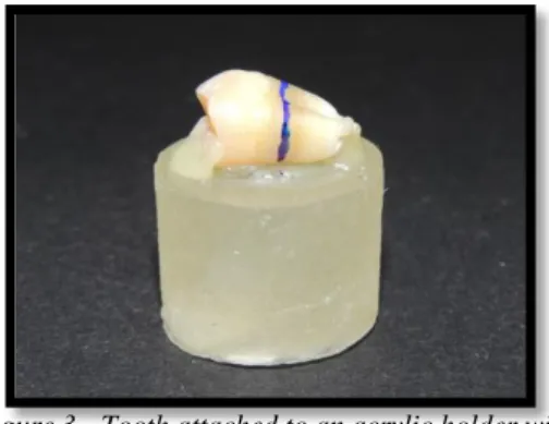
Daniela Rocha 7 Figure 3 - Tooth attached to an acrylic holder with
sticky wax.
Figure 4 - Roots cutting 1-2 mm below cementoenamel junction.
Figure 5, 6 and 7 – Removal of the pulp tissues.
1. The teeth were attached to an acrylic holder with sticky wax, parallel to the long axis of the tooth (Fig. 3);
2. The first cut was made parallel to the occlusal surface 1-2 mm below the cementoenamel junction to remove the roots (Fig. 4);
3. The crowns were detached from the acrylic holders and the pulp tissues were removed from the pulp chamber with a dentin curette and diamond burs (Fig. 5, 6 and 7). The crowns were attached again to the acrylic holder with sticky wax (Fig. 8).
Daniela Rocha 8 Figure 8 – The crown fixed to the acrylic holder.
Figure 9 – Mesiodistal crown cutting.
Figure 10 – Crowns glued with cyanoacrylate glue to acrylic holders.
3. The crowns were sectioned mesiodistally, parallel to the long axis of tooth, using a diamond disk at low speed, under constant water irrigation (Fig. 9), obtaining two enamel surfaces, a vestibular and a lingual/palatal.
4. The pulp chambers of the two pieces were then filled with cyanoacrylate glue (737 Black Magic Toughened adhesive, Permabond, Hampshire, UK) and the crown segments were fixed with the same glue to the acrylic holders, by the pulpal side (Fig. 10);
Daniela Rocha 9
5. With the purpose of creating a uniform smear layer obtained in similar conditions to those occurring in clinical situations, the enamel surface was cut with a cilindric high-speed diamond bur during 5 seconds under water irrigation (Pashley et al., 1988).
2.4 Distribution and treatment of the crown segments
The crown segments were kept in distilled water until the moment of treatment. The six crown segments were randomly assigned to one of the two adhesive groups. The order in which the crown segments were treated was random, to avoid possible bias due to any particular sequence of treatment.
The adhesion area was marked in the flattest area of the segments surface. The occlusal thirds of the vestibular and lingual surfaces were discharged due to their slope.
All the treatment procedures were performed by the same operator in the way that is followed described:
Group A – Scotchbond Universal Adhesive (3M ESPE Seefeld, Germany) as per manufacturer’s instructions – Total-etch (Etch-and-Rinse) technique on enamel (SBU TE E):
1. The enamel surface was slightly dried, without desiccate, but kept moist using a moist cotton pellet, so that the surface remained shiny and visibly moist.
2. The 32% phosphoric acid etching gel (Scotchbond Universal Etchant; 3M ESPE Seefeld, Germany) was applied to the marked enamel surface, during 15 seconds, and then rinsed with water and air-dried without desiccate. The excess of water was removed using a moist cotton pellet, so that the surface remained shiny and visibly moist.
3. The adhesive was applied, using a disposable microbrush, to the marked enamel surface, scrubbing lightly for 20 seconds.
4. The surface was then gently air-dried until it ceases to show any movement and the solvent was evaporated completely, forming a homogenous and slightly shiny film. Beginning with a soft blow of air from a distance of
Daniela Rocha 10 Figure 11 – Scotchbond Universal Adhesive.
approximately 10 cm, the air pressure was increased while decreasing distance, finishing at a distance of approximately 1-2 mm from the surface at maximum air pressure.
5. Finally, the surface was polymerized for 10 seconds.
Group B – Clearfil SE Bond Adhesive (Kuraray, Okayama, Japan) with pre-etching – Total-etch (Etch-and-Rinse) technique on enamel (CL TE E)*:
* - It is a modification of manufacturer´s instructions. Not recommended by
manufacturer.
1. The enamel surface was slightly dried, without desiccate, but kept moist using a moist cotton pellet, so that the surface remained shiny and visibly moist.
6. The 32% phosphoric acid etching gel (Scotchbond Universal Etchant; 3M ESPE Seefeld, Germany) was applied to the marked enamel surface, during 15 seconds, and then rinsed with water and air-dried without desiccate. The excess of water was removed using a moist cotton pellet, so that the surface remained shiny and visibly moist.
2. The primer was applied, using a disposable microbrush, to the marked enamel surface, and left in place for 20 seconds.
3. The adhesive was applied, using a disposable microbrush, to the marked enamel surface, scrubbing lightly for 20 seconds.
4. The surface was then gently air-dried, beginning with a soft blow of air from a distance of approximately 10 cm, the air pressure was increased while decreasing distance, finishing at a distance of approximately 1-2 mm from the surface at maximum air pressure.
Daniela Rocha 11 Figure 12 – Clearfil SE Bond plus the Scotchbond Universal etchant.
5. The adhesive was applied using a disposable microbrush, leaving a uniform, thin and even adhesive layer, removing the excess of adhesive with the same microbrush and with a soft blow of air.
6. Finally, the surface was polymerized for 10 seconds.
The adhesive polymerizes with visible light, so it was used a device with a lid to block natural light, and it was used at maximum of 3 minutes after placing it in the dispenser.
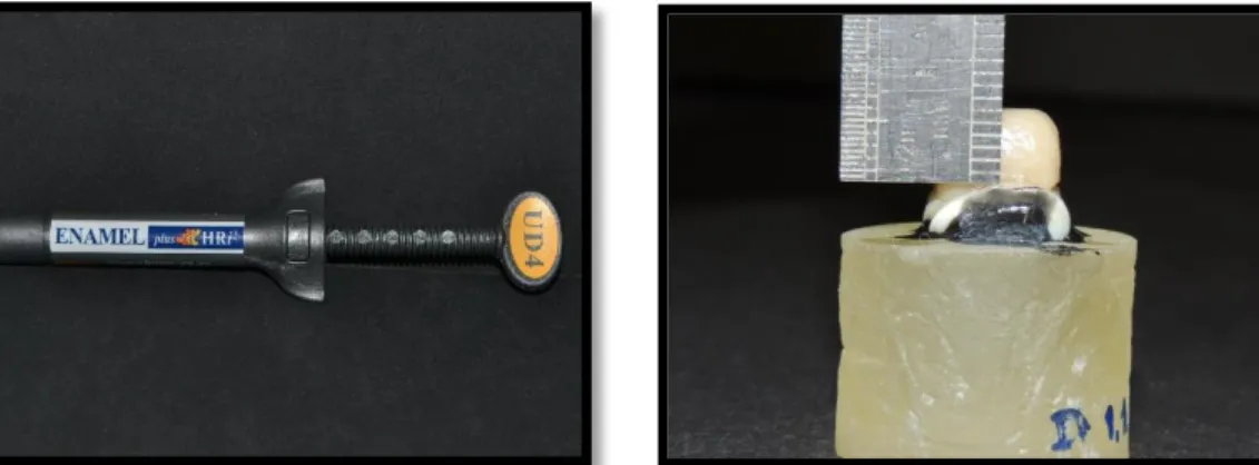
Resin composite build-ups were performed using a resin composite, ENAMEL Plus HRi (Micerium S.p.A. Avegno (GE) Italy; Fig. 13), shade UD4, in three increments of 2 mm each. Each increment was light cured for 20 seconds, according to the manufacturer's instructions, until reaching a height of 6 mm (Fig. 14). Additional light polymerization was performed on facial, lingual, mesial and distal surfaces of the composite build-up for 10 seconds each.
All light curing was performed with a light intensity of 600 mW/cm2 using a halogen light-activation unit (ELIPAR S10,3M ESPE Seefeld, Germany), with the 13 mm light guide held 1-2 mm from the treatment surface. The output of the curing light was periodically verified at >600 mW/cm2 with a radiometer (Curing Radiometer P/N 10503, USA) throughout the procedure.
Daniela Rocha 12 Figure 14 – Resin composite build-up.
Figure 13 – Resin composite ENAMEL Plus HRi
Material Manufacturer Components Lot code and expiring date Clearfil SE Bond Adhesive Kuraray, Okayama, Japan PRIMER: 10-MDP; HEMA;
Hydrophobic aliphatic dimethacrylate; dl-Camphorquinone; Water; Accelerators; Dyes; Others.
BOND:
10-MDP; Bis-GMA; HEMA;
Hydrophobic aliphatic methacrylate; dl-Camphorquinone; Colloidal silica; Initiators; Accelerators; Others.
Primer
Lot code: 2U0022 Expiring date: 07-2015 Bond: Lot code: 2T0039 Expiring date: 07-2015 Scotchbond Universal 3M ESPE Seefeld, Germany Bis-GMA; HEMA; (10-MDP)
Decamethylene dimethacrylate; Ethanol; Water; Silane treated silica;
2-propenoic acid, 2-methyl-, Reaction products with 1,10-
Decanediol and phosphorous oxide (p2o5); Copolymer of acrylic and itaconic acid; Dimethylaminobenzoat(-4);
Camphorquinone
(dimethylamino)ethyl methacrylate; Methyl ethyl ketone. Lot code: 490251 Expiring date: 09-2014 Ortophosphoric acid 32 % Scotchbond Universal Etchant; 3M ESPE Seefeld, Germany
Ortophosphoric acid (H3PO4) 32%; Thickener; colorant; deionized water
Lot code: 537103 Expiring date: 11-2015
Enamel plus HRi Composite UD4
Micerium S.p.A
Avegno (GE) Italy
Dymethachrylates; barium glass; Iteberium Trifluorethum; mixed oxides;
pre-polymers; aditives; catalysts; stabilizers; pigments; 1,4-Butandioldimethacrylate; Urethandimethacrylate (UDMA); Bis-GMA
Lot code: 2012 000 921 Expiring date: 12-2018
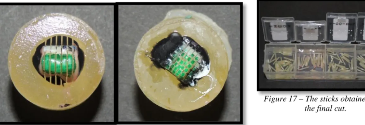
Daniela Rocha 13 Figure 15 and 16 – The teeth after being sectioned in both “x” and “y”
directions.
Figure 17 – The sticks obtained after the final cut.
2.5. Specimens preparation for the microtensile tests
All teeth were painted with different colors with waterproof ink. The exterior surface of the resin composite was also painted, in order to identify them.
The teeth were stored in distilled water in an incubator for 24 hours at 37 °C. Date and time of the restoration was registered.
Posteriorly, the teeth were longitudinally sectioned in both ―x‖ and ―y‖ (Fig. 15 and 16) directions with a slow-speed diamond disk (Diamond Wafering Blade - 10,2cmx0,3mm - Series 15HC, Buehler Ltd., Lake Bluff, IL, USA) under water irrigation, using a hard tissues microtome (IsometTM Precision Saw, Buehler Ltd. Ltd., Lake Buff, IL, EUA), to obtain sticks with a cross-sectional area of approximately 1 mm2.
A final cut is made at the base of the root, perpendicular to the long axis of the tooth, to separate the sticks from the acrylic supports.
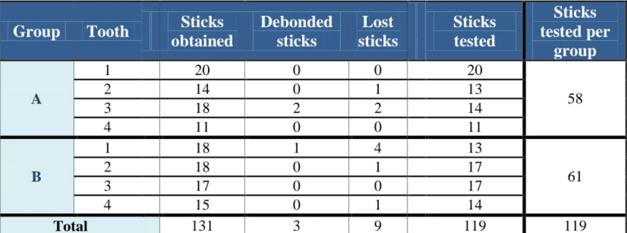
Debonded or lost sticks were registered. Debonded sticks were those separated in the adhesive interface during the cutting procedure. Lost sticks were those that were lost or fractured during test preparation.
The obtained sticks (Fig. 17) were kept in distilled water for a maximum of 24 hours, until the conclusion of the microtensile tests.
Daniela Rocha 14 Figure 19 – Instron 4502, universal testing machine. Figure 20 – The specimen attached to a
Geraldelli’s jig. Figure 18 – Specimens attached to Geraldelli’s jig.
Group Tooth Sticks obtained Debonded sticks Lost sticks Sticks tested Sticks tested per group A 1 20 0 0 20 58 2 14 0 1 13 3 18 2 2 14 4 11 0 0 11 B 1 18 1 4 13 61 2 18 0 1 17 3 17 0 0 17 4 15 0 1 14 Total 131 3 9 119 119
Table 2 – Characterization of sticks obtained, per group and per tooth.
2.6. Microtensile tests
The specimens were individually attached to a stainless-steel grooved Geraldelli´s jig (Fig. 18 and 20) with cyanoacrylate glue (737 Black Magic Toughened adhesive, Permabond, Hampshire, UK) and then submitted to a tension load using a universal testing machine (Instron 4502 H 3307, Norwood, MA, USA; - Fig. 19), at a crosshead speed of 1mm/min until fracture occurred.
A digital caliper (Ficher Darex®, France) was used to measure the sides of the bonding interface and calculate the bonding area in mm2. The load at fracture (KN) and the bonding surface area of the specimen were registered and µTBS calculated in MPa, by dividing the force imposed at time of fracture by the bond area (mm2).
Daniela Rocha 15
The failure modes were analyzed under a stereomicroscope at 10x and classified as adhesive (A) - failure occurring at the enamel-adhesive interface; cohesive when the failure occurred in enamel (CD); or in composite (CC); and mixed (M) - failure with composite and enamel/dentin at the interface.
2.7. Statistical Analysis
The statistical analysis of the results was performed through descriptive and inference methods.
The statistical analysis with the Kolmogorov-Smirnov test determined that p>0,05 (0,200) so we could use a parametric test. Also the Levene’s test for equality of variances determined that p>0,05 (0,383), so the samples/groups are homogenous, allowing the t-test interpretation be done in safe.
A paired-sample t-test was performed since the assumption of normality and equal variances among groups were valid.
Pretesting failures that occurred during specimen preparation were treated as left-censored data (Armstrong et al., 2010).
Daniela Rocha 16
3. Results
The number of microtensile beams (N), means, and standard deviations (SD) are shown in table 3. Clearfil SE (CL TE E) resulted in similar µTBS means (24,73 ± 13,38 MPa) in comparison to Scotchbond Universal (SBU TE E) (24,45 ± 10,88 MPa).
Variable =MPa
Group N Minimum Maximum Mean Standard
Deviation
Std error Mean
A - SBU TE E 58 3,90 45,36 24,4517 10,87830 1,42839
B - CL TE E 61 1,31 82,01 24,7379 13,37784 1,71286
Table 3 – Mean and SD of µTBS (MPa) per group.
In this graphic is shown the µTBS values for the two experimental groups.
Graphic 1 – Distribution of the µTBS (MPa) values for the two experimental groups.
There were not statistically significant differences among the two groups as revealed by the t-test (p < 0,05).
Daniela Rocha 17
t-test for Equality of Means
t df Sig. (2-tailed) Mean
Difference
MPa
Equal variances
assumed -,128 117 ,899 -,28627
Equal variances not
assumed -,128 114,288 ,898 -,28627
Table 4 – t-test
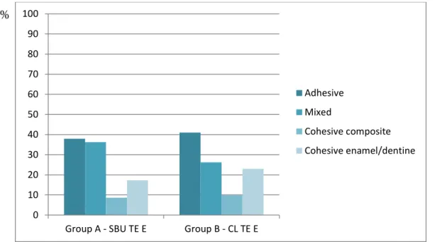
Table 5 and graphic 2 summarizes the types of failures obtained: in number and in percentage, per experimental group. 39,50% of the fractures were adhesive, without significative differences between groups, A (39,93%) and B (40,98%).
Type of failure
Group Adhesive Mixed
Cohesive composite Cohesive enamel/ dentine Total N % N % N % N % N % Group A - SBU TE E 22 37,93 21 36,21 5 8,62 10 17,24 58 100 Group B - CL TE E 25 40,98 16 26,23 6 9,84 14 22,95 61 100 Total 47 39,50 37 31,09 11 9,24 24 20,17 119 100
Table 5 – Characterization of the different types of failures per group.
Graphic 2 – Characterization of the different types of failures per group.
0 10 20 30 40 50 60 70 80 90 100
Group A - SBU TE E Group B - CL TE E
Adhesive Mixed
Cohesive composite Cohesive enamel/dentine
Daniela Rocha 18
4. Discussion
The clinical success of the adhesive restorations depends, not only, on the correct manipulation of the adhesive system, but also on the mode of application. There are differences between the self-etch mode application and the total-etch (Peumans et al., 2005), although self-etching systems have undergone a rapid evolution over the past few years. It becomes necessary to test the new adhesives introduced in the market, like Universal adhesives, and to assess if alterations in their mode of application change their bond strength.
The aim of this study was to analyse the microtensile bond strength of two adhesive systems, a universal and a self-etch one, and find out if the new universal adhesive system produced similar values to a self-etch adhesive widely studied.
After the experimental work and statistic analysis of the data, was possible to accept the null hypothesis and affirm, due to the obtained results, that there are no differences when comparing µTBS values of the universal and the self-etch adhesive.
µTBS test is one of the most used to study the most recent adhesive systems launched by diverse manufacturers (Armstrong et al., 2010), and for that reason was the method used in this study.
Clearfil SE Bond is now considered the ―gold standard‖ for all SE adhesives. In the laboratory, CSE tended to provide high dentin and enamel bond strengths (Brackett et al., 2008; Burrow et al., 2008; De Munck et al., 2005a; Mine et al., 2009; Perdigão et al., 2008; Van Landuyt et al., 2006; Van Landuyt et al., 2009). Likewise, an excellent clinical performance of CSE, with a 93 and 100% retention rate, respectively, at 3 years was reported in two studies (Turkun, 2003; Van Meerbeek et al., 2004). And one study (Peumans et al., 2010) resulted in a 98% retention rate at 8 years with or without separate enamel etching, another study (van Dijken, 2010) reported a retention rate on a yearly basis up to 8 years for CSE as compared with a 2-step etch-and-rinse adhesive. Also, a 5-year clinical study (Peumans et al., 2007) compared restorations bonded with CSE using recommended procedures compared with selective etching of the enamel margins with phosphoric acid. The results found that 36% of the restorations with recommended procedures had no enamel defects compared with 66% of the restorations having no defects when selective etching was used.
Daniela Rocha 19
adhesive (Scotchbond Universal) studied is very recent and insufficiently studied, at least on enamel. The universal adhesive was study on total-etch mode and the self-etch with a manufacturer’s modification, introducing a pre-etch phase.
The bond strength of these adhesive systems showed non-significant bond strength. These results are in agreement with data from a recent similar study (Freitas et al., 2012). So it seems that the bond strengths values are equal, when using a self-etch adhesive system or a universal one, on enamel. This was expected since both adhesives contain similar components, like 10-MDP, a functional phosphate monomer.
The presence of the molecule 10-MDP, which has chemical affinity for dental tissues (Yoshida et al., 2004), leads to an exposure of calcium and phosphate binding sites (Fukegawa et al., 2006), which are necessary for adhesion and to which the monomer binds in the second step. On enamel, the hydroxyapatite crystals are larger than in dentin, and the reactive surface is smaller, therefore acids have to be stronger to produce demineralization. MDP when interacting with hydroxyapatite forms a stable nano-layering at the adhesive interface (Inoue et al., 2005; Yoshida et al., 2012; Yoshihara et al., 2010). The hydrolysis of the ester bond from the acidic monomer results in a strong phosphoric acid that might increase the demineralization over time (Wang and Spencer, 2005).
Also SBU has polyalkenoic acid copolymer, besides MDP, in its formulation. Although both molecules may compete by binding to the calcium in hydroxyapatite (Yoshida et al., 2012), and they are usually associated with improved adhesive performance (Perdigao et al., 2013; Van Meerbeek et al., 2011), and that might be why the SBU had similar µTBS to CSE.
Besides acid concentration of acid used on the pre-etching, other factor that may play a role in the magnitude of enamel bond strengths are the orientation of the enamel prisms and the cohesive strength of the adhesive resin (Carvalho et al., 2000; Pashley and Tay, 2001). That is why in this study the enamel surfaces used where flattened parallel to the tooth axis, to standardize the orientation of enamel prisms.
Also, all adhesives tested were applied to bur-cut smear-layers prepared using high-speed diamond burs, used commonly for adhesive cavity preparation at the clinic. This procedure resulted in a uniform smear-layer with a roughness comparable to a smear layer created with SiC paper (Mine et al., 2010; Wahle and Wendt, 1993). Besides standardized bur-cut smear-layer formation, literature concerning the technique
Daniela Rocha 20
sensivity of the bonding procedure demonstrates that the performance of adhesive systems is significantly influenced by the technique variability of the operators, in particular procedures like etching, rinsing and drying (Giachetti et al., 2007; Giachetti et al., 2008; Miyazaki et al., 2000a). Therefore, all the bonding procedures in this study where made by the same operator.
Even though the manufacturer´s instructions are well-formulated, they are not very detailed and have some degree of ambiguity. For that reason an effort an effort was made to specify each step as much as possible, so that the protocol would become more consistent and standardized.
The specimen geometry has a significant influence on the homogeneity of stress concentration, which should be minimized. Due to the lack of consensus on specimen design and the relatively higher incidence of pre-testing failures during trimming, the stick-shaped specimens are simpler to prepare when compare to a dumbbell and hourglass geometry, and seem to introduce less flaws and stress during specimens preparation, so in this study was chosen to use stick/beam-like specimens (Armstrong et al., 2010; Ghassemieh, 2008; Pashley et al., 1999). Nonetheless, the stick shaped type of specimens present an higher standard deviation than the hourglass shaped ones (Ghassemieh, 2008).
In the present study the intensity all the light curing was controlled by a radiometer (Bala et al., 2005) to ensure that the intensity of the curing light was equal during all experimental work. Halogen light was used, since the literature indicates better results with this type of light (Pereira S., 2000).
Results of microtensile bond strength tests cannot easily be translated to clinical effectiveness of adhesive systems. Under intra-oral conditions, restorative interfaces are susceptible to degradation by saliva and other factors.
There are additional limitations when trying to extrapolate the in vitro results to the clinical performance of a material. An example of this, is the storage in water 24 hours before the testing, to simulate the intra-oral conditions (Huysmans et al., 1996). Another point observed is the high standard deviation obtained in all experimental work. Multiple imperfections may have occurred during the experimental part of the study. Bubbles or failures in the adhesive layer, lack of uniformity in the thickness of the adhesive, angulated traction forces (stick glued in the jig with a slight deviation, or not perfectly parallel to the Geraldelli’s jig, or problems standardizing the moisture on
Daniela Rocha 21
enamel, are all preponderant factors that may yield huge variations in standard deviation of the microtensile force values (Ghassemieh, 2008; Perdigao et al., 2002)3
From a clinical point of view, etching with phosphoric acid is problematic. Selective etching on enamel would be necessary, since etching dentine reduces the bond strength (Torii et al., 2002; Van Landuyt et al., 2006; Walter et al., 2011). Pronounced etching or over-etching of dentine leads to a total and deep demineralization of the collagenous structures, which have to be infiltrated by the adhesive system. Selective etching without affecting the dentine, however, is very difficult to achieve in the daily clinical practice.
Daniela Rocha 22
5. Conclusions
The results of the present study require the acceptance of the null hypothesis that there are no differences on the µTBS values when comparing a universal and a self-etch adhesive. Based on these results, it seems that for enamel, multi-mode universal adhesives yield similar adhesive performance than the gold standard two-step self-etching adhesives when the latest are also applied with selective enamel pre-self-etching. Further clinical studies are needed with these newest multi-mode universal adhesives, like Scotchbond Universal, especially because these materials are being increasingly used on patients without proof of clinical efficacy.
The need of selective etching on enamel is highlighted by the generally lower bond strengths found for SE systems to enamel compared with E&R systems (De Munck et al., 2003; De Munck et al., 2005b; Goracci et al., 2004; Loguercio et al., 2008). Selective etching without affecting dentine, however, is very difficult in daily clinical practice. Hence, a pre-treatment not leading to over-etching dentine but maintaining bond strength to both dentine and enamel would be desirable.
i
6. References
Armstrong S, Geraldeli S, Maia R, Raposo LH, Soares CJ, Yamagawa J (2010). Adhesion to tooth structure: a critical review of "micro" bond strength test methods. Dental materials : official publication of the Academy of Dental Materials 26(2):e50-62.
Asmussen E, Uno S (1992). Adhesion of restorative resins to dentin: chemical and physicochemical aspects. Operative dentistry Suppl 5(68-74.
Bala O, Olmez A, Kalayci S (2005). Effect of LED and halogen light curing on polymerization of resin-based composites. Journal of oral rehabilitation 32(2):134-140.
Bowen RL, Eick JD, Henderson DA, Anderson DW (1984). Smear layer: removal and bonding considerations. Operative dentistry Supplement 3(30-34.
Brackett WW, Tay FR, Looney SW, Ito S, Haisch LD, Pashley DH (2008). Microtensile dentin and enamel bond strengths of recent self-etching resins. Operative dentistry 33(1):89-95.
Buonocore MG (1955). A simple method of increasing the adhesion of acrylic filling materials to enamel surfaces. Journal of dental research 34(6):849-853.
Burrow MF, Kitasako Y, Thomas CD, Tagami J (2008). Comparison of enamel and dentin microshear bond strengths of a two-step self-etching priming system with five all-in-one systems. Operative dentistry 33(4):456-460.
Carvalho RM, Santiago SL, Fernandes CA, Suh BI, Pashley DH (2000). Effects of prism orientation on tensile strength of enamel. The journal of adhesive dentistry 2(4):251-257.
de Goes MF, Shinohara MS, Freitas MS (2014). Performance of a New One-step Multi-mode Adhesive on Etched vs Non-etched Enamel on Bond Strength and Interfacial Morphology. The journal of adhesive dentistry.
De Munck J, Van Meerbeek B, Satoshi I, Vargas M, Yoshida Y, Armstrong S et al. (2003). Microtensile bond strengths of one- and two-step self-etch adhesives to bur-cut enamel and dentin. American journal of dentistry 16(6):414-420.
ii
De Munck J, Van Landuyt K, Peumans M, Poitevin A, Lambrechts P, Braem M et al. (2005a). A critical review of the durability of adhesion to tooth tissue: methods and results. Journal of dental research 84(2):118-132.
De Munck J, Vargas M, Iracki J, Van Landuyt K, Poitevin A, Lambrechts P et al. (2005b). One-day bonding effectiveness of new self-etch adhesives to bur-cut enamel and dentin. Operative dentistry 30(1):39-49.
Erickson RL, Barkmeier WW, Kimmes NS (2009). Bond strength of self-etch adhesives to pre-etched enamel. Dental Materials 25(10):1187-1194.
Freitas MS, Shinohara MS, De Goes MF (2012). Effect of enamel pre-etching on bond strength of self-etching adhesives. Dental Materials 28, Supplement 1(0):e8-e9.
Fukegawa D, Hayakawa S, Yoshida Y, Suzuki K, Osaka A, Van Meerbeek B (2006). Chemical interaction of phosphoric acid ester with hydroxyapatite. Journal of dental research 85(10):941-944.
Ghassemieh E (2008). Evaluation of sources of uncertainties in microtensile bond strength of dental adhesive system for different specimen geometries. Dental materials : official publication of the Academy of Dental Materials 24(4):536-547.
Giachetti L, Russo DS, Bertini F, Pierleoni F, Nieri M (2007). Effect of operator skill in relation to microleakage of total-etch and self-etch bonding systems. Journal of Dentistry 35(4):289-293.
Giachetti L, Russo DS, Bambi C, Nieri M, Bertini F (2008). Influence of operator skill on microleakege of total-etch and self-etch bonding systems. Journal of Dentistry 36(1):49-53.
Goracci C, Sadek FT, Monticelli F, Cardoso PE, Ferrari M (2004). Microtensile bond strength of self-etching adhesives to enamel and dentin. The journal of adhesive dentistry 6(4):313-318.
Gordan VV, Vargas MA, Denehy GE (1998). Interfacial ultrastructure of the resin-enamel region of three adhesive systems. American journal of dentistry 11(1):13-16.
iii
Huysmans MC, van der Varst PG, Lautenschlager EP, Monaghan P (1996). The influence of simulated clinical handling on the flexural and compressive strength of posterior composite restorative materials. Dental materials : official publication of the Academy of Dental Materials 12(2):116-120.
Ibarra G, Vargas MA, Armstrong SR, Cobbb DS (2002). Microtensile bond strength of self-etching adhesives to ground and unground enamel. The journal of adhesive dentistry 4(2):115-124.
Inoue S, Koshiro K, Yoshida Y, De Munck J, Nagakane K, Suzuki K et al. (2005). Hydrolytic stability of self-etch adhesives bonded to dentin. Journal of dental research 84(12):1160-1164.
Kanemura N, Sano H, Tagami J (1999). Tensile bond strength to and SEM evaluation of ground and intact enamel surfaces. Journal of dentistry 27(7):523-530.
Loguercio AD, Moura SK, Pellizzaro A, Dal-Bianco K, Patzlaff RT, Grande RH et al. (2008). Durability of enamel bonding using two-step self-etch systems on ground and unground enamel. Operative dentistry 33(1):79-88.
Mena-Serrano A, Kose C, De Paula EA, Tay LY, Reis A, Loguercio AD et al. (2013). A new universal simplified adhesive: 6-month clinical evaluation. Journal of esthetic and restorative dentistry : official publication of the American Academy of Esthetic Dentistry [et al] 25(1):55-69.
Mine A, De Munck J, Cardoso MV, Van Landuyt KL, Poitevin A, Kuboki T et al. (2009). Bonding effectiveness of two contemporary self-etch adhesives to enamel and dentin. Journal of Dentistry 37(11):872-883.
Mine A, De Munck J, Vivan Cardoso M, Van Landuyt KL, Poitevin A, Kuboki T et al. (2010). Enamel-smear compromises bonding by mild self-etch adhesives. Journal of dental research 89(12):1505-1509.
Miyazaki M, Onose H, Moore BK (2000a). Effect of operator variability on dentin bond strength of two-step bonding systems. American journal of dentistry 13(2):101-104.
Miyazaki M, Sato M, Onose H (2000b). Durability of enamel bond strength of simplified bonding systems. Operative dentistry 25(2):75-80.
Myers CL, Rossi F, Cartz L (1974). Adhesive taglike extensions into acid-etched tooth enamel. Journal of dental research 53(2):435-441.
iv
Pashley DH, Tao L, Boyd L, King GE, Horner JA (1988). Scanning electron microscopy of the substructure of smear layers in human dentine. Arch Oral Biol 33(4):265-270.
Pashley DH, Carvalho RM, Sano H, Nakajima M, Yoshiyama M, Shono Y et al. (1999). The microtensile bond test: a review. The journal of adhesive dentistry 1(4):299-309.
Pashley DH, Tay FR (2001). Aggressiveness of contemporary self-etching adhesives: Part II: etching effects on unground enamel. Dental Materials 17(5):430-444.
Pashley DH, Tay FR, Breschi L, Tjäderhane L, Carvalho RM, Carrilho M et al. (2011). State of the art etch-and-rinse adhesives. Dental Materials 27(1):1-16.
Perdigao J, Lopes L, Lambrechts P, Leitao J, Van Meerbeek B, Vanherle G (1997). Effects of a self-etching primer on enamel shear bond strengths and SEM morphology. American journal of dentistry 10(3):141-146.
Perdigao J, Geraldeli S, Carmo AR, Dutra HR (2002). In vivo influence of residual moisture on microtensile bond strengths of one-bottle adhesives. Journal of esthetic and restorative dentistry : official publication of the American Academy of Esthetic Dentistry [et al] 14(1):31-38.
Perdigao J, Geraldeli S, Hodges JS (2003). Total-etch versus self-etch adhesive: effect on postoperative sensitivity. Journal of the American Dental Association (1939) 134(12):1621-1629.
Perdigao J (2007). New developments in dental adhesion. Dental clinics of North America 51(2):333-357, viii.
Perdigao J, Sezinando A, Monteiro PC (2012). Laboratory bonding ability of a multi-purpose dentin adhesive. American journal of dentistry 25(3):153-158.
Perdigao J, Sezinando A, Monteiro PC (2013). Effect of substrate age and adhesive composition on dentin bonding. Operative dentistry 38(3):267-274.
Perdigão J, Lopes MM, Gomes G (2008). In Vitro Bonding Performance of Self-etch Adhesives: II—Ultramorphological Evaluation. Operative dentistry 33(5):534-549.
v
Pereira S. PC, Mendes A. (2000). Avaliação da dureza superficial de uma resina composta híbrida em função da cor, tempo de exposição, intensidade de luz e profundidade do material. Jornal Brasileiro de Clínica Estética e Odontológica 4(23):63-67.
Peumans M, Kanumilli P, De Munck J, Van Landuyt K, Lambrechts P, Van Meerbeek B (2005). Clinical effectiveness of contemporary adhesives: A systematic review of current clinical trials. Dental Materials 21(9):864-881.
Peumans M, De Munck J, Van Landuyt K, Lambrechts P, Van Meerbeek B (2007). Five-year clinical effectiveness of a two-step self-etching adhesive. The journal of adhesive dentistry 9(1):7-10.
Peumans M, De Munck J, Van Landuyt KL, Poitevin A, Lambrechts P, Van Meerbeek B (2010). Eight-year clinical evaluation of a 2-step self-etch adhesive with and without selective enamel etching. Dental Materials 26(12):1176-1184.
Sano H, Shono T, Sonoda H, Takatsu T, Ciucchi B, Carvalho R et al. (1994). Relationship between surface area for adhesion and tensile bond strength--evaluation of a micro-tensile bond test. Dental materials : official publication of the Academy of Dental Materials 10(4):236-240.
Standardization IOf (2003). ISO/TR 11 405 Dental Materials - testing of adhesion to ttoh structure. Geneva, Switzerland: WHO, pp. 1-16.
Swift EJ, Jr., Perdigao J, Heymann HO (1995). Bonding to enamel and dentin: a brief history and state of the art, 1995. Quintessence international (Berlin, Germany : 1985) 26(2):95-110.
Tay FR, Gwinnett AJ, Pang KM, Wei SH (1996). Resin permeation into acid-conditioned, moist, and dry dentin: a paradigm using water-free adhesive primers. Journal of dental research 75(4):1034-1044.
Tay FR, Pashley DH (2002). Dental adhesives of the future. The journal of adhesive dentistry 4(2):91-103.
Tay FR, Pashley DH, King NM, Carvalho RM, Tsai J, Lai SC et al. (2004). Aggressiveness of self-etch adhesives on unground enamel. Operative dentistry 29(3):309-316.
Torii Y, Itou K, Nishitani Y, Ishikawa K, Suzuki K (2002). Effect of phosphoric acid etching prior to self-etching primer application on adhesion of resin composite to enamel and dentin. American journal of dentistry 15(5):305-308.
vi
Turkun SL (2003). Clinical evaluation of a self-etching and a one-bottle adhesive system at two years. Journal of dentistry 31(8):527-534.
van Dijken JW (2010). A prospective 8-year evaluation of a mild two-step self-etching adhesive and a heavily filled two-step etch-and-rinse system in non-carious cervical lesions. Dental materials : official publication of the Academy of Dental Materials 26(9):940-946.
Van Landuyt KL, Kanumilli P, De Munck J, Peumans M, Lambrechts P, Van Meerbeek B (2006). Bond strength of a mild self-etch adhesive with and without prior acid-etching. Journal of Dentistry 34(1):77-85.
Van Landuyt KL, Mine A, De Munck J, Jaecques S, Peumans M, Lambrechts P et al. (2009). Are one-step adhesives easier to use and better performing? Multifactorial assessment of contemporary one-step self-etching adhesives. The journal of adhesive dentistry 11(3):175-190.
Van Meerbeek B, Conn LJ, Jr., Duke ES, Eick JD, Robinson SJ, Guerrero D (1996). Correlative transmission electron microscopy examination of nondemineralized and demineralized resin-dentin interfaces formed by two dentin adhesive systems. Journal of dental research 75(3):879-888.
Van Meerbeek B, Yoshida Y, Lambrechts P, Vanherle G, Duke ES, Eick JD et al. (1998). A TEM study of two water-based adhesive systems bonded to dry and wet dentin. Journal of dental research 77(1):50-59.
Van Meerbeek B, De Munck J, Yoshida Y, Inoue S, Vargas M, Vijay P et al. (2003). Buonocore memorial lecture. Adhesion to enamel and dentin: current status and future challenges. Operative dentistry 28(3):215-235.
Van Meerbeek B, Kanumilli PV, De Munck J, Van Landuyt K, Lambrechts P, Peumans M (2004). A randomized, controlled trial evaluating the three-year clinical effectiveness of two etch & rinse adhesives in cervical lesions. Operative dentistry 29(4):376-385.
Van Meerbeek B, Yoshihara K, Yoshida Y, Mine A, J DM, K.L VL (2011). State of the art of self-etch adhesives. Dental Materials 27(1):17-28.
Wahle JJ, Wendt SL, Jr. (1993). Dentinal surface roughness: a comparison of tooth preparation techniques. J Prosthet Dent 69(2):160-164.
vii
Walter R, Swift EJ, Jr., Boushell LW, Braswell K (2011). Enamel and dentin bond strengths of a new self-etch adhesive system. Journal of esthetic and restorative dentistry : official publication of the American Academy of Esthetic Dentistry [et al] 23(6):390-396.
Wang Y, Spencer P (2005). Continuing etching of an all-in-one adhesive in wet dentin tubules. Journal of dental research 84(4):350-354.
Yoshida Y, Nagakane K, Fukuda R, Nakayama Y, Okazaki M, Shintani H et al. (2004). Comparative study on adhesive performance of functional monomers. Journal of dental research 83(6):454-458.
Yoshida Y, Yoshihara K, Nagaoka N, Hayakawa S, Torii Y, Ogawa T et al. (2012). Self-assembled Nano-layering at the Adhesive interface. Journal of dental research 91(4):376-381.
Yoshihara K, Yoshida Y, Nagaoka N, Fukegawa D, Hayakawa S, Mine A et al. (2010). Nano-controlled molecular interaction at adhesive interfaces for hard tissue reconstruction. Acta biomaterialia 6(9):3573-3582.
Zidan O, Hill G (1986). Phosphoric acid concentration: enamel surface loss and bonding strength. J Prosthet Dent 55(3):388-392.
viii
7. Appendix
I – Stereomicroscopy photographs of the different types of failures: A) Adhesive failures
Figure I and II – Adhesive Failures.
B) Cohesive failures
a. Enamel-Dentine b. Composite
ix
C) Mixed failures.
x
II – Tables
Group Kolmogorov-Smirnova Shapiro-Wilk
Statistic df Sig. Statistic df Sig.
MPa SBU TE E ,054 58 ,200
*
,979 58 ,423
CL TE E ,089 61 ,200* ,911 61 ,000
*. This is a lower bound of the true significance.
Table I: Shapiro-Wilk and kilmogorov.Smirnov normality tests
t-test for Equality of Means Std. Error
Difference
95% Confidence Interval of the Difference Lower Upper MPa Equal variances assumed 2,24188 -4,72619 4,15366 Equal variances not assumed 2,23029 -4,70433 4,13180
xi
III - Graphics
Graphic I: Normal distribution of the values for Group A: Scotchbond Universal Adhesive (3M ESPE Seefeld, Germany)on enamel
Graphic II: Normal distribution of the values for Group B: Clearfil SE Bond Adhesive (Kuraray, Okayama, Japan)on enamel
xii
IV – Manufacturer’s instructions
Scotchbond Universal Adhesive (3M ESPE Seefeld, Germany) as per manufacturer’s
instructions:
1. Selective Enamel Etching
a. Apply a commonly used phosphoric acid etching gel (about 35%), e.g., Scotchbond Universal Etchant to the prepared and unprepared (if present) tooth enamel and allow to react for 15 sec.
b. Rinse thoroughly with water and dry with water-free and oil-free air or with cotton pellets; do not overdry.
2. Total Etching Procedure
a. Apply a commonly used phosphoric acid etching gel (about 35%), e.g., Scotchbond Universal Etchant, to the prepared and unprepared (if present) tooth structure (enamel and dentin) and allow to react for 15 sec. b. Rinse thoroughly with water and dry with water-free and oil-free air or
with cotton pellets; do not overdry.
3. Use the disposable applicator to apply the adhesive to the entire tooth structure and rub it in for 20 sec. Avoid contact between the adhesive and the oral mucosa.
4. Subsequently direct a gentle stream of air over the liquid for about 5 sec until it no longer moves and the solvent has evaporated completely.
5. Harden the adhesive with a commonly used curing light for 10 sec.
Clearfil SE Bond Adhesive (Kuraray, Okayama, Japan) as per manufacturer’s
instructions:
1. Dispense the necessary amount of primer into a well of the mixing dish immediately before application
2. Apply primer to the entire cavity wall with a sponge or a disposable brush tip. Leave it in place for 20 seconds.
3. After conditioning the tooth surface for 20 seconds, evaporate the volatile ingredients with a mild oil-free air stream.
xiii
4. Dispense the necessary amount of bond into a well of the mixing dish.
5. Apply bond to the entire surface of the cavity with a sponge or a disposable brush tip.
6. After application, make the bond film as uniform as possible using a gentle oil-free air stream.