Anchorage evaluation of dental implants irradiated with
GaAIAs laser (830nm)
Bruno Pereira Campanha
1Daniel Humberto Pozza
2João Batista Blessmann Weber
3Marília Gerhardt de Oliveira
4Abstract
The aim of the present study was to evaluate titanium implants fixed with free rotation in rabbit’s tibia and irradiated with GaAlAs laser (4.3Joules/daily, 830nm wavelength, 12mW and 51 seconds) using a digital torquimeter. Thirty male rabbits were divided into two groups (Laser and Control) and subdivided according to their death day 15 (L1 and C1), 30 (L2 and C2) and 45 (L3 and C3) after surgery. A digital torquimeter evaluated the torque needed for removing the implants, breaking their union to the bone. The results were submitted to Student’s “t” test and ANOVA test, in order to validate these findings. It was observed an increase on removal torque values of irradiated implants both with 15 and 30 days after surgery, in comparison to control groups. With 45 days, there was no statistically significant difference between the Laser and Control groups values. It was concluded that Laser group showed a higher value in the removal torque when compared to non-irradiated implants.
Keywords: Dental implants- Laser- Osseointegration.
INTRODUCTION
The implantology is increasing the quality of people’s life, aiding them in situations of precarious buccal health conditions. In the past, without the use of implants, the rehabilitation possibilities with conventional prostheses, were finite and limited.1
If the osseointegration is no longer a doubt, the interface bone/implant continues to be researched. Surgical techniques are in constant evaluation and operative parameters thought. The implant texture and surface are altered and treated
in most different ways trying to achieve larger contact area.2
Today, the success of implants can be found as 94% in mandible and 92% in maxilla, and the problems still remain in implants mat failures.1
In agreement with Askary, Meffert and Griffin3 the initial stability is determined mainly
by mechanical properties of density and amount of bones (of maxilla and mandible) and influenced by the surgical technique, especially in bones of low quality. The authors report that the use of excessive
1 Doutor em Cirurgia e Traumatologia Bucomaxilofacial. Pontifícia Universidade Católica do Rio Grande do Sul - PUCRS. Porto Alegre. RS.
2 Doutor em Odontologia - Laser, Universiade Federal da Paraiba - UFPB/Universidade Federal da Bahia - UFBA - Salvador. BA
3 Doutor em Odontologia e Professor Adjunto da Pontifícia Universidade Católica do Rio Grande do Sul - PUCRS. Porto Alegre. RS. 4 Doutora em Odontologia e Professora Titular da Pontifícia Universidade Católica do Rio Grande do Sul - PUCRS. Porto Alegre. RS.
Correspondência para / Correspondence to:
Marília Gerhardt de Oliveira
Pontifícia Universidade Católica do Rio Grande do Sul (PUCRS) Avenida Ipiranga, 6681, Edifício 6, sala 209
90619-900. Porto Alegre - RS - Brazil
force by the surgeon during the operative action, mainly in bones of low quality, can cause on-extension in size of receiving bed of implant, with consequent decrease of primary stability.
The density of available bone has a great influence in the treatment of surfaces and in drawing of implant. The surgical approach as well as success of implants is more predictable in bones of large density.4
From the discovery of osseointegration, implantology is counting with growing indexes of success. Tinsley, Watson and Ogden 5 accomplished
a study in 39 Centers of Buccal Rehabilitation of United Kingdom. Fifty one percent of losses happened in an initial phase of cicatrization, that is, before the placement of prosthesis. The main failure cause was pointed as being the low quality of bone, mainly in maxilla, suggesting new studies to optimize the prognostic of implants that present larger tendency of loss.
Barewal and others 6 affirmed that
independently of quality of bone type, implants present a decrease of stability in the first month after surgery. They emphasize that this reduction in union bone/implant is more pronounced in implants in low quality bone.
Biomodulation of LLLT happens between doses of one and 10 joules/cm2.7 For Almeida-Lopes
and others 8, the biomodulation was, for a long
time, called incorrectly, biostimulation. The laser can be used to obtain antagonist effects amongst themselves, that therapy doesn’t just accelerate certain processes, but it also delays some others.
In 2001, Blay 9 accomplished a study to
determine if the osseointegration process of implants put in tibia of rabbits with high initial stability, suffers some alteration type when the area is irradiated with low power laser. The implants were inserted with initial torque of 40 newtons (N), when the implants obtained the initial stability resonance frequency were monitored. One of the groups was irradiated with 680nm laser and other with 830nm. Ten sessions were accomplished, with intervals of 48 hours, beginning immediately after surgical procedure. The density of energy was 4J/ cm2 through point, being 2 points on each side of
tibia. The values of the resonance frequency were statistically different among irradiated groups and no irradiated in two times of observation. The
authors concluded that after irradiation with low power laser, the implants with high initial stability, had osseointegration degree higher in relation to the group controls.
In 2004, Khadra and others10 evaluated
the action of GaAlAs laser (830nm, 150mW and 23J/cm2) on disks of titanium implanted in tibia
of rabbits. The animals were irradiated immediately after the surgery and in daily sessions in a total of 10 sessions. With 8 weeks of experiment the authors caused the death of animals. The physical union bone/implant was evaluated with the necessary tension to remove the implants and with microscopic analysis of the bone/implant contact area. The analysis of tension force showed advantagesin union of irradiated implants. The histomorphometric analysis showed larger area of bone/implant contact in irradiated groups. The authors suggested a favorable effect of laser in the healing and union of titanium implants.
The objective of this study was to evaluate through the removal torque of implants removal torque value the effect of laser (GaAlAs), 830nm wavelength, 12mW of potency, in dose of 4.3J/ cm2 and application time of 51 seconds, in
implants without initial stability in tibia of rabbits.
METHODS
The hypothesis of this study was that the value of removal torque of implants, installed in tibia of rabbits without initial stability, would be increased when submitted to proposed LLLT protocol.
The animal model was the rabbit (Oiyctolagus cuniculus) of New Zealand race. Thirty young adults animals, with approximately two months old, male, weighing among 1.5 – 2.0kg each, were distributed randomly in two groups - no irradiated and irradiated with low power laser, which were subdivided in three subgroups according with time elapsed among the surgery and the death, 15, 30 and 45 days.
The surgical procedures, maintenance, as well as death of animals were done in State Foundation of Production and Research in Health (FEPPS), Division of Production and Animal Experimentation - DPEA. The animals remained
in individual cages suspended, avoiding direct contact with feces and urine.
To accomplish the surgical procedure the animals were anesthetized with intra-peritoneal injection of Zoletil® 50 mg (5 mg/Kg) and Xilasin®
(3 mg/kg). The trichotomy was proceeded in the internal part of animal’s paw being followed of asepsis with iodoform diluted in alcohol 70%. Anesthetic solution with adrenaline 1:50.000 was injected, seeking local hemostasis. The incision of skin was accomplished with scalpel blade n° 15, following by incision of periosteum. The bone was exposed with Molt’s periostal elevator, after elevation of periosteum, the elevators were used to separate and support the implant area.
Then the gradual stagger of drills began, in a logical sequence established, in speed of 800 rotations for minute and 30N of torque. The implants were made by Conecção®.
Prostheses with special diameter of 2.5mm, length of 6mm and without surface treatment were used. This length was established seeking an monocortical anchorage and the perforation was accomplished until that drill touched the second cortex (with care of not perforating it), then the perforation was finished with a drill of 2.3mm of diameter, making possible a low intentional initial stability in the bone fixation of implant.
The implants were then inserted, through a manual key, supplied by the manufacturer. After obtain the initial stability, the implant was rotated in counterclockwise sense, allowing the implant freedom to rotate freely at bone site.
The suture was done with a 4.0 nylon, in anchored continuous way, following by demarcation of reference point for subsequent irradiation.
Immediately after the surgeries, the animals were submitted to the first session of GaAlAs laser with wavelength of 830 nm (THERA LASER®;
D.M.C Equipments LTDA São Carlos - SP), potency of 12mW, in continuous way and with a dose of 1.075J/cm2 per point, in 51 seconds.
An optic fiber with 0.02827cm2 of area
was used to irradiate the 4 points around each implant, in a total dose of 4.3J/cm2 by session.
The irradiation procedure was repeated every 48 hours, in a total of 7 application sessions (1, 3, 5, 7, 9, 11 and 13 days postoperative).
The control group was submitted to a false irradiation, under the same routine of irradiated animals. The sutures were removed after seven days in all groups.
The animals were killed in an atmosphere saturated with carbon dioxide, according with the observation time of 15, 38 and 45 days.
After death, the sites of implants were accessed and a carefully osteotomy was performed, leaving a safety margin of 20mm on each side of the implant. The material was kept in a solution of 10% neutral formalin by 24 hours. The pieces were included in acrylic resin, for subsequent measure with torquimeter.
The values of were obtained through an axial digital torquimeter (TSD 150; Torqueleader -MHH Engineering CO. LTD.), that promoted an
against-torque (anti-rotational move) in implants. Through the “peak” program of reading, the maximum torque for breaking the union bone/ implant was stored. The torquimeter used in this study, was calibrated at the laboratories of University of Engineering of PUCRS.
After tabulation of data averages obtaining and standard deviations of each group, the Student’s “t” test, was used for comparison between laser group and control group. The analysis of variances (ANOVA) was used for comparisons of the three subgroups laser and control in the different times of study.
RESULTS
The values of removal torques found in each group, are represented in tables and graphs below, according with the laser treatment and observation periods: 15, 30 and 45 days.
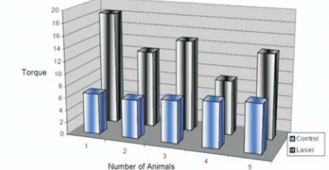
The Figure 1 presents the individual values of removal torques’ of L1 and C1 groups. The average removal torques for group L1 was of 13.62cNm while the average torque of group C1 was of 7.02cNm. When applying the Student “t” test, a result of 0.013 (p=0.013) was found for a significant level of 95% (p<0.05), indicating that torques’ values of groups the 15 day of experiment presents a statistically significant difference.
The Figure 2 presents the individual removal torques’ values of L2 and C2 groups. The average
L2 group removal torque was 24.84cNm while the average C2 group torque was 16.94 cNm. When the Student “t” test was applied a result of 0.030 (p=0,030) was found for a significant level of 95% (p<0.05), indicating that the values of the torques’ in 30 day of experiment have a statistically significant difference. .
The Figure 3 presents the individual removal torques’ values of L3 and C3 groups. The average L3 group removal torques was 27.4cNm while the average C3 group torque of was 23.5cNm. When
control), the ANOVA statistical test was applied. A statistically significant difference was observed among the torque averages found in the 15 and 30, 30 and 45 and 15 and 45 days of observation, for no irradiated groups (controls).
For laser groups a statistically significant difference was observed in torque averages between 15 and 30 days and 15 and 45 days of observation. Statistically significant difference wasn’t observed in averages among 30 and 45 days. The Figure 4 presents the evolution of removal torque’s averages of laser group and control group in the three appraised periods of time. .
DISCUSSION
In 1999 Al-Watban and Zhang11 tested
several protocols in laser therapy. The authors concluded that researches’ protocols should attempts
Figure 1 - Groups C1 x L1: Animals not irradiated com-pared to irradiated, in 15 days of observation.
Figure 2 - Groups C2 x L2: Animals not irradiated com-pared to irradiated, in 30 days of observation.
the Student “t” test was applied, a result of 0.215 (p=0.215) was found for a significant level of 95% (p<0.05), indicating that the torques’ values in 45 day of experiment don’t have a statistically significant difference. .
To verify the evolution of torque average in groups that received a same treatment (laser or
Figure 3 - Groups C3 x L3: Animals not irradiated com-pared to irradiated, in 45 days of observation.
Figure 4 - Torques' evolution of irradiated groups com-pared to no irradiated (C1, C2, C3 x L1, L2, L3), in 15, 30 and 45 days of observation..
to laser type choice evaluating the ideal laser for each tissue type and penetrabilition, measured by wavelength, to reach the target tissue. Once chosen the tissue and laser type, the dose should be adapted in search for best result.
For Khadra and others 10, Blay 9 and Lopes
and others 12 the applied wavelength in this study
is able to take radiation to bone tissue depth. According to Trelles and Mayayo 13, in hard
tissues low power laser irradiation was shown to increase vascularization and the number of trabecular in fractured tibias of mice.
The four points of irradiations around the implant used in this study, is in agreement with Lopes and others 12 that used irradiation around
of their implants and Campanha and others 14
that used irradiation around the wound accomplished in dorsal region of their animals, both in continuous way. The authors also respected an interval of 48 hours among sessions as in this study.
Pinheiro 15 used a 7 session protocol with
interval of 48 hours among the sessions, achieving good results in bone formation, giving support to this work.
The dose of 1.075J/cm2 by point, applied
in this study is in agreement with most laser protocols. Pinheiro 15 mention that doses between
1 and 5J/cm2 are more effective for soft and hard
tissues.
For Pinheiro 16 and Silva Júnior and others 17, the ideal potency should vary from 5 to 21mW,
when supplied with lower potencies by long time intervals.
Dortbudak, Haas and Mailath-Pokomy 18
believe that low-energy laser irradiation increases the number of viable osteocytes in the irradiated bone and this laser appears to produce highly reactive and vital peri-implant bone tissue, reducing healing times and enhancing osseointegration of the implants. Barewal and others 6 observed that
in the first month, the implants stability is the smallest during the period of bone healing, for all bone types and mainly in marrowy bones.
In this research it was opted to work with a willfully bad prognostic situation, in the attempt of evaluating the laser front to those situations where, clinically she has been reducing the chances
of success: bone of bad quality and implants with freedom rotational.
The L1 and L2 groups presented a statistically larger removal torque than C1 and C2 groups. With 15 and 30 days of experiment the groups that suffered laser irradiation, needed a larger torque for breaking the union with bone when compared to no irradiated groups. The L3 group didn’t present a statistically larger removal torque than the C3 group, meaning that with 45 days of experiment no statistically significant difference was identified among the irradiated group and no irradiated group.
Being the only variable among the groups, the laser should be responsible for these differences checked with the torquemeter. These discoveries agree with immense majority of works that mention the effects of laser light.10
Karu 19 affirms that action of laser’s is larger
in initial moments, moments of laser biological instability, as a trauma or wound in soft or hard tissue. The author believes that, in natural process of repair the organism search for a balance state, in the other words, as more dose of biological ba-lance (repair process’ final) as minor will be the laser’s action.
Agreeing with the author, in this study the best effects were obtained in initial moments (15 and 30 days), while the effects of laser light weren’t statistically significant with 45 days of experiment. Braverman and others 20 accomplished
wounds in the two sides of the same animal and irradiated only one side. Those authors found effects of laser light, as much for directly irradiated wounds as in no irradiated wounds when compared to other not irradiated to other not irradiated animals.
A good understanding of the treatment parameters and use of standardized protocols, are necessary for reproduction and comparison of results of studies.
The results showed that irradiated implants were more difficult to remove than not irradiated and the mean value of removal torque found in irradiated group was statistically higher than in control groups in initial osseointegration phase.
It is concluded that LLLT, carried out with the parameters of the present investigation, increases the bone/implant union in the initial osseointegration phase.
Avaliação da ancoragem de implantes dentários irradiados com laser
GaAlAs (830 nm)
Resumo
O objetivo desta pesquisa foi avaliar, através do uso de um torquímetro digital, se implantes de titânio, instalados com liberdade rotaciona em tíbias de coelhos e irradiados com laser de Arseneto de Gálio e Alumínio, na dose de 86 J/cm2, com comprimento de onda de 830 nm, potência de 12
mW e tempo de aplicação de 51 segundos, apresentam um valor de torque de remoção estatistica-mente maior que os valores obtidos nos grupos não irradiados (grupo controle). Para tanto, traba-lhou-se com trinta coelhos, da raça Nova Zelândia, machos, os quais foram divididos em dois grupos, Laser e Controle, e subdivididos de acordo com o dia de sua morte. Os grupos L1 e C1, L2 e C2, L3 e C3 foram mortos 15, 30 e 45 dias após a inserção do implante, respectivamente. Um torquímetro digital mensurou o torque necessário para o afrouxamento do implante, rompendo sua união com o osso. Os resultados foram, então, submetidos ao Teste “t” de Student e ao Teste ANOVA, a fim de validar esses achados. Observou-se, para um p=0,05, um aumento nos valores dos torques de remoção dos implantes irradiados com laser e controle (L1 e L2), tanto aos quinze quanto aos trinta dias após as cirurgias, em relação aos respectivos grupos controle (C1 e C2). Aos 45 dias, não mais foi observada diferença estatisticamente significante entre os valores encontrados nos grupos laser e controle (L1 e C1). Concluiu-se, nesse modelo animal e com o protocolo de irradiação utilizado neste estudo, que o laser, por ser a única variável entre os grupos, foi responsável pelo aumento do embricamento do implante ao osso, no mês inicial e mais critico da osseointegração. Palavras-chave: implantes dentários; laser; osseointegração.
6 BAREWAL, R.M. et al. Resonance frequency measurement of implant stability in vivo on implants with a sandblasted and acid-etched surface. Int. J. Oral Maxillofac. Implants,
Lombard, v.18, p.641-651, 2003.
7 REDDY, G.K. Photobiological basis and clinical role of low-intensity lasers in biology and medicine.
J. Clin. Laser Med. Surg., New York, v.22,
p.141-150, 2004.
8 ALMEIDA-LOPES, L. et al. Comparison of the low level laser therapy effects on cultured human gingival fibroblasts proliferation using different irradiance and same fluence. Lasers Surg. Med.,
New York, v.29, n.2, p.179-84, 2001.
9 BLAY, A. Efeitos da radiação laser em baixa intensidade no mecanismo de osseointegração de implantes: estudo “in vivo”. 2001. Dissertação
(Mestrado) – Faculdade de Odontologia, Univer-sidade de São Paulo, 2001.
REFERENCES
1 ASHLEY, E.T. et al. Ailing and failing endosseous dental implants: a literature review. J. Contemp. Dent. Pract., Cincinnati, v.4, n.2,
p.35-50, 2003.
2 WENNERBERG, A.; AIBREKTSSON, T.; KROL, J.J. A histomorphometric and removal torque study of screw-shaped titanium implants with three different surface topographies. Clin. Oral Implants Res., Copenhagen, v.6, p.24-30,
1995.
3 ASKARY, A.S.E.; MEFFERT, R.M.; GRIFFIN, T. Why do dental implants fail? Part I. Implant Dent., Baltimore, v.8, p.173-185, 1999.
4 MISCH, C.E. Implantes dentários contem-porâneos. 2ed. São Paulo: Santos; 2000.
5 TINSLEY, D.; WATSON, C.J.; OGDEN, A.R.A. Survey of U.K. centers on implant failures.
10 KHADRA, M. et al. Low-level laser therapy stimulates bone-implant interaction: an experimen-tal study in rabbits. Clin. Oral Implants Res.,
Copenhagen, v.15, p.325-332, 2004.
11 AL-WATBAN, F.A.H.; ZHANG, X.Y. The acceleration of wound healing is not attributed to laser skin transmission. Laser Ther., New
Rochelle, v.11, n.1, p.6-10, 1999.
12 LOPES, C.B. et al. Infrared laser light reduces loading time of dental implants: a Raman spectroscopic study. Photomed. Laser Surg.,
Larchmont, v.23, n.1, p.27-31, 2005.
13 TRELLES, M.A.; MAYAYO, E. Bone fracture consolidates faster with low power laser. Lasers Surg. Med., New York, v.7, p.36-45, 1987.
14 OLIVEIRA, M. G. de; PINHEIRO, A.L.B.; CAMPANHA, B.P. Polarised light (400 -2000nm): a description of the wound healing process using immunohistochemical analysis. In: LASERS IN DENTISTRY, 9, 2003, San Jose, CA. Proceedings of SPIE. Bellingham, 2003.
v.4950, p.144-149.
15 PINHEIRO, A.L.B. Effect of 830 nm Laser light on the repair of bone defects grafted with
inorganic bovine bone and decalcified cortical osseous membrane. J. Clin. Laser Med. Surg.,
New York, v.21, p.301-306, 2003.
16 PINHEIRO, A.L.B. Effects of low level laser therapy on malignant cells: in vitro study. J. Clin. Laser Med. Surg., New York, v.20, p.23-26,
2002.
17 PINHEIRO, A. L. B. et al. Biomodulatory effects of LLLT on bone regeneration. Laser Ther.,
New Rochelle, v.13, p.73-79, 2001.
18 DORTBUDAK, O.; HAAS, R.; MAILATH-POKOMY, G. Effect of low-power laser irradiation on bony implant sites. Clin. Oral Implants Res., Copenhagen, v.13, p.288-292,
2002.
19 KARU, T.I. Photobiological fundamentals of low power laser therapy. IEEE J. Quantum Electron., New York, v.10, p.1703-1717, 1987.
20 BRAVERMAMN, B. et al. Effect of Helium - Neon and infrared laser irradiation on wound healing in rabbits. Lasers Surg. Med., New York,
v.9, p.50-58, 1989.
Recebido em / Received: 12/01/2007 Aceito em / Accepted: 07/04/2007