Braz. J. of Develop.,Curitiba, v. 6, n. 3,p. 13180-13190 mar. 2020. ISSN 2525-8761
Development and evaluation of organic silicon nanoparticles
Desenvolvimento e avaliação de nanopartículas de silício orgânico
DOI:10.34117/bjdv6n3-255
Recebimento dos originais: 29/02/2020 Aceitação para publicação: 17/03/2020
Rosana Letícia da Rosa
Instituição: Universidade Estadual de Ponta Grossa
Mestre em Ciências Farmacêuticas pela Universidade Estadual de Ponta Grossa Endereço: Avenida General Carlos Cavalcanti, 4748, Uvaranas, Ponta Grossa Paraná- Brasil
CEP: 84030-900
E-mail: rosanaleticia@hotmail.com Luana Serbai
Instituição: Universidade Estadual de Ponta Grossa
Graduada em Farmácia pela Universidade Estadual de Ponta Grossa
Endereço: Avenida General Carlos Cavalcanti, 4748, Uvaranas, Ponta Grossa Paraná- Brasil CEP: 84030-900
E-mail: lserbai@hotmail.com Robson Schimandeiro Novak
Mestre em Ciências Farmacêuticas pela Universidade Estadual de Ponta Grossa Instituição: Universidade Estadual de Ponta Grossa
Endereço: Avenida General Carlos Cavalcanti, 4748, Uvaranas, Ponta Grossa Paraná - Brasil CEP: 84030-900
E-mail: robsonovak@hotmail.com Josiane de Fátima Padilha de Paula
Doutora em química com área de concentração em química orgânica- Polímeros, pela Universidade Federal do Paraná.
Instituição: Universidade Estadual de Ponta Grossa
Endereço: Avenida General Carlos Cavalcanti, 4748, Uvaranas, Ponta Grossa Paraná - Brasil CEP: 84030-900
E-mail: jopadilha@terra.com.br Ana Cristina Oltramari Toledo
Mestre em Botânica pela Universidade Federal do Paraná. Instituição: Universidade Estadual de Ponta Grossa
Endereço: Avenida General Carlos Cavalcanti, 4748, Uvaranas, Ponta Grossa Paraná- Brasil CEP: 84030-900
E-mail: acotoledo@hotmail.com Vanessa Von Mühlen de Carvalho
Braz. J. of Develop.,Curitiba, v. 6, n. 3,p. 13180-13190 mar. 2020. ISSN 2525-8761 Instituição: Universidade Estadual de Ponta Grossa
Endereço: Avenida General Carlos Cavalcanti, 4748, Uvaranas, Ponta Grossa Paraná - Brasil CEP: 84030-900
E-mail: vanessa_vmdc@hotmail.com Patricia Mathias Döll Boscardin
Doutora em Ciências Farmacêuticas pela Universidade Federal do Paraná Instituição: Universidade Estadual de Ponta Grossa
Endereço: Avenida General Carlos Cavalcanti, 4748, Uvaranas, Ponta Grossa Paraná- Brasil CEP: 84030-900
E-mail: pattydoll@globo.com ABSTRACT
Skin is an important component of outer beauty, it is the focus of many surgical and non-surgical procedures. Skin changes associated with intrinsic or extrinsic aging, such as wrinkles, sagging and pigmentation changes, can cause people to look for cosmetic procedures to improve the appearance of their skin. Studies demonstrate the importance of organic silicon in the production of type I collagen. The present work aimed to study the development of Silicium P® polymer nanoparticles. The suspension of nanoparticles was obtained by the technique of double emulsification-evaporation of the solvent where the physicochemical characterization was performed. These presented mean diameter of 562 nm, zeta potential of -37.2 mV and polydispersity index of 0.42. It was also obtained the infrared spectrum with Fourier transform where it showed that there were no molecular interactions that alter the chemical structures of the components of the formulation during the process of obtaining the nanoparticles.
Keywords: Collagen; Silicium P®; Skin aging; Nanoparticle. RESUMO
A pele é um componente importante da beleza externa, é o foco de muitos procedimentos cirúrgicos e não cirúrgicos. Alterações na pele associadas ao envelhecimento intrínseco ou extrínseco, como rugas, flacidez e alterações de pigmentação, podem levar as pessoas a procurar procedimentos cosméticos para melhorar a aparência da pele. Estudos demonstram a importância do silício orgânico na produção de colágeno tipo I. O presente trabalho teve como objetivo estudar o desenvolvimento de nanopartículas de polímero Silicium P®. A suspensão de nanopartículas foi obtida pela técnica de dupla emulsificação-evaporação do solvente onde foi realizada a caracterização físico-química. Estes apresentaram diâmetro médio de 562 nm, potencial zeta de -37,2 mV e índice de polidispersividade de 0,42. Também foi obtido o espectro infravermelho com transformada de Fourier, onde demonstrou que não houve interações moleculares que alteram as estruturas químicas dos componentes da formulação durante o processo de obtenção das nanopartículas.
Braz. J. of Develop.,Curitiba, v. 6, n. 3,p. 13180-13190 mar. 2020. ISSN 2525-8761 1. INTRODUCTION
One of the most important components of the skin is type I collagen. It is responsible for the physical and biological properties of the tissues, such as rigidity, stability and guarantees the structural integrity of these¹. Changes over the years in the skin can be quantitative and qualitative, leading to a decrease in the proteins of the dermis, as well as a reduction in the action of the fibroblasts. Consequently, there will be a decrease in type I collagen, as well as changes in the morphology and amount of elastic fibers2.
Thinking about improving the appearance of skin studies are being conducted in vivo and in vitro, where organic silicon is being inserted into the diet3. These studies demonstrate beneficial bone health as well as the synthesis of collagen type I4. In view of the importance of organic silicon in the production of collagen, new pharmaceutical forms are being considered for the administration of this component, since it is poorly absorbed when ingested as capsules or even through food5. Among these are the polymer nanoparticles.
Nanotechnology is being applied in the pharmaceutical area in order to increase the bioavailability of the drug, control the release at the specific site of action, decrease local toxicity, protect the molecule from extrinsic and / or intrinsic factors linked to the kinetics of the organism and also facilitates administration of drugs in a noninvasive way6.
Considering that the nanoparticles can increase the release and the therapeutic properties of the drugs and cosmetics, this work had as objective to develop and to evaluate the action of silicium P® (Monomethylsilanotriol) nanoparticles on 3T3 fibroblasts.
2. MATERIALS AND METHODS
The suspension of Silicium P® nanoparticles (NSiP) was prepared by the double emulsification-solvent evaporation technique7. Firstly, the polyvinyl alcohol (PVAL) was solubilized in ultra-purified water at a temperature of 40 °C under mechanical stirring, separately the poly (ε-caprolactone) (PCL) in chloroform was solubilized at room temperature under mechanical stirring. Silicium P®, hydrophilic active, was solubilized in ultra-purified water and to this solution was added Tween® 80. The nanoparticle suspension was obtained using a homogenizer (Ultra-Turrax) at a shaking of 22.000 rpm. First, the Silicium P® solution was emulsified in the PCL solution for 60 seconds. The emulsion formed (organic phase) was then poured onto the PVAL (aqueous phase) solution and the stirring was continued for 3 minutes. The emulsion formed was kept under magnetic stirring at room temperature for 4
Braz. J. of Develop.,Curitiba, v. 6, n. 3,p. 13180-13190 mar. 2020. ISSN 2525-8761 hours for the evaporation of the organic solvent. At the end of the process, a water / oil / water emulsion with a concentration of Silicium P® of 1 mg / mL was obtained.
After evaporation of the solvent using a refrigerated centrifuge (10,000 rpm / 10 min), the nanoparticles were washed 3 times with ultra-purified water to remove excess PVAL. The nanoparticles were resuspended and stored under refrigeration. The same procedure was used to obtain control nanoparticles (NC), where Silicium P® was not added.
The mean diameter, zeta potential and the polydispersity index (IPD) were determined using the laser autocorrelation spectrometer (MALVERN® Instruments, model Zetasizer Nanoseries ZS90). For the analysis, the sample was diluted in ultra-purified water to a final concentration of 0.02 mg / mL.
The morphological and surface evaluation of the nanoparticles was carried out in a scanning electron microscope with field emission MIRA3 (Tescan). The samples were metallized with gold on the SC7620 Mini Sputter Coater. 10 μl of the nanoparticle suspensions were added to the stubs and dried in an oven at 37 °C for 24 h. The micrographs were obtained after visualization of the samples employing acceleration voltages of 6kx to 21kx. The registration of the images occurred through the specific software.
The nanoparticles were analyzed by Fourier transform infrared spectroscopy in KBr-containing pellets using 4 mg of each sample and 196 mg of KBr spectroscopic grade (2% w / w) in the IRPrestige-21 (Shimadzu), with parameters of 64 scans.min-1 and resolution of 4 cm-1.
The effect of the nanoparticles on fibroblastic cells was evaluated by means of cell viability. To observe cell viability against different nanoparticle samples, the cells were plated in 96-well culture plates (0.10 x 10⁵ cells / well) and incubated for 24 hours. After the incubation period the supernatant was discarded and samples of different concentrations of NSi (50, 25, 12.5, 7.5, 5 and 2.25%) and NC (50, 25, 12.8 and 6.25%) were added to each well. In addition to the nanoparticles, different concentrations of Silicium P® (1000, 30, 25, 20 and 10 μL / mL) were tested.
Cell viability was determined after 24 hours of sample application using the MTT reduction method. The method analyzes mitochondrial activity by reducing the soluble salt 3- (4,5) dimethyl-2-thiazolyl) -2,5-diphenyl-2-tetrazole bromide. Salt metabolism results in the formation of insoluble crystals of violet coloration. The absence of the insoluble crystals indicates the non-reduction of the salt, due to cell death. Quantitative determination of cytotoxicity was performed by reading the absorbance spectrophotometer at the wavelength
Braz. J. of Develop.,Curitiba, v. 6, n. 3,p. 13180-13190 mar. 2020. ISSN 2525-8761 of 570 nm. Cell viability was determined by comparing the absorbance values obtained for treated and untreated cells with the samples (negative control).
3. RESULTS AND DISCUSSION
After preparation, the nanoparticles of silicon P® (NSi) had a mean diameter of 562 nm and the nanoparticles control 656.7 nm, a result of great interest, since particles with nanometric sizes are able to penetrate the layers of the skin or enter through of the hair follicle, releasing the encapsulated drug8. Mean diameter less than 600 nm is reported as suitable for topical application9. NSiP were monodisperse, since the IPD obtained was 0.42, studies report that IPD of less than 0.5 indicate a homogeneous solution10.
The PZ reflects the surface potential of the particles, which is influenced by the changes in the interface with the dispersing medium6,11 and is an indicator of the stability of the suspensions. One study reported that solutions with PZ near I30I mV are considered stable12. In this way, the NSiP can be considered stable, once they presented the PZ of -37.2, it can also be confirmed that the silicon did not change the stability characteristics of the nanoparticle suspension, where the Nc presented PZ of -35.1. The negative value is associated to PCL, due to the presence of the ester group at the end of the polymer chain 13.
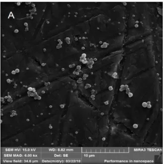
The morphological and surface evaluation of the nanoparticle suspension was performed by FEG. The nanoparticles of Silicium P® and control nanoparticles are identified in Fig. 1 and 2, respectively. NSi and NC presented a spherical shape and smooth surface, the similarity between the two indicates that the active does not influence the morphology and does not remain deposited under the surface of the nanoparticles.
The IVTF spectra of the NC and NSi are shown in figure 3. In both spectra it is possible to identify the functional groups present in the PCL, the presence of a band at 1721 cm -1, referring to the C = O stretch. The bands observed at 1171 cm -1 relate to stretching of the C-O bond. The symmetrical and asymmetric vibrations of the -CH2 group were observed at 2995 cm -1 and 2825 cm-1, respectively14. The band present in the two spectra at 3500 cm-1, refers to the alcohol function present in the poly (vinyl alcohol).
The spectral analysis of the nanoparticle suspensions indicated that the bands remained practically unchanged in the two spectra. It was suggested that there were no molecular interactions that altered the chemical structures of the components of the formulation during the process of obtaining the nanoparticles.
Braz. J. of Develop.,Curitiba, v. 6, n. 3,p. 13180-13190 mar. 2020. ISSN 2525-8761 The effect of Silicium P®, NC and NSi on fibroblast (3T3) lineage was evaluated by means of the MTT assay in the 24 hour period. For the evaluation of the effect of Silicium P® on fibroblastic cells, a stock solution with a concentration equal to 1000 μL.mL-1 was prepared from this solution, the following concentrations were obtained: 30, 25, 20 and 10 μL.mL-1. In Figure 4 it is possible to evaluate the effect of these concentrations of Silicium P® on the viability of 3T3 fibroblasts.
The cytotoxicity of a sample can be assessed by means of the cell viability obtained, according to ISO 10993-5: 1999 this occurs as follows: non-cytotoxic- cell viability> 90%, slightly cytotoxic- cell viability between 80 and 89 %, moderately cytotoxic- cell viability between 50 and 79% and severely cytotoxic- cell viability <50%.
From the results obtained with the Silicium P® cytotoxicity test, it can be concluded that the concentration of 1000 μL/mL can be considered moderately cytotoxic, since it reduced cell viability by 61%. At the concentrations of 30, 25, 20 and 10 μL/mL (1%), cell viability was observed above 85%, indicating a low cytotoxicity.
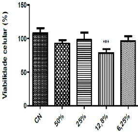
The NCs were tested at concentrations of 50, 25, 12.8 and 6.25% as shown in figure 5. The percentage of cell viability observed at the concentrations tested were 92, 98, 78 and 95%, respectively. Thus, with the exception of the 12.8% concentration, the other concentrations of NC may be considered non-cytotoxic.
The NSiP were tested at the concentrations of 50, 25, 12.5, 7.5, 5 and 2.5% as shown in figure 6. With the data obtained, it was possible to verify that the concentrations of 50% and 25% NSiP caused a greater reduction of viability cell phone. For both concentrations, cellular viability was observed around 60%, when compared to the negative control. At the concentrations of 12.5, 7.5, 5, 2.5%, a cell viability of 76, 81, 85 and 81%, respectively, was observed.
Considering the results obtained, concentrations of NSiP equal to or lower than 7.5% (Silicium P® concentration corresponding to 75 μL.mL-1) would be suitable for use, since according to the cytotoxicity test, this concentration may be considered slightly cytotoxic.
4. CONCLUSION
The double emulsification method followed by solvent evaporation was adequate to obtain Silicium P® nanoparticles, resulting in nanoparticles with a mean diameter of 562.55, low polydispersity index and negative zeta potential.
Braz. J. of Develop.,Curitiba, v. 6, n. 3,p. 13180-13190 mar. 2020. ISSN 2525-8761 The obtained nanoparticles have spherical shape and smooth surface. The spectra obtained in the infrared region indicated that no chemical bond was formed between Silicium P® and the polymer during the nanoencapsulation process.
The evaluation of the nanoparticles in 3T3 fibroblast cells, demonstrated a cytotoxic effect at the highest concentrations of nanoparticle suspensions tested. Its use being appropriate from the concentration equal to or below 7.5%. In an upcoming study, it would be appropriate to evaluate the production of type I collagen in Silicium P® nanoparticle-treated fibroblastic cells.
REFERENCES
1. VERHAEGEN, Pauline D. et al. Adaptation of the dermal collagen structure of human skin and scar tissue in response to stretch: an experimental study. Wound Repair and Regeneration, v. 20, n. 5, p. 658-666, 2012.
2. ZOUBOULIS, Christos C.; MAKRANTONAKI, Evgenia. Clinical aspects and molecular diagnostics of skin aging. Clinics in dermatology, v. 29, n. 1, p. 3-14, 2011.
3. LI, Zhaoping et al. Absorption of silicon from artesian aquifer water and its impact on bone health in postmenopausal women: a 12 week pilot study. Nutrition journal, v. 9, n. 1, p. 44, 2010.
4. REFFITT, D. M. et al. Orthosilicic acid stimulates collagen type 1 synthesis and osteoblastic differentiation in human osteoblast-like cells in vitro. Bone, v. 32, n. 2, p. 127-135, 2003. 5. UITTO, Jouni; BERNSTEIN, Eric F. Molecular mechanisms of cutaneous aging: connective tissue alterations in the dermis. In: Journal of Investigative Dermatology Symposium Proceedings. Elsevier, 1998. p. 41-44.
6. SCHAFFAZICK, Scheila Rezende et al. Caracterização e estabilidade físico-química de sistemas poliméricos nanoparticulados para administração de fármacos. Química nova. São Paulo. Vol. 26, n. 5 (2003), p. 726-737, 2003.
7. SOUTO, B. E.; SEVERINO, P.; SANTANA, H. M. Preparação de Nanopartículas Poliméricas a partir de Polímeros Pré-formados – Parte II. Polímeros, vol. 22, n. 1, p. 101-106, 2012.
8. SOARES, Margarida et al. Permeação cutânea: desafios e oportunidades. Journal of Basic and Applied Pharmaceutical Sciencies, v. 36, n. 3, 2016.
Braz. J. of Develop.,Curitiba, v. 6, n. 3,p. 13180-13190 mar. 2020. ISSN 2525-8761 9. VERMA, D. D., VERMA, S., BLUME, G., et al. Particle size of liposomes influences dermal delivery of substances into skin. International journal of pharmaceutics, v. 258, n. 1-2, p. 141-151, 2003.
10. AVADI, M. R. et al. Preparation and characterization of insulin nanoparticles using chitosan and Arabic gum with ionic gelation method. Nanomedicine: Nanotechnology, Biology, and Medicine, v. 6, p. 58–63, 2010.
11. MOSQUEIRA, Vanessa Carla Furtado et al. Poly (D, L‐lactide) nanocapsules prepared by a solvent displacement process: Influence of the composition on physicochemical and structural properties. Journal of pharmaceutical sciences, v. 89, n. 5, p. 614-626, 2000. 12. NEVES, A. R. et al. Novel resveratrol nanodelivery systems based on lipid nanoparticles to enhance its oral bioavailability. Int Journal of Nanomedicine, v. 8, p.177-187, 2013. 13. MORA-HUERTAS, C. E.; FESSI, H.; ELAISSARI, A. Polymer-based nanocapsules for drug delivery. International journal of pharmaceutics, v. 385, n. 1-2, p. 113-142, 2010. 14. PAVIA, D. L., LAMPMAN, M. G., KRIZ, S. G., et al. Introdução à Espectroscopia. 4ª ed. Cengage Learning, São Paulo, 2010.
FIGURE CAPTIONS
Figure 1. Morphological and surface aspects of Silicium P® nanoparticles. Acceleration Voltages: A: 6.00 kx. Rosa et al.
Figure 2. Morphological and surface aspects of control nanoparticles. Acceleration voltage 13,9kx. Rosa et.al.
Figure 3. IVTF spectra of the Silicium P® nanoparticles and control nanoparticles. Rosa et al. Figure 4. Effect of different concentrations of Silicium P® on the viability of 3T3 fibroblasts after 24 hours by the MTT reduction assay. Results are expressed as mean ± SEM (standard error of the mean) (n = 8). The statistical tests used were Anova, followed by Tukey's post-hoc test. *** P <0.0001 when compared with the control group. Rosa et al.
Figure 5. Effect of the control nanoparticle samples on the viability of 3T3 fibroblasts after 24 hours by the MTT reduction assay. Results are expressed as mean ± SEM (standard error of the mean) (n = 8). The statistical tests used were Anova, followed by Tukey's post-hoc test. *** P = 0.0037 when compared to the control group. Rosa et.al.
Figure 6. Effect of the Silicium P® nanoparticle samples on the viability of 3T3 fibroblasts after 24 hours by the MTT reduction assay. Results are expressed as mean ± SEM (standard
Braz. J. of Develop.,Curitiba, v. 6, n. 3,p. 13180-13190 mar. 2020. ISSN 2525-8761 error of the mean) (n = 8). The statistical tests used were Anova, followed by Tukey's post-hoc test. *** P <0.0001 when compared to the control group. Rosa et.al.
Figure 1. Morphological and surface aspects of Silicium P® nanoparticles. Acceleration Voltages: A: 6.00 kx.
Rosa et al.
Braz. J. of Develop.,Curitiba, v. 6, n. 3,p. 13180-13190 mar. 2020. ISSN 2525-8761
Figure 3. IVTF spectra of the Silicium P® nanoparticles and control nanoparticles. Rosa et al.
Figure 4. Effect of different concentrations of Silicium P® on the viability of 3T3 fibroblasts after 24 hours by
the MTT reduction assay. Results are expressed as mean ± SEM (standard error of the mean) (n = 8). The statistical tests used were Anova, followed by Tukey's post-hoc test. *** P <0.0001 when compared with the
Braz. J. of Develop.,Curitiba, v. 6, n. 3,p. 13180-13190 mar. 2020. ISSN 2525-8761
Figure 5. Effect of the control nanoparticle samples on the viability of 3T3 fibroblasts after 24 hours by the
MTT reduction assay. Results are expressed as mean ± SEM (standard error of the mean) (n = 8). The statistical tests used were Anova, followed by Tukey's post-hoc test. *** P = 0.0037 when compared to the control group.
Rosa et.al.
Figure 6. Effect of the Silicium P® nanoparticle samples on the viability of 3T3 fibroblasts after 24 hours by
the MTT reduction assay. Results are expressed as mean ± SEM (standard error of the mean) (n = 8). The statistical tests used were Anova, followed by Tukey's post-hoc test. *** P <0.0001 when compared to the