Braz. J. of Develop.,Curitiba, v. 6, n. 10, p. 82336-82356 oct. 2020. ISSN 2525-8761
Fungal community on skin tissue of amphibians collected in the Santarém
region, Pará, Brazil
Comunidade de fungos do tecido cutâneo de anfíbios coletados na região de
Santarém, Pará, Brasil
DOI:10.34117/bjdv6n10-604
Recebimento dos originais:01/10/2020 Aceitação para publicação:27/10/2020
Andreza da Silva Peixoto Graduating in Biological Sciences Federal University of Western Pará
Rua Vera Paz, s/n – Bairro Salé, 68040-255, Santarém – PA E-mail: [email protected]
Daniel de Sousa Guedes Graduating in Biological Sciences Federal University of Western Pará
Rua Vera Paz, s/n – Bairro Salé, 68040-255, Santarém – PA E-mail: [email protected]
Vanessa dos Santos Bentes Graduating in Biological Sciences Federal University of Western Pará
Rua Vera Paz, s/n – Bairro Salé, 68040-255, Santarém – PA E-mail: [email protected]
Nathan Sousa da Silva Graduating in Agrarian Sciences Federal University of Western Pará
Rua Vera Paz, s/n – Bairro Salé, 68040-255, Santarém – PA E-mail: [email protected]
Eveleise Samira Martins Canto Doctor in Biotechnology Federal University of Western Pará
Rua Vera Paz, s/n – Bairro Salé, 68040-255, Santarém – PA E-mail: [email protected]
Ricardo Alexandre Kawashita-Ribeiro Doctor in Ecology and Biodiversity Conservation
Federal University of Rondonópolis
Av. dos Estudantes, 5055 – Bairro Sagrada Família, 78735-910, Rondonópolis – MT E-mail: [email protected]
Braz. J. of Develop.,Curitiba, v. 6, n. 10, p. 82336-82356 oct. 2020. ISSN 2525-8761 Graciene do Socorro Taveira Fernandes
Doctor in Marine and Tropical Sciences Federal University of Western Pará
Rua Vera Paz, s/n – Bairro Salé, 68040-255, Santarém – PA E-mail: [email protected]
ABSTRACT
Among living beings, the Amphibia Class has unique characteristics. The cutaneous tissue of these animals has properties that provide ideal conditions for the establishment of microorganisms in their skin, which act in several important functions. The present study aimed to characterize the cultivable fungal microbiota of amphibians collected in the Santarém region, Western Pará, Brazil. Two species of toads were captured, totalling 25 individuals, being 15 specimens of Rhinella major and 10 individuals of R. marina. Collections were performed by active search at night, between the months of November 2017 to April 2019. Cutaneous samples were collected by smear in the dorsoventral region of the animal, using a swab soaked in sterile 0.85% saline solution with 10-3
dilution, and grown in Petri dishes containing Potato Dextrose Agar culture medium. The animals were euthanized and deposited in the herpetological collection of Federal University of Western Pará. For identification, isolation, morphological characterization and microculture of fungal colonies were used. In total, 75 fungal colonies representative of 11 genera were identified.
Penicillium and Aspergillus have been identified as the most representative genera in both
amphibian species. There were differences in the composition of the cutaneous mycobiota between
R. major and R. marina, but not related to seasonality, which may be linked to the environment they
inhabit, as well as to the physiological factors inherent to each species. The present work presents the taxa Absidia, Curvularia, Scopulariopsis and the group mycelia sterilia as new records of fungi associated with the skin tissue of adult anurans. Thus, it is suggested that further studies be carried out in Amazonian environments, with a larger sample size, as well as the use of molecular tools, for a better identification and understanding of the composition of the anuran skin microbiota.
Keywords: Anurans, Microbiota, Fungi, Amazonia. RESUMO
Entre os seres vivos, a Classe Amphibia possui características únicas, e precisam de mais estudos para fornecer informações sobre ecologia e interações com outros seres. O tecido cutâneo desses animais possui propriedades que fornecem condições ideais para o estabelecimento de microrganismos em sua pele, os quais atuam em diversas funções importantes. O presente estudo teve como objetivo caracterizar a microbiota fúngica cultivável de anfíbios coletados na região de Santarém, Oeste do Pará, Brasil. Foram capturadas duas espécies de anfíbios, totalizando 25 indivíduos, sendo 15 exemplares de Rhinella major e 10 indivíduos de Rhinella marina. O método de coleta foi de buscas ativas em horário noturno, entre os meses de novembro de 2017 a abril de 2019. O material cutâneo foi colhido por esfregaço na região dorsoventral do animal, utilizando um
swab embebido em solução salina 0,85% estéril até a diluição 10-3, e cultivado em placas de Petri contendo meio BDA. Os animais foram eutanasiados e posteriormente depositados na coleção de anfíbios da UFOPA. Utilizou-se o isolamento, a caracterização morfológica e o microcultivo das colônias fúngicas. Foram identificadas 75 colônias fúngicas representativas de 11 gêneros. Sendo os gêneros Penicillium e Aspergillus os mais representativos em ambas as espécies de anfíbios. Houve semelhanças e diferenças na composição da micobiota cutânea entre R. major e R. marina, o que pode estar ligado ao ambiente que habitam, bem como aos fatores fisiológicos inerentes a cada espécie. O presente trabalho apresenta os táxons Absidia, Curvularia, Scopulariopsis e o grupo mycelia sterilia como novos registros de fungos associados ao tecido cutâneo de anuros. Dessa
Braz. J. of Develop.,Curitiba, v. 6, n. 10, p. 82336-82356 oct. 2020. ISSN 2525-8761
forma, sugere-se que novos estudos sejam realizados em ambientes amazônicos, com um maior número amostral, assim como, a utilização de ferramentas moleculares, para a melhor identificação e entendimento da composição da microbiota cutânea dos anuros.
Palavras-chave: Anuros, Microbiota, Fungos, Amazônia.
1 INTRODUCTION
Amphibians have unique characteristics among all animals, therefore, they represent a valuable taxon for ecological and scientific purposes, since they bridge the evolutionary gap between land and aquatic animals (Xu and Lai, 2015). The amphibian skin performs multiple adaptive functions and possesses complex structures that make up an important set for the survival of these animals. In addition to the skin tissue acting on breathing, an important function in amphibians, it also acts as a first defensive barrier against external factors (Chuong et al., 2002; Huang et al., 2016).
In many animals, skin tissue is considered a vital component of the immune system’s structure against the establishment of pathogens and infections associated with the body, which activate innate and adaptive immune responses (Proksch, Brandner, and Jensen, 2008; Bos and Luiten, 2009). In amphibians, the skin tissue harboring various types of cells, glands, and diverse metabolic compounds from them (Pough, Janis, and Heiser, 2008; Vitt and Caldwell, 2013), the skin of these animals also hosts a microbial community related to the immune system, acting together to defend the organism (Lauer et al., 2007; Lam, Walke, Vredenburg, and Harris, 2010; Colombo, Scalvenzi, Benlamara, and Pollet, 2015; Holden et al., 2015).
The amphibian skin presents an ideal environment for the establishment of microorganisms, as it has optimal conditions for the permanence and survival of fungi and bacteria, such as humidity and nutrients from mucous secretions and other metabolic processes (Culp, Falkinham III, and Belden, 2007; Bletz, Perl, and Vences, 2017a). In addition to the host influencing the composition of the microbial community, factors such as skin renewal, stage of life and environment can alter and shape the microbiome of the cutaneous tissue of these animals (Cramp, McPhee, Meyer, Ohmer, and Franklin, 2014; Kueneman et al., 2014).
The microbiome, defined as the community of microorganisms that live in the organisms, act in many biological processes for their host, such as, acquisition of nutrients and energy; can affect its development and behavior; and act in protection against pathogens (Ezenwa, Gerardo, Inouye, Medina, and Xavier, 2012; Kelly and Mulder, 2012; Flórez, Biedermann, Engl, and Kaltenpoth, 2015).
Braz. J. of Develop.,Curitiba, v. 6, n. 10, p. 82336-82356 oct. 2020. ISSN 2525-8761
In Brazil, biological and ecological information is scarce for many species of amphibians (Peloso, Machado, and Becker, 2019). These species are inserted in three orders: Anura, Caudata and Gymnophiona, with anurans being the most abundant and the most studied among the others (Pough, Janis, and Heiser, 2008). This order includes frogs and toads, and 1,076 species are known in Brazil (Guerra, Jardim, Llusia, Márquez, and Bastos, 2020) distributed in 20 families and 90 genera (Segalla et al., 2016).
Rhinella Fitzinger, 1826 is a genus of toad of the Bufonidae family (Vitt and Caldwell, 2013;
Frost, 2020). The genus is composed of 92 species, naturally distributed in South and Central America, in open and forested areas (Frost, 2020), with 40 species occurring in Brazil (Segalla et al., 2016). Rhinella major Müller and Hellmich, 1936 is a medium-sized species ranging between 33 and 81 mm in length (Narvaes and Rodrigues, 2009), with nocturnal habits, and which lives and feeds almost exclusively in terrestrial environments, and is usually associated with anthropogenic environments (Hamann and González, 2015). Rhinella marina Linnaeus, 1758 is a large species reaching more than 225 mm in length, lives in forests and open areas, and is often found in disturbed areas and well adapted to urban life (AmphibiaWeb, 2020). Although this species is originally from the Americas, it was introduced in several countries (Acevedo, Lampo, and Cipriani 2016; González-Bernal, Greenlees, Brown, and Shine, 2016).
So far, the knowledge about the in Brazilian Amazon is dispersed in many publications, like taxonomic reviews, descriptions of new species or punctual fauna surveys (Ávila-Pires, Hoogmoed, and Vitt, 2007). There are no publications that address all set of amphibians, and also, there are no records regarding studies of amphibian mycobioma in the region. However, the cutaneous bacterial diversity of amphibians has been widely studied in different parts of the world, on different aspects, whether it is in the performance of these bacteria against pathogens (Rebollar, Martínez-Ugalde, and Orta, 2020), as sources of probiotics (Kueneman et al., 2016), or the composition of bacteria on the skin in relation to environmental factors (Varela, Lesbarrères, Ibáñez, and Green, 2018), revealing several phyla of bacteria to colonize the cutaneous tissue of these animals (McKenzie, Bowers, Fierer, Knight, and Lauber, 2012; Walke et al., 2015).
Regarding research involving fungi, there are studies on the species Batrachochytrium
dendrobatidis, a pathogen belonging to the phylum Chytridiomycota, which colonizes the skin of
amphibians causing chytridiomycosis, a disease responsible for the death and decline of populations of these animals in almost the entire world since the last century (Olson et al., 2013; Van Rooij, Martel, Haesebrouck, and Pasmans, 2015). Other species of fungi and the interactions between them
Braz. J. of Develop.,Curitiba, v. 6, n. 10, p. 82336-82356 oct. 2020. ISSN 2525-8761
and amphibians correspond to an important knowledge gap to be filled, given that free-living fungi are highly diverse and are known to play diverse ecological roles (Peay, Kennedy, and Talbot, 2016). Research that addresses the composition and interrelation of the cutaneous fungal microbiota of amphibians is still incipient, especially in the Amazon region (Kearns et al., 2017; Kueneman, Weiss, and McKenzie, 2017). Therefore, these studies are important to understand the biological and ecological process between amphibians and their microbiome, and have potential applications in the biotechnological industry and conservation.
Thus, the present study aimed to characterize the cultivable fungal microbiota of R. major and
R. marina collected in the Santarém region, Western Pará, Brazil.
2 MATERIALS AND METHODS
2.1 STUDY AREA AND ANIMAL COLLECTIONS
The study was carried out in the port area of the Federal University of Western Pará (UFOPA), at the Tapajós Unit (2º25'13.3” S, 54º44’26.7” W), located in the urban area of the municipality of Santarém, Pará, Brazil.
Amphibians were captured between the months of November 2017 and April 2019, comprising the rainy and drought seasons. The method of active search at night time was used, using a pair of sterile gloves for each animal, there was no distinction of sex. Subsequently, the specimens were transported in transparent zip-lock plastic bags to the Multidisciplinary Laboratory for Teaching Applied Biology (LaBio) at UFOPA.
2.2 MICROBIAL MATERIAL COLLECTION
At LaBio, the cutaneous material was collected by smearing the animal's dorsoventral region, using a swab soaked in sterile 0.85% saline solution, which was immediately placed in a test tube containing 9 mL of the same solution. Subsequently, the test tube was shaken vigorously, and serial dilutions of up to 10-3 were obtained. Then, the material was inoculated using the spread plate
technique, in petri dishes containing the culture medium Potato Dextrose Agar (PDA) Merck®, plus chloramphenicol, in duplicates.
After obtaining the smear, specimens were euthanized with intracranial injection of 2% lidocaine via foramen magnum (Sebben, 2007), followed by fixation with 10% formaldehyde and deposited in the UFOPA herpetological collection (LECAN) for further studies (License collection system: 52610-1; Animal Use Ethics Committee CEUA / UFOPA: 0320180023).
Braz. J. of Develop.,Curitiba, v. 6, n. 10, p. 82336-82356 oct. 2020. ISSN 2525-8761 2.3 OBTENTION AND IDENTIFICATION OF FUNGAL ISOLATES
After culture of inoculations, colony counts were recorded in colony forming units (CFU/mL). The filamentous fungal cultures were isolated in PDA medium plus chloramphenicol, and grown at room temperature (25 ºC ± 2) for about 7 to 10 days. Fungi purification occurred through successive cultures in PDA, until the properly isolated fungi were obtained. Cultures were preserved using the Castellani method (Capriles, Mata, and Middelveen, 1989).
The purified fungal isolates were morphologically characterized by being seeded at a central point in Petri dishes with PDA medium to analyze types of edges, growth radius, surface, texture, color, presence of pigmentation, among others. For the microscopic analysis, microculture technique was used (Riddell, 1950).
Fungal identification was performed to the lowest taxonomic level by comparing the morphological, macro and microscopic characteristics, and the available literature (Larone, 1993; Lacaz, Porto, Martins, Heins-Vaccari, and Melo, 2002).
2.4 STATISTICAL ANALYSES
To compare fungal composition between the rainy and dry seasons, a T test was used; and to compare the difference between the fungi genera, ANOVA was used, considering a significance level of α = 0.05.
3 RESULTS AND DISCUSSION
In total, 25 individuals were captured, of these, 15 specimens belong to the species Rhinella
major and 10 individuals to Rhinella marina.
Of filamentous fungi, 75 isolates were obtained, 47 of which were from Rhinella major and 28 from R. marina. Ten genera of fungi from R. major and 7 from R. marina were identified.
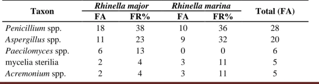
The most frequently genera found in the samples of the two anuran species were Penicillium spp. and Aspergillus spp. (Table 1).
Taxon Rhinella major Rhinella marina Total (FA)
FA FR% FA FR% Penicillium spp. 18 38 10 36 28 Aspergillus spp. 11 23 9 32 20 Paecilomyces spp. 6 13 0 0 6 mycelia sterilia 2 4 3 11 5 Acremonium spp. 2 4 3 11 5
Table 1. Frequency of filamentous fungi taxa isolated from the cutaneous microbiota of R. major and R. marina, collected in the port area of UFOPA, Tapajós Unit, Santarém, Pará, Brazil.
Braz. J. of Develop.,Curitiba, v. 6, n. 10, p. 82336-82356 oct. 2020. ISSN 2525-8761 Curvularia spp. 2 4 0 0 2 Scopulariopsis spp. 2 4 0 0 2 Cladosporium spp. 1 2 1 4 2 Mucor spp. 1 2 1 4 2 Trichoderma sp. 1 2 0 0 1 Bipolaris sp. 1 2 0 0 1 Absidia sp. 0 0 1 4 1 Total 47 28 75
Collections were made in the rainy season in November, February and April, and a total of 49 isolates of filamentous fungi were obtained. The collections in the dry season were carried out in June and October, and for this period, a total of 26 isolates of filamentous fungi were obtained.
There was no significant statistical difference in the composition of identified fungi of R.
major between the sampled seasonal periods (p = 0.05), where the number of fungi obtained in the
rainy season was n = 42 and in the dry season n = 5. When compared without the seasonality factor, the genera of fungi isolated from R. major showed a significant difference (p <0.05).
There was no significant difference in the number of fungi identified in R. marina, when compared between the two sampled seasonal periods (p = 0.18), being the number of fungi obtained in the rainy period n = 7 and in the dry season n = 21. When compared without the seasonality factor, the genera of fungi isolated from this species showed a significant difference (p <0.05). That is an interesting result, because both R. major and R. marina, have some similar morphological and behavioural characteristics, use the same substrate and the specimens were collected sintopically in the present study, and could be exposed to the same agents.
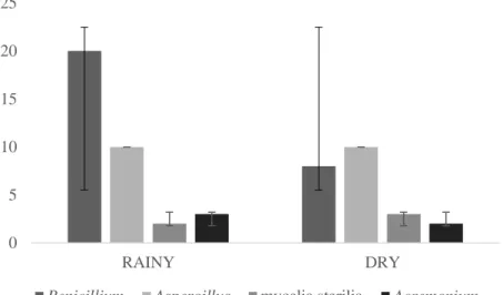
When comparing the composition of the genus of fungi for the same seasonal periods between the species R. major and R. marina there was no significant difference, in the rainy season (p = 0.06) and in the dry season (p = 0.15). However, the genus Penicillium was more frequent in the rainy season. Figure 1 shows the distribution of most common taxa found in both anuran species when comparing the two seasonal periods.
Braz. J. of Develop.,Curitiba, v. 6, n. 10, p. 82336-82356 oct. 2020. ISSN 2525-8761
Figure 1. Distribution of the most common taxa present in both species, R. major and R. marina in different seasonal periods (rainy and dry), collected in the port area of UFOPA, Tapajós Unit, Santarém, Pará, Brazil
The predominant genera among the identified fungi were Penicillium and Aspergillus with greater frequency, in both amphibian species. These results may be related to the proximity of amphibians to the soil, as there are reports of these fungi present in these environments (Souchie, Azcón, Barea, Saggin-Júnior, and Silva, 2005; Douglas and Amuzie, 2017; Kearns et al., 2017). In addition to being considered environmental fungi, Aspergillus and Penicillium, are also characterized as toxin-producing fungi, which can cause damage to human and animal health (Freire, Vieira, Guedes, and Mendes, 2007; Zain, 2011). However, the present study did not carry out studies related to the production of toxins by these organisms.
Penicillium and Aspergillus have been reported in the skin, mouth, intestines and eggs of
different species of free-living and captive frogs, as well as in amphibians used in gastronomy (Petrisko et al., 2008; Mendoza, Aguirre- Rojas, Sarria, and Giraldo, 2012; Douglas and Amuzie, 2017; Kearns et al., 2017; Ogoanah, Okafor, and Eyong, 2017). The occurrence of these fungi in amphibians is not uncommon, and to date, no cases have been reported that evidence their relationship to animal infections.
According to Kearns et al. (2017) Penicillium species found in amphibians and in the environment may be a potential candidate for use in therapy as probiotics against B. dendrobatidis. The use of fungal probiotics can cause the production of antimicrobial metabolites and lead to the induction of host defenses against parasitic fungi that are not part of the animal's indigenous microbiota (Kearns et al., 2017; Banik, Halder, Ghosh, and Mondal, 2019).
Genus Paecilomyces was among the three most frequent in the amphibian mucosa, and is composed of species that occur in a wide variety of environments, and even incorporates many
0 5 10 15 20 25 RAINY DRY
Braz. J. of Develop.,Curitiba, v. 6, n. 10, p. 82336-82356 oct. 2020. ISSN 2525-8761
species that produce substances with biological activity (Li, Xu, Liu, and Zhang, 2020). Some species of this genus, such as P. lilacinus and P. carneus, have antimicrobial and antioxidant activity (Abdel-Wareth, Ghareeb, Abdel-Aziz, and El-Hagrassi, 2019; Sheeba, Ali, and Anuradha, 2019). These species, and P. variotii, can also act as biocontrol agents (Li, Xu, Liu, and Zhang, 2020). However, other species of Paecilomyces can cause diseases in humans and other vertebrates, in addition to being cited as entomopathogenic (Liang, Han, Chu, and Liu, 2005).
The occurrence of Paecilomyces in anurans was reported in a survey with the cutaneous microbiota of Dendropsophus columbianus (Hylidae) in Colombia (Mendoza et al., 2012) carried out in a pasture-forestal zone with livestock activity and comparing the soil fungal samples, hooves of cattle and skin of D. columbianus, being this fungal genus also found in the soil samples.
The mycelia sterilia group generically includes non-sporulated fungal isolates that are grouped as morphospecies, based on similarities in the characteristics of the colonies (Lacap, Hyde and Liew, 2003). The fungi of the mycelia sterilia complex were classified as environmental without clinical importance, however, this scenario has changed in recent years, as some studies have identified this group as infectious agents in humans (Pounder et al., 2007; Singh et al., 2013; Garcia-Hermoso et al., 2015). With regard to amphibians, this fungal group has been registered infecting eggs of species of the families Centrolenidae and Phyllomedusidae in Central America (Villa, 1979).
Acremonium genus includes species with a worldwide distribution, being commonly found in
the environment, such as soil and decomposing plant material, and there are reports of some species present in food (Das, Saha, Dar, and Ramachandran, 2010; Klimko, Khostelidi, Melekhina, Gornostaev, and Semelev, 2016). Most species representing the Acremonium genus are saprophytic and non-pathogenic, however, there are pathogenic species affecting plants and insects, some of which are especially considered as causative agents in opportunistic infections in humans and other mammals (Summerbell, 2003; Perdomo et al., 2011; Ballhausen et al., 2016).
The occurrence of Acremonium in anurans is reported in the species Dendrobates auratus (Dendrobatidae), which is characterized as part of the cutaneous microbiota of the studied species (Kearns et al., 2017). This genus has also been found in amphibians of the order Caudata, in aquatic salamanders diagnosed with microbial infections in United States and Slovenia (Nickerson et al., 2011; Bizjak-Mali et al., 2018).
The genus Curvularia is composed of environmental species, isolated from air, soil, decomposing material, and fresh water (Manamgoda, Cai, McKenzie, Chukeatirote, and Hyde, 2012a; Manamgoda, Rossman, Castlebury, Chukeatirote, and Hyde, 2015). There are also reports worldwide of plant and animal pathogens, including humans (Manamgoda et al., 2012b; Da Cunha
Braz. J. of Develop.,Curitiba, v. 6, n. 10, p. 82336-82356 oct. 2020. ISSN 2525-8761
et al., 2013; Krizsán et al., 2015). Curvularia also houses several species with biological activities that produce several secondary metabolic compounds with antibacterial, antifungal and antioxidant action, among others (Khiralla, Spina, Saliba, and Laurain-Mattar, 2019).
The presence of Curvularia in anurans is not reported in any study to date as part of the cutaneous microbiota of these animals. However, the occurrence in injured tissues of the salamander
Cryptobranchus alleganiensis bishopi (Cryptobranchidae) has been recorded (Nickerson et al.,
2011). According to Reavill (2001) this genus may be one of the causative agents of chromomycosis in amphibians.
Scopulariopsis comprises a genus of fungi with wide geographical distribution in natural
environments such as soil, air, plant debris, and decomposing animals (Sandoval-Denis et al., 2013; Pérez-Cantero and Guarro, 2020). This genus includes saprobic, colonizing species and infection agents in mammals, including humans and insects (Iwen, Schutte, Florescu, Noel-Hurst, and Sigler, 2012; Sandoval-Denis et al., 2013).
Regarding the occurrence of Scopulariopsis in anurans, this genus was not found in any study as part of the microbiome of these animals, being reported for the first time in the present study.
Cladosporium, is a very heterogeneous genus of hyphomycetes that inhabit the most diverse
environments, occurring in a wide ecological range, being found in soil, air, plant debris, plants and food (Bensch, Braun, Groenewald, and Crous, 2012). This genus groups endophytic, phytopathogenic, saprobic, fungicidal and pathogenic species of humans and animals (Crous, Braun, Schubert, and Groenewald, 2007; Sandoval-Denis et al., 2016).
The genus Cladosporium is presented in some studies as belonging to the amphibian microbiome (Nickerson et al., 2011; Mendoza et al., 2012; Kearns et al., 2017; Ogoanah et al., 2017; Medina et al., 2019) making it a relatively common genus occurring in these animals. However, this genus is a major cause of chromomycosis in amphibians, which can affect both free-living and captive animals (Densmore and Green, 2007; Kim, Eom, Park, and Ra, 2008).
The genus Mucor groups several species that are found in multiple environments such as in soil, air, dust, supplies and in food, being it either raw or processed (Hermet, Méheust, Mounier, Barbier, and Jany, 2012). There are saprobic species, some described as endophytes and others as opportunistic pathogens in animals, including humans (Hoffmann et al., 2013).
Among the species of the genus Mucor, the best known specie to colonize amphibians is M.
amphibiorum, characterized as a pathogenic agent of these animals, both free-living and captive,
causing the infection known as mucormycosis (Reavill, 2001; Connolly, 2015). Despite this, the genus has been isolated from animals with a healthy organism (Douglas and Amuzie, 2017;
Braz. J. of Develop.,Curitiba, v. 6, n. 10, p. 82336-82356 oct. 2020. ISSN 2525-8761
Ogoanah et al., 2017), which leads to the assumption that other species of the genus can colonize amphibians without causing damage to the host.
Trichoderma is a very diverse genus with species distributed all over the world. They are
commonly found in environments such as soil, decaying wood, plants and other fungi (Schuster and Schmoll, 2010; Atanasova, 2014). The direct influence of Trichoderma on human health is multifaceted. These fungi are potential sources of medicinal compounds, some are mycotoxin producers and others are human pathogens (Mukherjee, Horwits, Singh, Mukherjee, and Schmoll, 2013; Sandoval-Denis et al., 2014).
Occurrence of Trichoderma in amphibians has been previously reported in some studies (Petrisko et al., 2008; Mendoza et al., 2012; Ogoanah et al., 2017) and at the present time no species of the genus has been identified as a potential pathogenic agent to anurans.
The genus Bipolaris includes various species with a worldwide distribution and are found in association with various hosts, as epiphytes or pathogens (Khiralla et al., 2019). Most of the
Bipolaris species are not only saprobic and plant pathogens, but some of the saprobic species are
potentially capable of infecting humans and animals (Da Cunha et al., 2012).
Genus Absidia comprises species that inhabit soil, air, and saprobic species present in organic matter (Hoffmann, Discher, and Voigt, 2007; Benny, 2008). There are reports of other species that are facultative parasites of other fungi or plants, and opportunistic pathogens that mainly cause mycoses in humans and animals (Thirion-Delalande, Guillot, Jensen, Crespeau, and Bernex, 2005; Constantinides, Misra, Nassab, and Wilson, 2008; García-Pajares et al., 2012).
Absidia, Curvularia, Scopulariopsis and the mycelia sterilia group have not been reported so
far in any work of cutaneous microbiome of adult anurans, making this the first work to present the occurrence of these taxa in the species R. major and R. marina.
There were some similarities and differences in the composition of the cutaneous mycobiota of R. major and R. marina, which may be linked to environmental variables such as temperature and disturbance levels to which they are subject, since the environment, as well as the physiological factors of each species, also play an important role in the composition of microorganisms from amphibian skin (Kueneman et al., 2014; Bletz et al., 2017b).
Several genera of fungi found in this study as part of the cutaneous microbiota of R. major and R. marina are recognized as environmental microorganisms commonly found in soil, water and air. The horizontal transmission of symbiotic microorganisms to the host is influenced by the host’s mucous layer that acts as a filter, selecting microorganisms, and free-living microorganisms themselves can be attracted to the hosts by metabolites produced by them (Bright and Bulgheresi,
Braz. J. of Develop.,Curitiba, v. 6, n. 10, p. 82336-82356 oct. 2020. ISSN 2525-8761
2010). Thus, both the mechanism of horizontal transmission, as well as vertical transmission, can be one of the elements to understand why there are microorganisms that are commonly found both in the cutaneous microbiota of amphibians and in the surrounding environment, and why there are microorganisms that can be found only in the cutaneous tissue of these animals, being little or not found in the environment where the amphibian is inserted (Walke et al., 2014; Medina et al., 2019). It is important to note that although some identified fungal genera are reported as pathogenic for animals and humans, no evidence was found that these microorganisms were causing any harm to the animals used in this study. Likewise, because these animals live in environments that are directly influenced by anthropic activities, the relationship between the environment and the microbiome of these animals should be considered, which can be colonized by opportunistic microorganisms and coexist in mutualism until biotic and / or abiotic changes occur (Assis, 2012; Rebollar and Harris, 2019).
In addition, Penicillium, Acremonium, Curvularia and Absidia present species with antimicrobial properties, which, together with the amphibian host's immune system, can promote resistance of these animals against the actions or establishment of microorganisms with pathogenic potentials (Hoffmann, 2010; Kearns et al., 2017; Khan, 2017; Khiralla et al., 2019).
In the present study, it was demonstrated that there were no significant differences in the fungal community of R. major and R. marina between the rainy and dry periods, however new studies need to be carried out with a larger sample to verify the influence of the seasonal periods effects on the fungal microbiota of these animals. Although studies with bacteria prove the influence of seasonal periods on bacterial composition, for fungal communities there are still many knowledge gaps (Varela, Lesbarrères, Ibáñez, and Green, 2018). There is also a need to sample natural pristine environments and other species, with different biological aspects, to detect possible differences.
Therefore, the importance of studies that clarify the fungal microbiota of amphibians in Amazonian environments is emphasized, as these have varied habitats and diverse biotic and abiotic influence, considering that the microbiota present in these animals can reveal important ecological relationships and consequently promote strategies related to the preservation of species such as amphibians, which are considered environmental bioindicators.
4 CONCLUSION
The present work presents the taxa Absidia, Curvularia, Scopulariopsis and the mycelia sterilia group as new records of skin-associated fungi in adult anurans, increasing knowledge about the microbiome of this group.
Braz. J. of Develop.,Curitiba, v. 6, n. 10, p. 82336-82356 oct. 2020. ISSN 2525-8761
According to the experimental conditions studied, it was possible to grow 75 fungal colonies present in the species R. major and R. marina, with no statistically significant differences in the composition of fungal taxa in the studied seasonal periods.
It is suggested that further studies be carried out in Amazonian environments, with a larger sample size, other species and environments, as well as the use of molecular tools, for a better identification and understanding of the composition of anuran skin microbiota.
ACKNOWLEDGEMENTS
We thank Federal University of Western Pará that made it possible to grant a Pro-TCC scholarship, which helped in the realization of this work. To the technical team and students of the Multidisciplinary Laboratory for Teaching Applied Biology and the Bacteriology Laboratory at UFOPA.
REFERENCES
ABDEL-WARETH, M. T. A.; GHAREEB, M. A.; ABDEL-AZIZ, M. S.; EL-HAGRASSI, A. M. Snailicidal, antimicrobial, antioxidant and anticancer activities of Beauveria bassiana, Metarhizium
anisopliae and Paecilomyces lilacinus fungal extracts. Egyptian Journal of Aquatic Biology and
Fisheries, v. 23, n. 2, p. 195-212, 2019.
ACEVEDO, A. A.; LAMPO, M.; CIPRIANI, R. The cane or marine toad, Rhinella marina (Anura, Bufonidae): two genetically and morphologically distinct species. Zootaxa, v. 4103, n. 6, p. 574-586, 2016.
AMPHIBIAWEB. Rhinella marina: South American Cane Toad. Accessible at http://amphibiaweb.org/species/229. University of California, Berkley, USA. Accessed in July 30, 2020.
ASSIS, A. B. Microbiota, secreções cutâneas e microclima: consequências para os anfíbios. Revista da Biologia, v. 8, p. 45-48, 2012.
ATANASOVA, L. Ecophysiology of Trichoderma in genomic perspective. In: GUPTA, V.; SCHMOLL, M.; HERRERA-ESTRELLA, A.; UPADHYAY, R. S.; DRUZHININA, I.; TUOHY, M. (Ed.), Biotechnology and Biology of Trichoderma (p. 25-40). Elsevier, 2014.
ÁVILA-PIRES, T. C. S.; HOOGMOED, M. S.; VITT, L. J. Herpetofauna da Amazônia. In: NASCIMENTO, L. B.; OLIVEIRA, M. E. (Ed.), Herpetologia no Brasil II (p. 13-43). Belo Horizonte, MG: Sociedade Brasileira de Herpetologia, 2007.
BALLHAUSEN, B. D.; GEISWEID, K.; HARTMANN, K.; HIRSCHBERGER, J.; MAJZOUB, M.; SCHULZ, B. Systemic Acremonium species infection in a dog. Tierärztliche Praxis Ausgabe K: Kleintiere/Heimtiere, v. 44, n. 06, p. 424-428, 2016.
Braz. J. of Develop.,Curitiba, v. 6, n. 10, p. 82336-82356 oct. 2020. ISSN 2525-8761
BANIK, A.; HALDER, S. K.; GHOSH, C.; MONDAL, K. C. Fungal Probiotics: Opportunity, Challenge, and Prospects. In: YADAV, A. N.; SINGH, S.; MISHRA, S.; GUPTA, A. (Ed.), Recent Advancement in White Biotechnology Through Fungi (p. 101-117). Cham, SWI: Springer, 2019. BENNY, G. L. Methods used by Dr. RK Benjamin, and other mycologists, to isolate zygomycetes. Aliso: A Journal of Systematic and Evolutionary Botany, v. 26, n. 1, p. 37-61, 2008.
BENSCH, K.; BRAUN, U.; GROENEWALD, J. Z.; CROUS, P. W. The Genus Cladosporium. Studies in Mycology, v. 72, p. 1-401, 2012.
BIZJAK-MALI, L.; ZALAR, P.; TURK, M.; BABIČ, M. N.; KOSTANJSEK, R.; GUNDE-CIMERMAN, N. Opportunistic fungal pathogens isolated from a captive individual of the European blind cave salamander Proteus anguinus. Diseases of Aquatic Organisms, v. 129, n. 1, p. 15-30, 2018.
BLETZ, M. C.; PERL, R. B.; VENCES, M. Skin microbiota differs drastically between co-occurring frogs and newts. Royal Society Open Science, v. 4, n. 4, p. 170107, 2017a.
BLETZ, M. C.; ARCHER, H.; HARRIS, R. N.; MCKENZIE, V. J.; RABEMANANJARA, F. C.; RAKOTOARISON, A.; VENCES, M. Host ecology rather than host phylogeny drives amphibian skin microbial community structure in the biodiversity hotspot of Madagascar. Frontiers in Microbiology, v. 8, n. 1530, 2017b.
BOS, J. D.; LUITEN, R. M. Skin Immune System. In: STOCKFLETH, E.; ULRICH, C. (Ed.), Skin Cancer after Organ Transplantation (p. 45-62). Springer US, 2009.
BRIGHT, M.; BULGHERESI, S. A complex journey: transmission of microbial symbionts. Nature Reviews Microbiology, v. 8, n. 3, p. 218-230, 2010.
CAPRILES, C. H.; MATA, S.; MIDDELVEEN, M. Preservation of fungi in water (Castellani): 20 years. Mycopathologia, v. 106, n. 2, p. 73-79, 1989.
CHUONG, C. M., NICKOLOFF, B. J., ELIAS, P. M., GOLDSMITH, L. A., MACHER, E., MADERSON, P. A., SUNDBERG, J. P.; TAGAMI, H.; PLONKA, P. M.; THESTRUP-PEDERSON, K.; BERNARD, B. A.; SCHÖDER, J. M.; DOTTO, P.; CHANG, C. M.; WILLIAMS, M. L.; FEINGOLD, K. R.; KING, L. E.; KLINGMAN, A. M.; REES, J. L.; CHRISTOPHERS, E. What is the 'true' function of skin?. Experimental Dermatology, v. 11, n. 2, p. 159-187, 2002.
COLOMBO, B. M.; SCALVENZI, T.; BENLAMARA, S.; POLLET, N. Microbiota and mucosal immunity in amphibians. Frontiers in Immunology, v. 6, n. 111, 2015.
CONNOLLY, J. H. Mucormycosis in the platypus and amphibians caused by Mucor
amphibiorum. Microbiology Australia, v. 36, n. 2, p. 83-87, 2015.
CONSTANTINIDES, J.; MISRA, A.; NASSAB, R.; WILSON, Y. Absidia corymbifera fungal infection in burns: a case report and review of the literature. Journal of Burn Care & Research, v. 29, n. 2, p. 416-419, 2008.
Braz. J. of Develop.,Curitiba, v. 6, n. 10, p. 82336-82356 oct. 2020. ISSN 2525-8761
CRAMP, R. L.; MCPHEE, R. K.; MEYER, E. A.; OHMER, M. E.; FRANKLIN, C. E. First line of defence: the role of sloughing in the regulation of cutaneous microbes in frogs. Conservation Physiology, v. 2, n. 1, p. cou012, 2014.
CROUS, P. W.; BRAUN, U.; SCHUBERT, K.; GROENEWALD, J. Z. Delimiting Cladosporium from morphologically similar genera. Studies in Mycology, v. 58, p. 33-56, 2007.
CULP, C. E.; FALKINHAM III, J. O.; BELDEN, L. K. Identification of the natural bacterial microflora on the skin of eastern newts, bullfrog tadpoles and redback salamanders. Herpetologica, v. 63, n. 1, p. 66-71, 2007.
DA CUNHA, K. C.; SUTTON, D. A.; FOTHERGILL, A. W.; CANO, J.; GENÉ, J.; MADRID, H.; DE HOOG, S.; CROUS, P. W.; GUARRO, J. Diversity of Bipolaris species in clinical samples in the United States and their antifungal susceptibility profiles. Journal of Clinical Microbiology, v. 50, n. 12, p. 4061-4066, 2012.
DA CUNHA, K. C.; SUTTON, D. A.; FOTHERGILL, A. W.; GENÉ, J.; CANO, J.; MADRID, H.; DE HOOG, S.; CROUS, P. W.; GUARRO, J. In vitro antifungal susceptibility and molecular identity of 99 clinical isolates of the opportunistic fungal genus Curvularia. Diagnostic Microbiology and Infectious Disease, v. 76, n. 2, p. 168-174, 2013.
DAS, S.; SAHA, R.; DAR, S. A.; RAMACHANDRAN, V. G. Acremonium species: a review of the etiological agents of emerging hyalohyphomycosis. Mycopathologia, v. 170, n. 6, p. 361-375, 2010.
DENSMORE, C. L.; GREEN, D. E. Diseases of amphibians. Ilar Journal, v. 48, n. 3, p. 235-254, 2007.
DOUGLAS, S. I.; AMUZIE, C. C. Microbiological quality of Hoplobatrachus occipitalis (Amphibia, Anura) used as meat. International Journal of Current Microbiology and Applied Sciences, v. 6, n. 6, p. 3192-3200, 2017.
EZENWA, V. O.; GERARDO, N. M.; INOUYE, D. W.; MEDINA, M.; XAVIER, J. B. Animal behavior and the microbiome. Science, v. 338, n. 61041, p. 98-199, 2012.
FLÓREZ, L. V.; BIEDERMANN, P. H.; ENGL, T.; KALTENPOTH, M. Defensive symbioses of animals with prokaryotic and eukaryotic microorganisms. Natural Product Reports, v. 32, n. 7, p. 904-936, 2015.
FREIRE, F. D. C. O.; VIEIRA, I. G. P.; GUEDES, M. I. F.; MENDES, F. N. P. Micotoxinas: importância na alimentação e na saúde humana e animal. Embrapa Agroindústria Tropical, v. 48, 2007.
FROST, D. R. Amphibian Species of the World: An Online Reference. Version 6.1 Electronic Database accessible at https://amphibiansoftheworld.amnh.org/index.php. American Museum of Natural History, New York, USA, 2020.
GARCIA‐HERMOSO, D.; ALANIO, A.; CABARET, O.; OLIVI, M.; FOULET, F.; CORDONNIER, C.; COSTA, J-M.; BRETAGNE, S. High diversity of non‐sporulating moulds in
Braz. J. of Develop.,Curitiba, v. 6, n. 10, p. 82336-82356 oct. 2020. ISSN 2525-8761
respiratory specimens of immunocompromised patients: should all the species be reported when diagnosing invasive aspergillosis?. Mycoses, v. 58, n. 9, p. 557-564 2015.
GARCÍA-PAJARES, F.; SÁNCHEZ-ANTOLÍN, G.; ALVÁREZ, C. A.; RUBIALES, B. M.; NÚÑEZ-RODRÍGUEZ, H.; DEL VAL, L. S.; RUIZ-ZORRILLA, R.; BARRERA, A.; GÓMEZ-NIETO, A.; HERRERO, I. P.; GARCÍA, A. V.; CARO-PATÓN, A. Cutaneous mucormycosis infection by Absidia in two consecutive liver transplant patients. Transplantation Proceedings, v. 44, n. 6, p. 1562-1564, 2012.
GONZÁLEZ-BERNAL, E.; GREENLEES, M. J.; BROWN, G. P.; SHINE, R. Toads in the backyard: why do invasive cane toads (Rhinella marina) prefer buildings to bushland?. Population Ecology, v. 58, n. 2, p. 293-302, 2016.
GUERRA, V.; JARDIM, L.; LLUSIA, D.; MÁRQUEZ, R.; BASTOS, R. P. Knowledge status and trends in description of amphibian species in Brazil. Ecological Indicators, v. 18, n. 2020, p. 106754, 2020.
HAMANN, M. I.; GONZÁLEZ, C. E. Helminth parasites in the toad Rhinella major (Bufonidae) from Chaco region, Argentina. Acta Herpetologica, v. 10, n. 2, p. 93-101, 2015.
HERMET, A.; MÉHEUST, D.; MOUNIER, J.; BARBIER, G.; JANY, J.-L. Molecular systematics in the genus Mucor with special regards to species encountered in cheese. Fungal Biology, v. 116, n. 6, p. 692–705, 2012.
HOFFMANN, K.; DISCHER, S.; VOIGT, K. Revision of the genus Absidia (Mucorales, Zygomycetes) based on physiological, phylogenetic, and morphological characters; thermotolerant
Absidia spp. form a coherent group, Mycocladiaceae fam. nov. Mycological Research, v. 111, n.
10, p. 1169-1183, 2007.
HOFFMANN, K. Identification of the genus Absidia (Mucorales, Zygomycetes): a comprehensive taxonomic revision. In: GHERBAWY, Y.; VOIGT, K. (Ed.), Molecular Identification of Fungi (p. 439-460). Berlin: Springer, 2010.
HOFFMANN, K.; PAWŁOWSKA, J.; WALTHER, G.; WRZOSEK, M.; DE HOOG, G. S.; BENNY, G. L.; KIRK, P. M.; VOIGT, K. The family structure of the Mucorales: a synoptic revision based on comprehensive multigene-genealogies. Persoonia: Molecular Phylogeny and Evolution of Fungi, v. 30, p. 57-76, 2013.
HOLDEN, W. M.; HANLON, S. M.; WOODHAMS, D. C.; CHAPPELL, T. M.; WELLS, H. L.; GLISSON, S. M.; MCKENZIE, V. J.; KNIGHT, R.; PARRIS, M. J.; ROLLINS-SMITH, L. A. Skin bacteria provide early protection for newly metamorphosed southern leopard frogs (Rana
sphenocephala) against the frog-killing fungus, Batrachochytrium dendrobatidis. Biological
Conservation, v. 187, p. 91-102, 2015.
HUANG, L.; LI, J.; ANBOUKARIA, H.; LUO, Z.; ZHAO, M.; WU, H. Comparative transcriptome analyses of seven anurans reveal functions and adaptations of amphibian skin. Scientific Reports, v. 6, n. 1, p. 1-11, 2016.
Braz. J. of Develop.,Curitiba, v. 6, n. 10, p. 82336-82356 oct. 2020. ISSN 2525-8761
IWEN, P. C.; SCHUTTE, S. D.; FLORESCU, D. F.; NOEL-HURST, R. K.; SIGLER, L. Invasive
Scopulariopsis brevicaulis infection in an immunocompromised patient and review of prior cases
caused by Scopulariopsis and Microascus species. Medical Mycology, v. 50, n. 6, p. 561-569, 2012.
KEARNS, P. J.; FISCHER, S.; FERNÁNDEZ-BEASKOETXEA, S.; GABOR, C. R.; BOSCH, J.; BOWEN, J. L.; TLUSTY, M. F.; WOODHAMS, D. C. Fight fungi with fungi: antifungal properties of the amphibian mycobiome. Frontiers in Microbiology, v. 8, n. 2494, 2017.
KELLY, D.; MULDER, I. E. Microbiome and immunological interactions. Nutrition Reviews, 70(suppl_1), p. S18-S30, 2012.
KHAN, N. T. Cephalosporin C Production from Acremonium chrysogenum. Enzyme Engineering, v. 6, n. 1, 2017.
KHIRALLA, A.; SPINA, R.; SALIBA, S.; LAURAIN-MATTAR, D. Diversity of natural products of the genera Curvularia and Bipolaris. Fungal Biology Reviews, v. 33, n. 2, p. 101-122, 2019.
KIM, S.; EOM, A. H.; PARK, D.; RA, N. Y. Detection of infectious fungal diseases of frogs inhabiting in Korea. Mycobiology, v. 36, n. 1, p. 10-12, 2008.
KLIMKO, N. N.; KHOSTELIDI, S. N.; MELEKHINA, Y. E.; GORNOSTAEV, D. A.; SEMELEV, V. N. Acremonium Pneumonia Successfully Treated in Patient with Acute Myeloid Leukemia: A Case Report. Journal of Bacteriology & Mycology: Open Access, v. 2, n. 5, p. 116-119, 2016.
KRIZSÁN, K.; PAPP, T.; MANIKANDAN, P.; SHOBANA, C. S.; CHANDRASEKARAN, M.; VÁGVÖLGYI, C.; KREDICS, L. Clinical Importance of the Genus Curvularia. In: RAZZAGHI-ABYANEH, M.; SHAMS-GHAHFAROKHI, M.; RAI, M. (Ed.), Medical Mycology: Current Trends and Future Prospects (p. 147-204). CRC Press, 2015.
KUENEMAN, J. G.; PARFREY, L. W.; WOODHAMS, D. C.; ARCHER, H. M.; KNIGHT, R.; MCKENZIE, V. J. The amphibian skin‐associated microbiome across species, space and life history stages. Molecular Ecology, v. 23, n. 6, p. 1238-1250, 2014.
KUENEMAN, J. G.; WEISS, S.; MCKENZIE, V. J. Composition of micro-eukaryotes on the skin of the cascades Frog (Rana cascadae) and patterns of correlation between skin microbes and
Batrachochytrium dendrobatidis. Frontiers in Microbiology, v. 8, n. 2350, 2017.
KUENEMAN, J. G.; WOODHAMS, D. C.; HARRIS, R.; ARCHER, H. M.; KNIGHT, R.; MCKENZIE, V. J. Probiotic treatment restores protection against lethal fungal infection lost during amphibian captivity. Proceedings of the Royal Society B: Biological Sciences, v. 283, n. 1839, 20161553, 2016.
LACAP, D. C.; HYDE, K. D.; LIEW, E. C. Y. An evaluation of the fungal 'morphotype' concept based on ribosomal DNA sequences. Fungal Diversity, v. 12, p. 53-66, 2003.
LACAZ, C. S.; PORTO, E.; MARTINS, J. E. C.; HEINS-VACCARI, E. M.; MELO, N. T. Tratado de Micologia Médica. São Paulo: Sarvier, 2002.
Braz. J. of Develop.,Curitiba, v. 6, n. 10, p. 82336-82356 oct. 2020. ISSN 2525-8761
LAM, B. A.; WALKE, J. B.; VREDENBURG, V. T.; HARRIS, R. N. Proportion of individuals with anti-Batrachochytrium dendrobatidis skin bacteria is associated with population persistence in the frog Rana muscosa. Biological Conservation, v. 143, n. 2, p. 529-531, 2010.
LARONE, D. H. Medically Important Fungi: A Guide to Identification. Washington, D.C.: American Society for Microbiology, 1993.
LAUER, A.; SIMON, M. A.; BANNING, J. L.; ANDRÉ, E.; DUNCAN, K.; HARRIS, R. N. Common cutaneous bacteria from the eastern red-backed salamander can inhibit pathogenic fungi. Copeia, v. 2007, n. 3, p. 630-640, 2007.
LI, X. Q.; XU, K.; LIU, X. M.; ZHANG, P. A Systematic Review on Secondary Metabolites of
Paecilomyces Species: Chemical Diversity and Biological Activity. Planta Medica, 2020.
LIANG, Z. Q.; HAN, Y. F.; CHU, H. L.; LIU, A. Y. Studies on the genus Paecilomyces in China I. Fungal Diversity, v. 20, p. 83-101, 2005.
MANAMGODA, D. S.; CAI, L.; MCKENZIE, E. H. C.; CHUKEATIROTE, E.; HYDE, K. D. Two new Curvularia species from northern Thailand. Sydowia, v. 64, n. 2, p. 255-266, 2012a.
MANAMGODA, D. S.; CAI, L.; MCKENZIE, E. H. C.; CROUS, P. W.; MADRID, H.; CHUKEATIROTE, E.; SHIVAS, R. G.; TAN, Y. P.; HYDE, K. D. A phylogenetic and taxonomic re-evaluation of the Bipolaris-Cochliobolus-Curvularia complex. Fungal Diversity, v. 56, n. 1, p. 131-144, 2012b.
MANAMGODA, D. S.; ROSSMAN, A. Y.; CASTLEBURY, L. A.; CHUKEATIROTE, E.; HYDE, K. D. A taxonomic and phylogenetic re-appraisal of the genus Curvularia (Pleosporaceae): human and plant pathogens. Phytotaxa, v. 212, n. 3, p. 175-198, 2015.
MCKENZIE, V. J.; BOWERS, R. M.; FIERER, N.; KNIGHT, R.; LAUBER, C. L. Co-habiting amphibian species harbor unique skin bacterial communities in wild populations. The ISME Journal, v. 6, n. 3, p. 588-596, 2012.
MEDINA, D.; HUGHEY, M. C.; WALKE, J. B.; BECKER, M. H.; PONTARELLI, K.; SUN, S.; BADGLEY, B.; BELDEN, L. K. Amphibian skin fungal communities vary across host species and do not correlate with infection by a pathogenic fungus. Environmental Microbiology, v. 21, n. 8, p. 2905-2920, 2019.
MENDOZA, Á. M.; AGUIRRE-ROJAS, L.; SARRIA, M.; GIRALDO, A. Hongos dérmico saprófitos de Dendropsophus columbianus (Hylidae) en Caloto, Colombia. Boletín Científico, v. 16, n. 1, p. 33-40, 2012.
MUKHERJEE, P. K., HORWITZ, B. A., SINGH, U. S., MUKHERJEE, M., & SCHMOLL, M.
Trichoderma in agriculture, industry and medicine: an overview. In: MUKHERJEE, P. K.;
HORWITZ, B. A.; SINGH, U. S.; MUKHERJEE, M.; SCHMOLL, M. (Ed.), Trichoderma Biology and Applications (p. 1-9). Boston: CAB International, 2013.
Braz. J. of Develop.,Curitiba, v. 6, n. 10, p. 82336-82356 oct. 2020. ISSN 2525-8761
NARVAES, P.; RODRIGUES, M. T. Taxonomic revision of Rhinella granulosa species group (Amphibia, Anura, Bufonidae), with description of a new species. Arquivos de Zoologia, v. 40, n. 1, p. 1-73, 2009.
NICKERSON, C. A.; OTT, C. M.; CASTRO, S. L.; GARCIA, V. M.; MOLINA, T. C.; BRIGGLER, J. T.; PITT, A. L.; TAVANO, J. J.; BYRAM, J. K.; BARRILA, J.; NICKERSON, M. A. Evaluation of microorganisms cultured from injured and repressed tissue regeneration sites in endangered giant aquatic Ozark hellbender salamanders. PLoS ONE, v. 6, n. 12, p. e28906, 2011.
OGOANAH, O. S.; OKAFOR, A. E.; EYONG, M. M. Effect of different preparatory methods on microbial load of the edible frog Hoplobatrachus occipitalis from Aguleri, Anambra State, Nigeria. NISEB Journal, v. 17, n. 3, p. 91-95, 2017.
OLSON, D. H.; AANENSEN, D. M.; RONNENBERG, K. L.; POWELL, C. I.; WALKER, S. F.; BIELBY, J.; GARNER, T. W. J.; WEAVER, G.; The Bd Mapping Group.; FISHER, M. C. Mapping the global emergence of Batrachochytrium dendrobatidis, the amphibian chytrid fungus. PloS ONE, v. 8, n. 2, p. e56802, 2013.
PEAY, K. G.; KENNEDY, P. G.; TALBOT, J. M. Dimensions of biodiversity in the Earth mycobiome. Nature Reviews Microbiology, v. 14, n. 7, p. 434, 2016.
PELOSO, P. L. V.; MACHADO, I.; BECKER, G. Fotografia da conservação na herpetologia: um ensaio sobre anfíbios ameaçados de extinção no Brasil. Herpetologia Brasileira, v. 8, n. 1, p. 26-29, 2019.
PERDOMO, H.; SUTTON, D. A.; GARCÍA, D.; FOTHERGILL, A. W.; CANO, J.; GENÉ, J.; SUMMERBELL, R. C.; RINALDI, M. G.; GUARRO, J. Spectrum of clinically relevant
Acremonium species in the United States. Journal of Clinical Microbiology, v. 49, n. 1, p.
243-256, 2011.
PETRISKO, J. E.; PEARL, C. A.; PILLIOD, D. S.; SHERIDAN, P. P.; WILLIAMS, C. F.; PETERSON, C. R.; BURY, R. B. Saprolegniaceae identified on amphibian eggs throughout the Pacific Northwest, USA, by internal transcribed spacer sequences and phylogenetic analysis. Mycologia, v. 100, n. 2, p. 171-180, 2008.
PÉREZ-CANTERO, A.; GUARRO, J. Current knowledge on the etiology and epidemiology of
Scopulariopsis infections. Medical Mycology, v. 58, n. 2, p. 145-155m 2020.
POUGH, F. H.; JANIS, C. M.; HEISER, J. B. A Vida dos Vertebrados. São Paulo: Atheneu, 2008.
POUNDER, J. I.; SIMMON, K. E.; BARTON, C. A.; HOHMANN, S. L.; BRANDT, M. E.; PETTI, C. A. Discovering potential pathogens among fungi identified as nonsporulating molds. Journal of Clinical Microbiology, v. 45, n. 2, p. 568-571, 2007.
PROKSCH, E.; BRANDNER, J. M.; JENSEN, J. M. The skin: an indispensable barrier. Experimental Dermatology, v. 17, n. 12, p. 1063-1072, 2008.
REAVILL, D. R. Amphibian skin diseases. Veterinary Clinics of North America: Exotic Animal Practice, v. 4, n. 2, p. 413-440, 2001.
Braz. J. of Develop.,Curitiba, v. 6, n. 10, p. 82336-82356 oct. 2020. ISSN 2525-8761
REBOLLAR, E. A.; HARRIS, R. Ecology of amphibian-microbial symbioses. Frontiers in Microbiology, v. 10, p. 766, 2019.
REBOLLAR, E. A.; MARTÍNEZ-UGALDE, E.; ORTA, A. H. The amphibian skin microbiome and its protective role against chytridiomycosis. Herpetologica, v. 76, n. 2, p. 167-177, 2020.
RIDDELL, R. W. Permanent stained mycological preparations obtained by slide culture. Mycologia, v. 42, n. 2, p. 265-270, 1950.
SANDOVAL-DENIS, M.; SUTTON, D. A.; FOTHERGILL, A. W.; CANO-LIRA, J.; GENÉ, J.; DECOCK, C. A.; DE HOOG, G. S.; GUARRO, J. Scopulariopsis, a poorly known opportunistic fungus: spectrum of species in clinical samples and in vitro responses to antifungal drugs. Journal of Clinical Microbiology, v. 51, n. 12, p. 3937-3943, 2013.
SANDOVAL-DENIS, M.; SUTTON, D. A.; CANO-LIRA, J. F.; GENÉ, J.; FOTHERGILL, A. W.; WIEDERHOLD, N. P.; GUARRO, J. Phylogeny of the clinically relevant species of the emerging fungus Trichoderma and their antifungal susceptibilities. Journal of Clinical Microbiology, v. 52, n. 6, p. 2112-2125, 2014.
SANDOVAL-DENIS, M.; GENÉ, J.; SUTTON, D. A.; WIEDERHOLD, N. P.; CANO-LIRA, J. F.; GUARRO, J. New species of Cladosporium associated with human and animal infections. Persoonia: Molecular Phylogeny and Evolution of Fungi, v. 36, p. 281-298, 2016.
SCHUSTER, A.; SCHMOLL, M. Biology and biotechnology of Trichoderma. Applied Microbiology and Biotechnology, v. 87, n. 3, p. 787-799, 2010.
SEBBEN, A. Microdissecação fisiológica a fresco: uma nova visão sobre a anatomia de anfíbios e répteis. In: NASCIMENTO, L. B.; OLIVEIRA, M. E. (Ed.), Herpetologia no Brasil II (p. 311-325). Belo Horizonte, MG: Sociedade Brasileira de Herpetologia, 2007.
SEGALLA, M. V.; CARAMASCHI, U.; CRUZ, C. A. G.; GRANT, T.; HADDAD, C. F. B.; LANGONE, J. A.; GARCIA, P. C. A. Brazilian amphibians: List of species. Herpetologia Brasileira, v. 5, n. 2, p. 34-46, 2016.
SHEEBA, H., ALI, M. S.; ANURADHA, V. Bioactive compounds and antimicrobial activity of fungal crude extract from medicinal plants. Journal of Pharmaceutical Sciences and Research, v. 11, n. 5, p. 1826-1833, 2019.
SINGH, P. K.; KATHURIA, S.; AGARWAL, K.; GAUR, S. N.; MEIS, J. F.; CHOWDHARY, A. Clinical significance and molecular characterization of nonsporulating molds isolated from the respiratory tracts of bronchopulmonary mycosis patients with special reference to basidiomycetes. Journal of Clinical Microbiology, v. 51, n. 10, p. 3331-3337, 2013.
SOUCHIE, E. L.; AZCÓN, R.; BAREA, J. M.; SAGGIN-JÚNIOR, O. J.; SILVA, E. M. R. Solubilização de fosfatos em meios sólido e líquido por bactérias e fungos do solo. Pesquisa Agropecuária Brasileira, v. 40, n. 11, p. 1149-1152, 2005.
Braz. J. of Develop.,Curitiba, v. 6, n. 10, p. 82336-82356 oct. 2020. ISSN 2525-8761
SUMMERBELL, R. C. Aspergillus, Fusarium, Sporothrix, Piedraia, and their relatives. In: HOWARD, D. H. (Ed.), Pathogenic Fungi in Humans and Animals (p. 237-498). New York: CRC Press, 2003.
THIRION‐DELALANDE, C.; GUILLOT, J.; JENSEN, H. E.; CRESPEAU, F. L.; BERNEX, F. Disseminated acute concomitant aspergillosis and mucormycosis in a pony. Journal of Veterinary Medicine Series A, v. 52, n. 3, p. 121-124, 2005.
VAN ROOIJ, P.; MARTEL, A.; HAESEBROUCK, F.; PASMANS, F. Amphibian chytridiomycosis: a review with focus on fungus-host interactions. Veterinary Research, v. 46, n. 1, p. 137, 2015.
VARELA, B. J.; LESBARRÈRES, D.; IBÁÑEZ, R.; GREEN, D. M. Environmental and host effects on skin bacterial community composition in panamanian frogs. Frontiers in Microbiology, v. 9, n. 298, 2018.
VILLA, J. Two fungi lethal to frog eggs in Central America. Copeia, v. 1979, n. 4, p. 650-655, 1979.
VITT, L. J.; CALDWELL, J. P. Herpetology: An Introductory Biology of Amphibians and Reptiles. Academic Press, 2013.
WALKE, J. B.; BECKER, M. H.; LOFTUS, S. C.; HOUSE, L. L.; CORMIER, G.; JENSEN, R. V.; BELDEN, L. K. Amphibian skin may select for rare environmental microbes. The ISME Journal, v. 8, n. 11, p. 2207-2217, 2014.
WALKE, J. B.; BECKER, M. H.; HUGHEY, M. C.; SWARTWOUT, M. C.; JENSEN, R. V.; BELDEN, L. K. Most of the dominant members of amphibian skin bacterial communities can be readily cultured. Applied and Environmental Microbiology, v. 81, n. 19, p. 6589-6600, 2015.
XU, X.; LAI, R. The chemistry and biological activities of peptides from amphibian skin secretions. Chemical Reviews, v. 115, n. 4, p. 1760-1846, 2015.
ZAIN, M. E. Impact of mycotoxins on humans and animals. Journal of Saudi Chemical Society, v. 15, n. 2, p. 129-144, 2011.