(Annals of the Brazilian Academy of Sciences) ISSN 0001-3765
www.scielo.br/aabc
Understanding the mechanisms of glutamine action in critically ill patients
GISELE P. OLIVEIRA1, CRISTINA M. DIAS1, PAOLO PELOSI2 and PATRICIA R.M. ROCCO1 1Laboratório de Investigação Pulmonar, Instituto de Biofísica Carlos Chagas Filho
Universidade Federal do Rio de Janeiro, Centro de Ciências da Saúde, Av. Carlos Chagas Filho, s/n Cidade Universitária, Ilha do Fundão, 21941-902 Rio de Janeiro, RJ, Brasil
2Department of Ambient, Health and Safety, University of Insubria
Villa Toeplitz Via G.B. Vico, 46 21100, Varese, Italy
Manuscript received on May 22, 2009; accepted for publication on July 7, 2009
ABSTRACT
Glutamine (Gln) is an important energy source and has been used as a supplementary energy substrate. Furthermore, Gln is an essential component for numerous metabolic functions, including acid-base homeostasis, gluconeogenesis, nitrogen transport and synthesis of proteins and nucleic acids. Therefore, glutamine plays a significant role in cell homeostasis and organ metabolism. This article aims to review the mechanisms of glutamine action during severe illnesses. In critically ill patients, the increase in mortality was associated with a decreased plasma Gln concentration. During catabolic stress, Gln consumption rate exceeds the supply, and both plasma and skeletal muscle pools of free Gln are severely reduced. The dose and route of Gln administration clearly influence its effectiveness: high-dose parenteral appears to be more beneficial than low-dose enteral administration. Experimental studies reported that Gln may protect cells, tissues, and whole organisms from stress and injury through the following mechanisms: attenuation of NF (nuclear factor)-κB activation, a balance between pro- and anti-inflammatory cytokines, reduction in neutrophil accumulation, improvement in intestinal integrity and immune cell function, and enhanced of heat shock protein expression. In conclusion, high-doses of parenteral Gln (>0.50 g/kg/day) demonstrate a greater potential to benefit in critically ill patients, although Gln pathophysiological mechanisms requires elucidation.
Key words:heat shock protein, apoptosis, cytokines, glutamine.
INTRODUCTION
Glutamine is an amino acid that has received consider-able attention during the past 10 years. It has been shown to be beneficial for the metabolically stressed patient, es-pecially the critically ill patients. During acute illnesses patients experience nutritional depletion that is corre-lated to low plasma and low mucosal glutamine concen-trations (Oudemans-van Straaten et al. 2001). Such de-ficiencies are common among hospitalized patients and are associated with an increased risk of developing in-fectious complications, organ failure, and death (Roth et al. 1982, Planas et al. 1993, Oehler et al. 2002).
Correspondence to: Profa. Dra. Patricia Rieken Macedo Rocco E-mail: [email protected]
Glutamine has many essential metabolic functions in the organ. This amino acid is an energy substrate for most cells, especially for enterocytes and lymphocytes; it is also a precursor for nucleotide, glutamate, and, in particular, for glutathione synthesis, an important cellu-lar antioxidant (Oba et al. 2004). It plays a central role in nitrogen transport within the body, and is the most important substrate for renal ammoniagenesis.
2007) in critical illnesses. Patients receiving high-dose parenteral glutamine presented the highest beneficial ef-fects (Déchelotte et al. 2006, Tang et al. 2007).
Experimental studies in the current literature indi-cate that glutamine use may prevent the occurrence of lung injury, tissue metabolic dysfunction, and reduce mortality after injury (Doruk et al. 2005, Déchelotte et al. 2006, Morrison et al. 2006, Peng et al. 2006, Tang et al. 2007). The present review will focus on the effects of glutamine during critical illnesses.
METABOLISM AND CATABOLISM OF GLUTAMINE IN NORMAL CONDITIONS
Glutamine is the most abundant free amino acid in the body and commonly known as a nonessential amino acid due to the ability of most cells to produce it (Darmaun et al. 1986, Boza et al. 2000, Labow et al. 2001, Yeh et al. 2005). Glutamine is present in the plasma at levels around 0.6 mM and in the intracellular space at levels around 2 and 20 mM (Darmaun et al. 1986). It also serves as a metabolic intermediate, contributing carbon and nitrogen for the synthesis of other amino acids, nu-cleic acids, fatty acids, and proteins (Newsholme et al. 1986, Boza et al. 2000). Glutamine through glutamate is a glutathione precursor, a tripeptide consisting of glu-tamate, glycine, and cysteine, with intracellular antiox-idant capacity (Oba et al. 2004). Thus, its functions within the cell are generally separated into four cate-gories: 1) its role in nitrogen transport; 2) its importance in maintaining the cellular redox state; 3) its position as a metabolic intermediate; and 4) its role as an energy source. Although some tissues use glutamine for one pathway more than others, glutamine metabolism occurs in all cells (Newsholme et al. 1986).
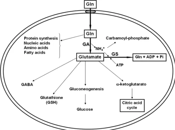
Glutamine is synthesized by the cytosolic glutamine synthetase (GS) in many tissues, but degraded by mito-chondrial glutaminase (GA) and utilized in high amounts by other tissues that do not synthesize it (Labow et al. 1998, 2001, Karinch et al. 2001). Thus, glutamine meta-bolism is controlled by glutamine synthetase and glu-taminase (Labow et al. 1998, 2001) (Fig. 1) as follow:
Glutamate + NH+ 4 + ATP
glutamine synthetase
−−−−−−→ Glutamine + ADP + Pi + H+
Glutamine + H2O
glutaminase
−−−−−−−→ Glutamate + NH+4
Glutamine catabolism is initiated by the removal of an amine group to form glutamate (Oba et al. 2004). This can occur through a number of cytosolic transam-inase enzymes that use theγ-amino nitrogen of gluta-mine in a variety of metabolic synthesis. However, the rate at which these reactions utilize Gln depends ulti-mately upon the metabolic demand for the reaction prod-ucts and is, therefore, not appropriate for the control of glutamine homeostasis (Labow et al. 2001). The mito-chondrial enzyme glutaminase catalyzes the hydrolysis of theγ-amino group of Gln to form glutamate and am-monia (Labow et al. 2001, Oba et al. 2004). Amam-monia can be used to form carbamoyl phosphate or can diffuse from the mitochondria and the cell itself. Furthermore, glutamate can formα-ketoglutarate and, thus, enter into the citric acid cycle (Oba et al. 2004). Therefore, the catabolism of glutamine through glutaminase can be in-creased without the production of excessive amounts of specific metabolites (Fig. 1).
The synthesis of glutamine from glutamate is me-diated by the enzyme glutamine synthetase. Thus, the regulated expression of this enzyme plays a key role in an organ’s overall glutamine production rate. In con-trast to the many enzymes that utilize glutamine as a substrate, only glutamine synthetase is responsible for de novo synthesis of glutamine. In this line, glutamine synthetase catalyses the formation of Gln from gluta-mate and ammonia in the cytoplasm (Labow et al. 1999, 2001) (Fig. 1). Because both of these substrates are relatively abundant, the rate of glutamine formation is highly dependent upon the activity of glutamine syn-thetase (Labow et al. 2001).
METABOLISM AND CATABOLISM OF GLUTAMINE DURING CRITICAL ILLNESS
Fig. 1 – Metabolism and anabolism of glutamine (Gln). Glutamine is synthesized by the action of glutamine synthetase (GS) and degraded by mitochondrial glutaminase (GA). Glutamine can be synthesized by most tissues in glutamate and ammonia. Ammonia can be used to form carbamoyl phosphate. Glutamate can formα-ketoglutarate, glucose in the liver and kidneys, gluthatione in most of cells, and Gamma-aminobutyric acid (GABA) in neurons.
metabolism, and they are part of the acute stress re-sponse (Mezzarobba et al. 2003). They are considered the primary mediators of glutamine synthetase expres-sion during stress, and act on the lung and skeletal muscle in a rapid and direct glucocorticoid receptor-me-diated manner (Abcouwer et al. 1995). The transcrip-tional response of the rat glutamine synthetase gene to glucocorticoid has been characterized and shown to be attributable to two genetic regions. Each of these re-gions gives large glucocorticoid induction of transcrip-tion to the glutamine synthetase promoter, as well as het-erologous promoter in a glucocorticoid-dependent fash-ion (Abcouwer et al. 1995).
Despite a large transcriptional response, glutamine synthetase protein levels do not always increase with GS mRNA levels, which suggests that the post-transcrip-tional control mechanism also regulates glutamine syn-thetase expression (Labow et al. 1998, 1999, 2001). Studies demonstrated that the presence of glutamine in the medium regulates glutamine synthetase expression
via a post-transcriptional mechanism, where the rate of glutamine synthetase protein degradation is diminished and its activity is augmented in the presence of low glu-tamine concentration (Labow et al. 1998).
Although the skeletal muscle is the main source of Gln in normal conditions, skeletal muscles and lungs work together to maintain the circulating glutamine pool during critical illnesses (Buttrose et al. 1987, Labow et al. 2001).
increases glutamine synthetase protein stability in the lung, but without significant augment in GS mRNA (El-gadi et al. 1998). The combined effects of Gln depletion and glucocorticoids hormones can act synergistically to increase glutamine synthetase protein expression, allow-ing the lung to adjust GS activity to meet actual Gln demand (Labow et al. 1998).
GLUTAMINE AND THE EXPRESSION OF HEAT SHOCK PROTEINS
Glutamine’s beneficial effects on critical illnesses may result from enhanced heat shock proteins (HSP) expres-sion (Singleton et al. 2005b, Morrison et al. 2006) ex-pressed by leucocytes (Oehler et al. 2002), monocytes (Eliasen et al. 2006), and granulocytes (Ganter et al. 2006). The heat shock proteins are a group of proteins essential to cellular survival under stressful conditions. These are a family of highly conserved proteins belong-ing to multigene families rangbelong-ing in molecular size from 10 to 105 kDa – different weights are used for identifi-cation – , and found in all major cellular compartments (Fleshner et al. 2004, Ganter et al. 2006).
The stress-inducible HSP70 and HSP72 are indu-cible forms of the stress protein, which may confer cellu-lar protection (Oehler et al. 2001). Treatment of sepsis-induced acute lung injury with an adenovirus overex-pressing HSP72 limits nuclear factor (NF)κB (a crucial transcription factor regulating the expression of many pro-inflammatory cytokines and immunoregulatory mo-lecules) activation and prevents lung injury (Weiss et al. 2007). There is evidence that Gln can enhance HSP70 and 72 expression in lung macrophages and epithelial cells (Singleton et al. 2005c) leading to marked pro-tection against sepsis-induced injury (Singleton et al. 2005b, Singleton and Wischmeyer 2007). The cellular functions of intracellular HSP70 and HSP72 are respon-sible for limiting protein aggregation, facilitating pro-tein refolding, and chaperoning propro-teins. These cellular functions serve to improve cell survival in the face of a broad array of cellular stressors (Oehler et al. 2001).
It has been recognized that HSP72 is also found in extracellular space (eHSP72) where it exerts immuno-modulatory function on innate and acquired immunity (Wright et al. 2000). In fact, during the early phase of ALI, there was an activation of the stress protein
re-sponse (SPR) and a release of HSP72 into the alveolar spaces. HSP72 may serve as a marker of stress pro-tein response activation in the distal air spaces of ALI patients, while SPR activation may protect the alveolar epithelium against oxidative stress (Ganter et al. 2006). Initially, studies reported that eHSP72 was only released as a result of necrotic/lytic cell death, but it is now rec-ognized that elevated eHSP72 may be found in the ab-sence of necrosis (Ganter et al. 2006). In this con-text, Ganter and colleagues (2006) showed a preserved alveolar fluid clearance and high levels of eHSP72 in pulmonary edema fluid of ALI patients, suggesting that the high level of eHSP72 is not related to cell injury in distal air spaces. It seems that, under physical or psychological acute stress, eHSP72 may exacerbate in-flammatory process (Asea et al. 2000) or stimulate the release of endogenous eHSP72 into the blood via an a1-adrenergic receptor-mediated mechanism facilitating the innate immunity (Campisi et al. 2003).
Glutamine starvation reduces the HSP70 expres-sion since it decreases HSP70 mRNA expresexpres-sion (Elia-sen et al. 2006). As plasma glutamine depletion occurs during systemic inflammation, the impaired HSP70 ex-pression under these conditions is likely to have delete-rious effects on the survival and function of leucocytes, and may contribute to the immunosuppression observed in these patients. However, Gln administration may re-store the expression of HSP protecting tissues against injury (Singleton et al. 2005a, b, c, Morrison et al. 2006), improving survival in animals with abdominal sepsis (Singleton et al. 2005a, b). Recently, Singleton and Wischmeyer (2007), using knockout mice, demon-strated that the beneficial effects of glutamine on sur-vival, lung injury, and the inflammatory response fol-lowing cecal ligation and puncture surgery depended on the expression of HSP70.
demonstrated that the molecular mechanism of Gln mediated HSP70 expression appears to be dependent on O-GlcNac pathway activation and subsequent O-glyco-sylation and phosphorylation of key transcription fac-tors, HSF-1 and Sp1, required for HSP70 induction (Singleton and Wischmeyer 2008).
MODULATION OF IMMUNE SYSTEM
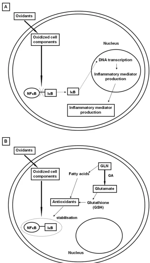
Glutamine is known to modulate immune cell function and production of cytokines. It may be mediated via at-tenuation of multiple pathways of inflammation, such as NF-κB, kinases proteins, inhibition of increases in iNOS expression (Singleton et al. 2005a), attenuating the in-teractions between polymorphonuclear lymphocytes and endothelium, and decreasing neutrophil infiltration into tissues (Doruk et al. 2005, Yeh et al. 2005). Two path-ways that can explain Gln’s effects are: a) enhancement of MAPK phosphatase (MKP-1) expression, responsi-ble for halting the production of pro-inflammatory cy-tokines, acting as an important negative regulator to in-flammatory stimuli (Camps et al. 2000), b) inhibition of phosphorylation and degradation of IκB-α, an inhibitory protein that is bound to NF-κB, avoiding its translating to the nucleus (Zingarelli et al. 2003, Weiss et al. 2007) (Fig. 2).
Glutamine infusion can result in enhanced tissue glutathione levels, partly responsible in avoiding the ac-tivation of NF-κB, and increase antioxidant capacity (Belmonte et al. 2007, Humbert et al. 2007). Whereas HSP70 protects cells against oxidative stress by the re-pair or removal of damage proteins, the second major protection factor of mammalian cells, the antioxidant glutathione, reacts directly with ROS in order to prevent oxidative damage. A significant correlation between re-duced glutamine supply and diminished intracellular glutathione was observed in cultured peripheral blood mononuclear cells (Roth et al. 2002) and in critically pa-tients (Wernerman et al. 1999). Doruk and colleagues (2005) showed reduced glutathione levels in the dia-phragm of rats submitted to cecal ligation and puncture surgery, which was reversed with Gln administration.
Glutamine supplementation also promotes balanced Th1/Th2 response during sepsis, decreasing IL-6 secre-tion in non-hepatic organs, while reducing intra-lympho-cyte IL-4 and enhancing IFN-αexpressions (Yeh et al.
2005). O’Leary and colleagues (2007) showed in rats with abdominal sepsis that parenteral nutrition with glu-tamine recovered serum levels of IL-6 and IL-10.
It is known that glutamine is an important fuel for lymphocytes and macrophages (Parry-Billings et al. 1990). Macrophages and neutrophils are involved in the early, non-specific host defense responses, and play an important role in the pathophysiology and/or protection against sepsis. In fact, this amino acid is required for the expression of lymphocyte cell surface markers, such as CD25, CD45RO, and CD17 (Roth et al. 2002). Ad-ditionally, septic rats pretreated with glutamine showed reduced neutrophil infiltration in diaphragm muscle, pre-venting biochemical and histopathological changes (Do-ruk et al. 2005). It has been demonstrated that Gln sup-plementation benefits the human leukocyte antigen-DR expression, essential molecule for antigen presentation, on monocytes in trauma and surgery patients (Boelens et al. 2002).
Another interesting question is the intestinal per-meability. As glutamine is an important fuel for the en-terocyte, intestinal consumption is important for main-taining the integrity of the intestinal barrier with subse-quent prevention of bacterial translocation and, through stimulation of the gut-associated immune system, pre-vention of gut barrier atrophy. It is proposed that a de-rangement of the gut mucosal barrier function, which oc-curs during critical illnesses, results in an amplification of the general inflammatory response predisposing patients to multiple organ failure (Ziegler et al. 1988, van Der Hulst et al. 1998). The intestinal mucosa provides a bar-rier between bacteria and bacterial products in the intesti-nal lumen and body’s circulation and organs. Prophylac-tic treatment with glutamine may minimize changes in intestinal permeability and bacteria translocation caused by endotoxemia in rats receiving total parenteral nutri-tion (Ding and Li 2003).
Fig. 2 – Nuclear factor (NF)κB activation pathway. Panel A: NF-κB is sequestered in the cytoplasm by its interaction with a member of the
inhibitory kappa B (IκB) family. After an injurious stimuli, IκB-αis phosphorylated and degraded. Thus, NF-κB is transported to the cell nucleus
where it binds to DNA in the promoter regions of the target genes, transcripting many proinflammatory molecules that are responsible to the generation of inflammatory cascade during critical illnesses. Panel B: glutamine (Gln) may inhibit IKB-αphosphorylation and degradation in
APOPTOSIS
Apoptosis can be induced by a range of environmental, physical or chemical stress. Studies have established that the survival-promoting effects of HSP70 can be partly attributed to the suppression of apoptosis (Huang et al. 2001). The reduced HSP70 expression in glutamine-starving cells, together with their impaired antioxidant capacity, is thus likely to make them more sensitive to the induction of apoptosis.
Evidence showed that cells in the presence of glu-tamine are not sensitive to Fas ligand (Ko et al. 2001). JNK/SAPK pathway is involved in the apoptosis pro-cess increased by Fas stimulation. JNK/SAPK induced by Fas ligand is mediated by ASK1 (a critical protein kinase in apoptosis), which is activated after Fas ligand treatment only in the absence of Gln. Thus, Gln may suppress apoptosis signal-regulating kinase (ASK-1) and JNK/SAPK activation by Fas ligand (Ko et al. 2001). Furthermore, L-glutamine potentiation of HSP72 is as-sociated with increased gut epithelial resistance to apop-totic injury, and reduced HSP72 may be associated with increased caspase activity in glutamine-deficient (Rope-leski et al. 2005). In fact, Gln induces autophagy under stressed conditions, and prevents apoptosis under heat stress through its regulation of the mTOR and p38 MAP kinase pathways (Sakiyama et al. 2009).
Glutathione (GSH) metabolism is also closely re-lated to apoptotic processes of immune cells (Chang et al. 1999). The increase of intracellular GSH is suffi-cient to reduce Fas-triggered increase in apoptotic cells. An overexpression of Bcl-2, an anti-apoptotic protein, causes redistribution of glutathione to the nucleus, there-by altering nuclear redox and blocking caspase activity (Chang et al. 2002).
GLUTAMINE AND THE DEVELOPMENT OF ACUTE LUNG INJURY/ACUTE RESPIRATORY DISTRESS SYNDROME
Critically ill patients are at high risk of glutamine deple-tion (Planas et al. 1993) and subsequent complicadeple-tions, such as the developing of acute lung injury/acute respi-ratory distress syndrome (ARDS). Therapeutic interven-tions to improve outcomes from ALI/ARDS have met with limited success (Phua et al. 2009).
There are many experimental studies investigating
the effects and the mechanisms responsible for gluta-mine’s beneficial effects in ALI (Singleton et al. 2005a, b, c, Morrison et al. 2006, Singleton and Wischmeyer 2007). However, in these studies, glutamine was admin-istered before (Ding and Li 2003, Doruk et al. 2005) or few hours (Singleton et al. 2005a, b) after the induc-tion of lung injury. In this context, Gln could prevent neutrophil recruitment and infiltration (Yeh et al. 2005, Peng et al. 2006), protect the alveolar barrier (Single-ton et al. 2005b), and attenuate inflammatory lung in-jury, leading to survival improvement (Singleton et al. 2005b). The mechanisms underlying the majority of these findings may be related to Gln’s ability to induce heat shock protein (HSP) expression, which is known to enhance cell survival in the face of injury and attenu-ate the systemic inflammatory response (Singleton and Wischmeyer 2007), besides attenuating NF-κB pathway (Doruk et al. 2005, Yeh et al. 2005), buffering oxida-tive stress via glutathione generation (Oba et al. 2004), protecting gut barrier, and providing substrate for the appropriate division of immune cells (Oba et al. 2004).
Related to clinical trials, the studies have not inves-tigated the development of ALI/ARDS as an end-point.
CLINICAL STUDIES
Evidence shows that nutritional support improves clin-ical outcomes in critclin-ical illnesses (Ziegler et al. 2005, Déchelotte et al. 2006, Juange et al. 2007). In this con-text, the concept of immunonutrition emerges: some nu-trients are given in supranormal amounts to achieve a “pharmacological” effect (Déchelotte et al. 2006). Many nutrients are potentially considered immunonutrients, such asω-3 fatty acids, nucleotides, arginine, and glu-tamine.
The same is also true for the free glutamine export from muscle, which suffers no alteration over time (Vesali et al. 2002). Furthermore, critically ill patients present plasma glutamine depletion, which is an independent factor additional to the APACHE II score for mortality prediction in ICU patients at admission (Oudemans-van Straaten et al. 2001).
Therefore, if a conditional deficiency occurs, crit-ically ill patients should obtain some benefit with its exogenous replacement (Roth et al. 1982, Planas et al. 1993, Oudemans-van Straaten et al. 2001, Oehler et al. 2002). In fact, studies have demonstrated that Gln sup-plemented nutrition reduced the clinical complications of these patients (Wischmeyer et al. 2001, Ziegler et al. 2005, Déchelotte et al. 2006, Tang et al. 2007).
The option of Gln delivery via enteral or parenteral route is a subject of ongoing debate. The recent ICU guidelines have been consistent with their recommen-dations concerning the supplementation of Gln in par-enteral nutrition (Heyland et al. 2003, Kreymann 2006). One study performed on patients with burns (Wisch-meyer et al. 2001) reported reduced infections compli-cation with the use of parenteral glutamine independent of the nutrition administration method. Total parenteral nutrition supplemented with alanil-glutamine dipeptide in ICU patients was associated with a reduced rate of infection, including the incidence of pneumonia, but it did not improve survival (Déchelotte et al. 2006). Sur-gical patients treated with parenteral nutrition containg alanil-glutamine dipeptide demonstrated reduced in-fectious morbidity rate (Estívariz et al. 2008, Fuentes-Orozco et al. 2008), as well as a significantly higher serum HSP70 level (Ziegler et al. 2005), which is corre-lated with a decrease in ICU length of stay. Patients af-ter portal hypertension surgery receiving total parenaf-teral nutrition supplemented with Gln presented an improve-ment in immune function and preservation of intestinal integrity (Tang et al. 2007) (Table I). These good results achieved with parenteral glutamine are due to adequate systemic delivery in the most severe patients.
However, clinical efficacy of glutamine supplemen-tation was not demonstrated in some reports (Hall et al. 2003, Schulman et al. 2005, Juang et al. 2007), prob-ably because of the heterogeneous patients populations studied, the use of different doses of glutamine, and the
lack of a predictable physiologic end-point for gluta-mine supplementation previously identified in animals studies. Additionally, factors related to the clinical man-agement need to be taken into account.
Although the guidelines about enteral/parenteral nutrition recommend the enteral route to delivery im-munonutrients, so far there have not been sufficient data to support enteral glutamine supplementation in surgical or critically ill patients (Schulman et al. 2005, Juang et al. 2007). The addition of Gln to standard enteral feeds or to an immunomodulatory formula did not improve survival in intensive care unit patients (Hall et al. 2003, Schulman et al. 2005). In a retrospective case-control study, enteral glutamine supplementation was not asso-ciated with a change in the cumulative rate of infectious complications in burn intensive care patients (Juang et al. 2007). In a prospective study developed in a surgi-cal trauma intensive care unit, there were no differences among the groups receiving enteral glutamine supple-mentation or standard feedings related to the acquisition of lung infections (Schulman et al. 2006) (Table I). One paper has demonstrated reduced mortality in adult burn patients given enteral glutamine supplements (Garrel et al. 2003). The controversial results may come from the patient population, as well as the amount and route of Gln administration (Table I). Indeed, burn patients may benefit the most from improved gut barrier function, in-cluding the competence of the gut immune system, since mortality rate is associated with systemic infection in this population.
TABLE I
Effect of enteral and parenteral glutamine in critical illnesses.
Authors Year Study design Patients Glutamine dose Results
Wischmeyer et al. 2001 Prospective, double-blind, randomized
Burn intensive care unit (n=26)
0.57 g/kg parenteral
Reduced bacteremic episodes
Garrel et al. 2003 Double-blind, randomized clinical trial
Burn center (n=41) 26 g/day enteral
Reduced blood infections and decreased mortality
Hall et al. 2003 Prospective, randomized
General ICU (n=363) 20 g/l supplemented enteral nutrition
Did not reduce the incidence of infectious
Schulman et al. 2005 Prospective Surgical and trauma (n=185)
0.6 g/kg enteral
Did not improve survival and the rate of infectious Ziegler et al. 2005 Randomized,
controlled, double-blind
Surgical unit (n=29) 0.50 g/kg parenteral
Decreased ICU length of stay
Déchelotte et al. 2006 Prospective, double-blind, controlled, randomized Multiple trauma, complicated surgery, pancreatitis (n=114) 0.50 g/kg parenteral
Reduced rate of infectious
Schulman et al. 2006 Prospective Surgical trauma intensive care unit (n=185)
0.60 g/kg enteral
Did not influence the acquisition of characteristics of infectious
Juang et al. 2007 Retrospective case-control
Burn intensive care unit (n=32)
0.52 g/kg enteral
Did not reduce infectious complications
Tang et al. 2007 Prospective, randomized and controlled Portal hypertension surgery (n=42) 0.30 g/kg parenteral
Improved immune function and intestinal integrity
Estívariz et al. 2008 Double-blind, randomized, controlled Surgical patients (n=59) 1.5 g/kg/d parenteral
Reduced infectious in SICU patients
Fuentes-Orozco et al. 2008 Prospective, randomized Severe acute pancreatitis (n=44) 0.50 g/kg parenteral Decreased infectious morbidity rate
effect on mortality. In critically ill patients, glutamine supplementation may be associated with a reduction in complication and mortality rates (Novak et al. 2002), but the greatest benefit was observed in patients receiving parenteral Gln.
The critical illness polyneuropathy (CIP) occurs mostly in patients who have systemic inflammatory re-sponse syndrome (SIRS), sepsis, or multiple organ dys-function syndrome (MODS). CIP manifests especially with the weakness of the respiratory muscles, leading to problems in weaning from mechanical ventilation in the intensive care unit (Tennila et al. 2000). A prospec-tive randomizing study in ICU patients using different doses of parenteral glutamine evaluated their effects on
muscle (Tjäder et al. 2004) and showed that, in spite of all Gln doses being able to normalize plasma glutamine concentrations, Gln did not attenuate the depletion of muscle free glutamine frequently observed in ICU pa-tients. Therefore, glutamine administration may not af-fect skeletal muscle turnover.
CONCLUSIONS
Several controlled studies suggest that glutamine sup-plementation has beneficial effects on the clinical out-come of critically ill patients. These results may be explained by the glutamine’s influences on the inflam-matory response, oxidative stress, apoptosis modulation, and the integrity of gut barrier. High-dose parenteral (>0.50 g/kg/day) glutamine appears to present the great-est potential for benefit in critically ill patients. Addi-tional studies should be performed to determine whether the outcomes are derived from the parenteral or enteral route, or if any specific group of patients could benefit from this therapy.
ACKNOWLEDGMENTS
We would like to express our gratitude to Mr. Andre Benedito da Silva and Mrs. Jaqueline Lima do Nasci-mento for their skillful technical assistance. This work is supported by Programa de Apoio a Núcleos de Excelên-cia (PRONEX-FAPERJ), Conselho Nacional de Desen-volvimento Científico e Tecnológico (CNPq), Fundação Carlos Chagas Filho de Amparo à Pesquisa do Estado do Rio de Janeiro (FAPERJ), Instituto do Milênio Ino-vação e Desenvolvimento de Fármacos e Medicamentos (IM-INOFAR).
RESUMO
A glutamina (Gln) é uma importante fonte de energia e tem sido usada como substrato energético suplementar. Além disso, a Gln é um componente essencial para numerosas funções metabólicas tais como: homeostase ácido-base, gliconeogê-nese, transporte de nitrogênio e síntese de proteínas e ácidos nucléicos. Portanto, a glutamina desempenha um papel im-portante na homeostase celular e no metabolismo dos órgãos. Esse artigo objetiva rever os mecanismos de ação da gluta-mina na doença grave. Em pacientes criticamente enfermos, o aumento da mortalidade foi associado com uma diminuição de Gln plasmática. Durante o estresse catabólico, o consumo de Gln excede a oferta, e a quantidade de glutamina livre no plasma e músculo esquelético encontra-se reduzida. A dose e via de administração da Gln claramente influencia sua eficá-cia: alta dose por via parenteral parece ser mais benéfica do que uma dose baixa administrada por via enteral. Estudos ex-perimentais relataram que Gln protege as células, tecidos, e
todo o organismo do estresse através dos seguintes mecanis-mos: atenuação na ativação do fator nuclear (NF)-κB, balanço entre citocinas pró- e anti-inflamatórias, redução no acúmulo de neutrófilos, melhora na integridade intestinal e função imune celular, e aumento da expressão da proteína de choque térmico. Em conclusão, o uso de glutamina em altas doses e por via pa-renteral (>0,50 g/kg/dia) demonstrou ser benéfica em pacien-tes criticamente enfermos, embora os mecanismos fisiopatoló-gicos necessitam ser melhor elucidados.
Palavras-chave:proteína de choque término, apoptose, citoci-nas, glutamina.
REFERENCES
ABCOUWERSF, BODEBPANDSOUBAWW. 1995.
Gluco-corticoids regulate rat glutamine synthetase expression in a tissue-specific manner. J Surg Res 59: 59–65.
ANDREASENAS, PEDERSEN-SKOVSGAARDT, MORTEN -SENOH, VANHALLG, MOSELEYPLANDPEDERSEN
BK. 2009. The effect of glutamine infusion on the inflam-matory response and HSP70 during human experimental endotoxemia. Crit Care 13: R7.
ASEAA, KRAEFTSK, KURT-JONESEA, STEVENSONMA, CHENLB, FINBERGRW, KOOGCANDCALDERWOOD
SK. 2000. HSP70 stimulates cytokine production through a CD14-dependant pathway, demonstrating its dual role as a chaperone and cytokine. Nat Med 6: 435–442. BELMONTELET AL. 2007. Effects of glutamine
supplemen-tation on gut barrier, glutathione content and acute phase response in malnourished rats during inflammatory shock. World J Gastroenterol 13: 2833–2840.
BOELENSPG, HOUDIJKAP, FONKJC, NIJVELDTRJ, FER -WERDA CC, VON BLOMBERG-VAN DER FLIERBM, THIJS LG, HAARMAN HJ, PUYANA JC AND VAN
LEEUWEN PA. 2002. Glutamine-enriched enteral
nu-trition increases HLA-DR expression on monocytes of trauma patients. J Nutr 132: 2580–2586.
BOZAJJ, MOENNOZD, BOURNOTCE, BLUMS, ZBINDEN
I, FINOT PAANDBALLEVREO. 2000. Role of
gluta-mine on the de novo purine nucleotide synthesis in Caco-2 cells. Eur J Nutr 39: 38–46.
BUTTROSEM, MCKELLARDANDWELBOURNETC. 1987.
Gut-liver interaction in glutamine homeostasis: portal am-monia role in uptake and metabolism. Am J Physiol 252: E746–750.
CAMPISIJ, LEEM THAND FLESHNER M. 2003.
danger signal to the immune system. Cell Stress Chaper-ones 8: 272–286.
CAMPSM, NICHOLSAANDARKINSTALLS. 2000. Dual specificity phosphatases: a gene family for control of MAP kinase function. FASEB J 14: 6–16.
CHANGWK, YANGKDANDSHAIOMF. 1999. Lympho-cyte proliferation modulated by glutamine: involved in the endogenous redox reaction. Clin Exp Immunol 117: 482–488.
CHANGWK, YANGKD, CHUANGH, JANJTANDSHAIO
MF. 2002. Glutamine protects activated human T cells from apoptosis by up-regulating glutathione and Bcl-2 lev-els. Clin Immunol 104: 151–160.
DARMAUND, MATTHEWSDEANDBIERDM. 1986. Glu-tamine and glutamate kinetics in humans. Am J Physiol 251: E117–126.
DÉCHELOTTEPET AL. 2006. L-alanyl-L-glutamine dipep-tide-supplemented total parenteral nutrition reduces infec-tious complications and glucose intolerance in critically ill patients: the French controlled, randomized, double-blind, multicenter study. Crit Care Med 34: 598–604. DINGLAANDLIJS. 2003. Effects of glutamine on intestinal
permeability and bacterial translocation in TPN-rats with endotoxemia. World J Gastroenterol 9: 1327–1332. DORUKN, BUYUKAKILLIB, ATICIS, CINELL, TAMERL,
AVLAND, BILGINE ANDORALU. 2005. The effect
of preventive use of alanyl-glutamine on diaphragm mus-cle function in cecal ligation and puncture-induced sepsis model. JPEN J Parenter Enteral Nutr 29: 36–43. ELGADI KM, LABOW BI, ABCOUWER SF AND SOUBA
WW. 1998. Sepsis increases lung glutamine synthetase expression in the tumor-bearing host. J Surg Res 78: 18– 22.
ELIASENMM, BRABEC M, GERNERC, POLLHEIMER J, AUERH, ZELLNER M, WEINGARTMANNG, GAROF, ROTHE ANDOEHLER R. 2006. Reduced stress
toler-ance of glutamine-deprived human monocytic cells is as-sociated with selective down-regulation of HSP 70 by de-creased mRNA stability. J Mol Med 84: 147–158. ESTÍVARIZCFET AL. 2008. Efficacy of parenteral nutrition
supplemented with glutamine dipeptide to decrease hos-pital infectious in critically ill surgical patients. JPEN J Parenter Enteral Nutr 32: 389–402.
FLESHNER M, CAMPISIJ, AMIRILANDDIAMONDDM. 2004. Cat exposure induces both intra- and extra-cellular HSP72: the role of adrenal hormones. Psychoneuroen-docrinology 29: 1142–1152.
FUENTES-OROZCO C, CERVANTES-GUEVARA G, MUCI
-ÑO-HERNÁNDEZI, LÓPEZ-ORTEGAA, AMBRIZ-GON
-ZÁLEZG, GUTIÉRREZ-DE-LA-ROSAJL, GÓMEZ-HER
-RERAE, HERMOSILLO-SANDOVAL JM ANDGONZÁ -LEZ-OJEDAA. 2008. L-alanyl-L-glutamine-supplement-ed parenteral nutrition decreases infectious morbidity rate in patients with severe acute pancreatitis. JPEN J Parenter Enteral Nutr 32: 403–411.
GAMRIN L, ESSÉNP, FORSBERGAM, HULTMANE AND
WERNERMAN J. 1996. A descriptive study of skeletal
muscle metabolism in critically ill patients: free amino acids, energy-rich phosphates, protein, nucleic acids, fat, water, and electrolytes. Crit Care Med 24: 575–583. GANTER MT, WARE LB, HOWARD M, ROUX J, GART
-LANDB, MATTHAY MA, FLESHNER M AND PITTET
JF. 2006. Extracellular heat shock protein 2 is a marker of the stress protein response in acute lung injury. Am J Physiol Lung Cell Mol Physiol 291: L354–361. GARREL D, PATENAUDE J, NEDELEC B, SAMSON L,
DORAIS J, CHAMPOUX J, D’ELIA M AND BERNIER
J. 2003. Decreased mortality and infectious morbidity in adult burn patients given enteral glutamine supplements: a prospective, controlled, randomized clinical trial. Crit Care Med 31: 2444–2449.
GOETERSC, WENNA, MERTESN, WEMPEC, VANAH, STEHLEP ANDBONE HG. 2002. Parenteral L-alanyl-L-glutamine improves 6-month outcome in critically ill patients. Crit Care Med 30: 2032–2037.
GRIFFITHS RD, JONES C ANDPALMER TE. 1997. Six-month of critically ill patients given glutamine-supple-mented parenteral nutrition. Nutrition 13: 295–302.
GROVER Z, TUBMANRANDMCGUIREW. 2007. Gluta-mine supplementation for young infants with severe gas-trointestinal disease. Cochrane Database Syst Rev 24: CD005947.
HALL JC, DOBB G, HALL J, DESOUZAR, BRENNANL
ANDMCCAULEY R. 2003. A prospective randomized trial of enteral glutamine in critical illness. Intensive Care Med 29: 1710–1716.
HEYLANDDK, DHALIWALR, DROVERJW, GRAMLICHL ANDDODEK P. 2003. Canadian Critical Care Clinical Practice Guidelines Committee. Canadian clinical prac-tice guidelines for nutrition support in mechanically venti-lated, critically ill adult patients. JPEN J Parenter Enteral Nutr 27: 355–373.
HEYLAND DK, DHALIWALR, DAYAG, MUSCEDERE J,
Critical Care Trials Group. Reducing deaths due to ox-idative stress (The REDOXS Study): Rationale and study design for a randomized trial of glutamine and antioxidant supplementation in critically-ill patients. Proc Nutr Soc 65: 250–263.
HUANGL, MIVECHINFANDMOSKOPHIDISD. 2001. In-sights into regulation and function of the major stress-induced hsp70 molecular chaperone in vivo: analysis of mice with targeted gene disruption of the hsp70.1 or hsp-70.3. Mol Cell Biol 21: 8575–8591.
HUMBERT B, NGUYEN P, MARTINL, DUMON H, VAL -LETTEG, MAUGÈREPANDDARMAUND. 2007. Effect of glutamine on glutathione kinetics in vivo in dogs. J Nutr Biochem 18: 10–16.
JUANGP, FISH DN, JUNGR AND MACLAREN R. 2007. Enteral glutamine supplementation in critically ill patients with burn injuries: a retrospective case-control evaluation. Pharmacotherapy 27: 11–19.
KARINCHAM, PANM, LINCM, STRANGERANDSOUBA
WW. 2001. Glutamine metabolism in sepsis and infec-tion. J Nutr 131(Suppl 9): 2535S–2538S.
KOYG, KIMEY, KIM T, PARK H, PARK HS, CHOI EJ ANDKIMS. 2001. Glutamine-dependent antiapoptotic interaction of human glutaminyl-t RNA synthetase with apoptosis signal-regulating kinase 1. J Biol Chem 276: 6030–6036.
KREYMANNKG. 2006. German Society for Nutritional Me-dicine and European Society for Parenteral and Enteral Nutrition. ESPEN Guidelines on Enteral Nutrition: In-tensive care. Clin Nutr 25: 210–223.
LABOW BI, ABCOUWER SF, LINCMANDSOUBA WW. 1998. Glutamine synthetase expression in the rat lung is regulated by protein stability. Am J Physiol 275: l877– 1886.
LABOWBI, SOUBAWWANDABCOUVERSF. 1999.
Glu-tamine synthetase expression in muscle is regulated by transcriptional and posttranscriptional mechanisms. Am J Physiol 276: 1136–1145.
LABOWBI, SOUBAWWANDABCOUWERSF. 2001.
Mech-anisms governing the expression of the enzymes of glu-tamine metabolism – Glutaminase and Glutaminase Syn-thetase. J Nutr 131: 2467S–2474S.
LEWISMI, BODINESC, KAMAGARN, XUX, DAXAND
FOURNIERM. 2006. Effect of severe short-term malnu-trition on diaphragm muscle signal transduction pathways influencing protein turnover. J Appl Physiol 100: 1799– 1806.
MEZZAROBBAV, TORRENTA, LEYDIERI, ALLESS, BRA
-JONB, MIGNONM, ATTAIXDANDMEYNIAL-DENIS
D. 2003. The role of adrenal hormones in the response of glutamine synthetase to fasting in adult and old rats. Clin Nutr 22: 569–575.
MORRISONAL, DINGESM, SINGLETONKD, ODOMSK, WONGHRANDWISCHMEYERPE. 2006. Glutamine’s
protection against cellular injury is dependent on heat shock factor-1. Am J Physiol Cell Physiol 290: C1625– 1632.
NEWSHOLMEP, CURI R, GORDON S ANDNEWSHOLME
EA. 1986. Metabolism of glucose, glutamine, long-chain fatty acids and ketone bodies by murine macrophages. Biochem J 239: 121–125.
NOVAKF, HEYLANDDK, AVENELLA, DROVERJWAND
SUX. 2002. Glutamine supplementation in serious ill-ness: a systematic review of the evidence. Crit Care Med 30: 2022–2029.
OBAM, BALDWINRLANDBEQUETTEBJ. 2004. Oxidation of glucose, glutamate, and glutamine by isolated ovine enterocytes in vitro is decreased by the presence of other metabolic fuels. J Anim Sci 82: 479–486.
OEHLERR, PUSCHE, ZELLNERM, DUNGELP, HERGOV
-ICSN, HOMONCIKM, ELIASENMM, BRABECMAND
ROTHE. 2001. Cell type-specific variations in the induc-tion of hsp70 in human leukocytes by feverlike whole body hyperthermia. Cell Stress and Chaperones 6: 306–315. OEHLER R, PUSCHE, DUNGELP, ZELLNERM, ELIASEN
MM, BRABECMANDROTHE. 2002. Glutamine deple-tion impairs cellular stress response in human leucocytes. Br J Nutr 87(Suppl): S17–21.
O’LEARYMJ, XUEA, SCARLETTCJ, SEVETTEA, KEEAJ
ANDSMITH RC. 2007. Parenteralversusenteral nutri-tion: effect on serum cytokines and the hepatic expression of mRNA of suppressor of cytokine signaling proteins, insulin-like growth factor-1 and the growth hormone re-ceptor in rodent sepsis. Crit Care 11: R79.
OUDEMANS-VANSTRAATENHM, BOSMANRJ, TRESKES
M, VAN DER SPOEL HJ AND ZANDSTRA DF. 2001. Plasma depletion and patient outcome in acute ICU ad-missions. Intensive Care Med 27: 84–90.
PARRY-BILLINGS M, EVANS J, CALDER PC AND NEW
-SHOLME EA. 1990. Does glutamine contribute to im-munosuppression after major burns? Lancet 336: 523– 525.
PENGZY, HAMIELCR, BANERJEEA, WISCHMEYERPE, FRIESERSANDWISCHMEYERP. 2006. Glutamine
expression following inflammatory cytokine-induced in-jury is dependent on heat shock factor-1 expression. JPEN J Parenter Enteral Nutr 30: 400–406.
PHUAJET AL. 2009. Has mortality from acute respiratory
distress syndrome decreased over time? A systematic re-view. Am J Respir Crit Care Med 179: 220–227. PIRKKALAL, NYKANENPANDSISTONENL. 2001. Roles
of the heat shock transcription factors in regulation of the heat shock response and beyond. FASEB J 15: 1118– 1131.
PLANASM, SCHWARTZS, ARBOSMAANDFARRIOLM.
1993. Plasma glutamine levels in septic patients. JPEN J Parenter Enteral Nutr 17: 299–300.
ROPELESKI MJ, RIEHM J, BAERKA, MUSCH MWAND
CHANG EB. 2005. Anti-apoptotic effects of
L-Gluta-mine-mediated transcriptional modulation of the heat shock protein 72 during heat shock. Gastroenterology 129: 170–184.
ROTH E, FUNOVICS J, MUHLBACHER F, SCHEMPER M, MAURITZWANDFRITSCHA. 1982. Metabolic disor-ders in severe abdominal sepsis: Glutamine deficiency in skeletal muscle. Clin Nutr 1: 25–41.
ROTHE, OEHLERR, MANHARTN, EXNERR, WESSNER
B, STRASSER E ANDSPITTLER A. 2002. Regulative potential of glutamine-relation to glutathione metabolism. Nutrition 18: 217–221.
SAKIYAMAT, MUSCHMW, ROPELESKIMJ, TSUBOUCHI
H AND CHANG EB. 2009. Glutamine increases auto-phagy under Basal and stressed conditions in intestinal epithelial cells. Gastroenterology 136: 924–932. SCHULMANASET AL. 2005. Does the addition of glutamine
to enteral feeds affect patient mortality? Crit Care Med 33: 2501–2506.
SCHULMANASET AL. 2006. Does enteral glutamine sup-plementation decrease infectious morbidity? Surg Infect (Larchmt) 7: 29–35.
SINGLETONKDANDWISCHMEYERPE. 2007. Glutamine’s protection against sepsis and lung injury is dependent on heat shock protein 70 expression. Am J Physiol Regul Integr Comp Physiol 292: R1839–1845.
SINGLETONKDANDWISCHMEYERPE. 2008. Glutamine induces heat shock protein expression via O-glycosylation and phosphorylation of HSF-1 and Sp1. JPEN J Parenter Enteral Nutr 32: 371–376.
SINGLETON KD, BECKEY VE AND WISCHMEYER PE. 2005a. Glutamine prevents activation of NF-κB and
stress kinase pathways, attenuates inflammatory cytokine release, and prevents acute respiratory distress syndrome (ARDS) following sepsis. Shock 24: 583–589.
SINGLETONKD, SERKOVAN, BECKEY VEANDWISCH -MEYERPE. 2005b. Glutamine attenuates lung injury and improves survival after sepsis: Role of enhanced heat shock protein expression. Crit Care Med 33: 1206–1213. SINGLETON KD, SERKOVA N, BANERJEE A, MENG
X, GAMBONI-ROBERTSON F ANDWISCHMEYER PE. 2005c. Glutamine attenuates endotoxin-induced lung metabolic dysfunction: potential role of enhanced heat shock protein 70. Nutrition 21: 214–223.
TANGZF, LING YB, LIN N, HAO ZAND XURY. 2007. Glutamine recombinant human growth hormone protects intestinal barrier function following portal hypertension surgery. World J Gastroenterol 13: 2223–2228.
TENNILAA, SALMIT, PETTILAV, ROINERO, VARPULAT
ANDTAKKUNENO. 2000. Early signs of critical illness
polyneuropathy in ICU patients with systemic inflamma-tory response syndrome or sepsis. Intensive Care Med 26: 1360–1363.
TJÄDERI, ROOYACKERSO, FORSBERGAM, VESALIRF,
GARLICK PJ AND WERNERMAN J. 2004. Effects on skeletal muscle of intravenous glutamine supplementa-tion to ICU patients. Intensive Care Med 30: 266–275.
TUBMAN TR, THOMPSON SWAND MCGUIREW. 2005. Glutamine supplementation to prevent morbidity and mor-tality in preterm infants. Cochrane Database Syst Rev 25: CD001457.
VANDERHULSTRR, VONMEYENFELDTMF, VANKREEL
BK, THUNNISSENFB, BRUMMERR, ARENDSJWAND
SOETERS PB. 1998. Gut permeability, intestinal
mor-phology, and nutritional depletion. Nutrition 14: 1–6. VESALI RF, KLAUDE M, ROOYACKERS OE, TJADER I,
BARLEHANDWERNERMANJ. 2002. Longitudinal pat-tern of glutamine/glutamate balance across the leg in long-stay intensive care unit patients. Clin Nutr 21: 505–514.
WEISSYG, BROMBERGZ, RAJN, RAPHAELJ, GOLOUBI
-NOFFP, BEN-NERIAHYANDDEUSTCHMANCS. 2007.
Enhanced heat shock protein 70 expression alters proteaso-mal degradation of IkappaB kinase in experimental acute respiratory distress syndrome. Crit Care Med 35: 2128– 2138.
WERNERMAN J, LUO JL AND HAMMARQVIST F. 1999.
WISCHMEYER PE, LYNCH J, LIEDEL J, WOLFSON R, RIEHM J, GOTTLIEBL ANDKAHANAM. 2001. Glu-tamine administration reduces Gram-negative bacteremia in severely burned patients: A prospective, randomized, double-blind trialversusisonitrogenous control. Crit Care Med 29: 2075–2080.
WRIGHT BH, CORTONJM, EL-NAHAS AM, WOOD RF ANDPOCKLEYAG. 2000. Elevated levels of circulant-ing heat shock protein 70 (HSP70) in peripheral and renal vascular disease. Heart Vessels 15: 18–22.
WUBJ, KINGSTONREANDMORIMOTORI. 1986. Human
HSP70 promoter contains at least two distinct regulatory domains. Proc Natl Acad Sci USA 83: 629–633. YEHCL, HSUCS, YEHSLANDCHENWJ. 2005. Dietary
glutamine supplementation modulates Th1/Th2 cytokine and interleukin-6 expressions in septic mice. Cytokine 31: 329–334.
ZHENGYM, LIF, ZHANGMMANDWUXT. 2006.
Glu-tamine dipeptide for parenteral nutrition in abdominal surgery: a meta-analysis of randomized controlled trials. World J Gastroenterol 12: 7537–7541.
ZHENGYM, LIF, QIB, LUOB, SUNH, LIUSANDWUX. 2007. Application of perioperative immunonutrition for gastrointestinal surgery: a meta-analysis of randomized controlled trials. Asia Pac J Clin Nutr 16 Suppl 1: 253– 257.
ZIEGLER TR, SMITH RJ, O’DWYER ST, DEMLING RH
ANDWILMOREDW. 1988. Increased intestinal
perme-ability associated with infection in burn patients. Arch Surg 123: 1313–1319.
ZIEGLERTR, OGDENLG, SINGLETONKD, LUOM, FER
-NANDEZ-ESTIVARIZ C, GRIFFITH DP, GALLOWAYJR ANDWISCHMEYERPE. 2005. Parenteral glutamine in-creases serum heat shock protein 70 in critically ill pa-tients. Intensive Care Med 31: 1079–1086.