Clinical Update
I
fCurrent and Heart Rate Control
Luiz Antonio Machado César
Instituto do Coração do Hospital das Clínicas – FMUSP – São Paulo, SP, Brazil
Mailing address: Luiz Antonio Machado César •
Rua Constantino de Souza, 1580 - 04605-004 – São Paulo, SP - Brazil E-mail: [email protected]
Manuscript received May 15, 2006; revised manuscript received October 5, 2006; accepted October 5, 2006.
Key words
Heart rate, arrhytmia.
Introduction
Understanding the mechanisms underlying the spontaneous depolarization of cardiac cell membranes, either those of the sinoatrial node or those that may have this ability, such as Purkinje fiber cells in the His bundle, has always been important, because it is the spontaneous depolarization during phase 4 of the action potential that allows these cells to determine a heartbeat, that is to say, act as the heart’s natural pacemaker. Moreover, it is through this pathway that the autonomic nervous system regulates heart rate (HR)1. That
a depolarizing ionic current existed in the Purkinje fiber cells was already known; however, this was erroneously interpreted as a pure K+ current2 and thought to disappear during the
whole time course of the action potential to be reactivated when the membrane potential reached its minimum value in phase 4.When the If current was discovered in the sinoatrial
node, there was doubt as to whether two different currents allowing the existence of a heart’s natural pacemaker could be present. But these doubts were dispelled in 1981, when it was demonstrated that both currents were, in fact, identical3,4.
Thus, it became understood that the heart cell membrane has the ability to depolarize spontaneously and, ever since, this current has been studied with great interest for its ionic, kinetic, and modulatory components5.
Molecular biology has made it possible to identify protein subunits, which are the channels through which this ionic current flows; by cloning these subunits, new insights were gained. A new class of ion channels was described: the hyperpolarization-activated cyclic nucleotide-gated (HCN) family, the members of which can be cloned6-8. This new
family comprises four isoforms distributed in several cardiac and neuronal cells (including retinal cells), the latter being responsible for controlling neuronal excitability9. Because
sodium currents are more important in phase 1 of cell depolarization, the fact that there is a current of this type in phase 4, activated upon membrane hyperpolarization, earned it the name of funny current. Hence, it became known as If, I for current andf for funny. Except for electrophysiologists, most cardiologists were not familiar with this current, until a drug that is both safe and effective for humans was developed to block it. This drug has successfully passed Phase 2 clinical trials. It is interesting, therefore, to review the electrophysiological
mechanisms involved in cell depolarization in order to understand how the If works, how it can be blocked, and what studies have been done on this blockade.
If current
The depolarization of sinoatrial nodal cells is voltage-dependent, occurring in phase 1 of membrane depolarization after the voltage threshold has been reached (about -60mV), when calcium channels open (Figure 1). During this process, sodium channels, which are activated at potential levels much closer to zero, also open, unlike the sodium channels in Purkinje cells, which are activated at rather lower levels (between –90 and –70 mV) and trigger their phase 1 of rapid depolarization. During phases 2 and 3 of repolarization several channels are activated, basically with influx of potassium into the intracellular space and efflux of sodium and calcium to the extracellular space, causing the membrane potential to return to its resting electrochemical gradient. In phase 4, there is a slow, gradual depolarization up to the threshold in which calcium channels are reactivated and depolarization occurs. The If current, which depends on sodium and potassium ion channels, accounts for this spontaneous membrane depolarization (Figures 2 A and B). Of course this current is affected by several stimuli that act on these ion channels, such as the sympathetic and parasympathetic (Figure 3). Thus, beta-1 receptor stimulation increases the If current, whereas vasovagal stimulation, through muscarinic cholinergic nerve terminals reduces it7. This current, unlike
other known currents, is activated from a threshold of -40 to 50mV and reaches maximal activation between 100 and -110 mV, and it allows ions to enter the cell. It activates slowly upon membrane hyperpolarization (phase 4), and the more negative the membrane potential difference is, the faster the ionic flow. The time constant of the If current is one second at -55 mV, shortening by 0.5 seconds at -75 mV and rapidly
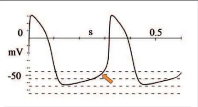
Fig. 1 - Action potential of the sinoatrial node cells. Depolarizing current depends basically on calcium influx. In phase 4, the potential difference decreases progressively until the threshold is reached (-50 mV), when depolarization occurs (arrow).
Clinical Update
Luiz Antonio Machado César If Current and heart rate control
Arq Bras Cardiol 2007; 88(4) : e96-e99
deactivating after membrane depolarization, between +15 and +30 mV, during the action potential plateau (Figure 2B). Several interferences in this current functioning have already been described, but the most important are the following: 1) modulation of the If current by the sympathetic system, changing its flow velocity and, thereby, the frequency of membrane depolarization (Figure 3)8, 2) the blockade of
If channels alters the rate of cell membrane’ spontaneous
diastolic depolarization.
If current blockade
The existence of an ionic current implies the presence of channels that allow ions to pass through the cell membrane. The blockade of these channels that carry If current was demonstrated in xrabbit sinoatrial node cells11-13. The degree
of this blockade and, therefore, its capacity to decrease the frequency of membrane depolarization, vary according to the membrane action potential. Particularly with zatebradine12, a
reasonably specific blocker of the If current, the more negative
the voltage, exactly when ion influx is higher, the greater the blockage. Therefore, the greater the ion influx the greater its ability to reduce depolarization frequency and vice-versa. It has also been demonstrated that this blockage only occurs when the channels are open. By decreasing depolarization frequency of the sinoatrial node this agent decreases heart rate.
Ivabradine
This molecule, a benzocycloalkane derivative, has a high degree of specificity for If current inhibition and exclusively
lowers HR significantly (Figure 4). In addition to being much more selective than zatebradine and other If current blockers, it exerts its effect at a much lower concentration13-15. Similar
to zatebradine, ivabradine acts at the intracellular side of the membrane15 and requires open channels to exert its blocking
action. At near-to-zero voltages, when channels are closed, no effect is observed with this drug. Experimentally, ivabradine action has shown to be much more potent in the presence of outward currents than in the presence of inward currents.
Fig. 2 - (A) Action potential of sinoatrial node cell showing (orange line) when the If current is activated. (B) Between the first two vertical lines, If current behavior when the membrane is clamped (action potential of -75, -65, -55 e -45 mV) can be noted. The greater the negative gradient, the greater is this current flow, measured
in fractions on an ampere (pA). The last line (right), orange-colored, shows the If current behavior during action potential (Adapted from DiFrancesco4)
A) B)
I
f current
I
f current
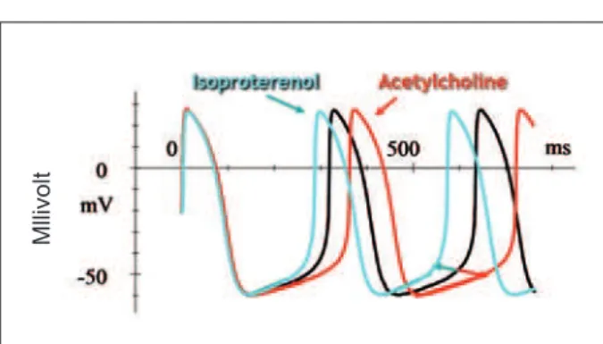
Fig. 3 - Preparation of rabbit sinoatrial cells. The white line refers to the membrane action potential. Under sympathetic stimulation, the curve shifts to the left, with increased frequency of spontaneous depolarization. Under parasympathetic stimulation, the opposite occurs. Note that only phase 4 of spontaneous depolarization is affected, without any other change in the
curve. (Adapted 8,10).
Ml
li
vo
lt
Therefore, it is clear that ivabradine block is not only voltage-dependent or channel-state voltage-dependent.
The effect of ivabradine has sparked interest for use in humans, because HR reduction is the primary aim in some situations, particularly in patients with coronary disease, since heart rate is the major determinant of myocardial oxygen uptake. Moreover, the possibility of achieving cardiovascular protection by lowering HR also brought perspectives for its use, especially because, unlike beta-blockers, ivabradine is devoid of inotropic or any other hemodynamic systemic effect. On the other hand, its effect remains unchanged even under adrenergic stimulation, although it has no effect on β -adrenergic receptors. In experimental models using isolated rat atrial cells, HR was reduced by up to –34% and in rabbits, by -24%. In these studies, almost no variation was found in either duration or amplitude of cell membrane action potential. All
Clinical Update
Luiz Antonio Machado César If Current and heart rate control
Arq Bras Cardiol 2007; 88(4) : e96-e99
findings in the sinoatrial node and Purkinje cells corroborate the absence of any effect by ivabradine to cause calcium channel blockade. Furthermore, in experimental studies using dogs undergoing treadmill exercise, ivabradine did not affect the natural increase in cardiac output and contractility during exercise nor was there change in coronary vasodilation capacity, unlike propranolol, used as a comparator in these experiments16. Given these results, a clinical program was
developed to evaluate the effect of this drug in humans, in compliance with the stringent requirements currently applied to studies of any new drug.
Clinical trials in stable angina – After Phase I trials17, a
large Phase II trial confirmed that ivabradine lowers HR both at rest and during exercise18. In this study, 360 patients with
stable angina and documented CAD were randomly assigned, in a double-blind fashion, to receive one of three doses of ivabradine (2.5mg, 5mg, or 10mg twice daily) or placebo. The study’s primary endpoint was to assess, by using exercise tolerance test, the time to onset of ST-segmentdepression on electrocardiogram (ECG) and time to limiting angina at the trough of drug activity, that is, 12 hours post-dose. As secondary endpoints, the following parameters were assessed: HR and double product, both at rest and at peak exercise, ECG data four hours post-medication, plus angina frequency and sublingual nitrate consumption. The study design comprised a two-week treatment in one of the four above-mentioned groups followed by a two- or three-month open-label
follow-up phase during which all patients received ivabradine 10 mg twice daily. Drug safety was assessed through routine laboratory tests, the frequency of adverse events, and vital signs, in addition to 24-hour ECG Holter monitoring. During the two-week treatment period, ivabradine 5 and 10 mg BID increased significantly the time to onset of ST-segment depression by 44 s e 46 s, respectively, compared to 9 s for the placebo group (p < 0.005). The dose effect was also significant (p = 0.005), but the effect of the 2.5 mg dose did not differ from that of the placebo. In the two- to three-month open-label extension phase, those who were initially in the placebo group also experienced a significant decrease in the number of angina attacks and ischemia and an increase in time to limiting angina, with p < 0.001.The benefits observed were associated with the degree of HR reduction induced by ivabradine. Additionally, the tolerability profile was good, and the only major adverse event was visual disturbance reported by up to 27% of the patients in the ivabradine 10 mg BID group, mostly changes in light intensity perception, described as mild and transient. Other studies, which have already been concluded but are still to be published, compared ivabradine with two common antianginal drugs, the β-blocker atenolol19
and the calcium-channel antagonist amlodipine20. In both
studies, ivabradine was shown to be as effective as atenolol and amlodipine in controlling anginal symptoms and increasing the time to ischemia during exercise testing. Two clinical trials are currently underway: one to assess both the efficacy and safety of ivabradine in patients receiving background therapy with atenolol and the other to assess the effect of HR lowering in patients with heart failure secondary to ischemic heart disease or dilated cardiomyopathy, in order to check whether the additional HR reduction may have a beneficial effect on cardiovascular events and death. Based on all this information and knowing that a reasonably large number of individuals with contraindication, or much more frequently, intolerance to
β-blockers, such as patients with chronic pulmonary disease, asthma, peripheral vascular insufficiency, diabetes mellitus, gastrointestinal changes, and keratitis, as well as intolerance to calcium antagonists that slow heart rate, such as patients with obstipation, lower-extremity edema, and hypotension, among others, it can be concluded that ivabradine is a very attractive option as an antianginal agent, particularly because it lowers HR without exerting hemodynamic effects and has a very good tolerability profile.
References
1. Brown HF, Di Francesco D, Noble SJ. How does adrenaline accelerate the heart? Nature. 1979; 280: 235-6.
2. Noble D, Tsie RW. The kinetics and rectifier properties of the slow potassium current in calf Purkinje fibres. J Physiol. 1968; 195: 185-214.
3. DiFrancesco D. A new interpretation of the pacemaker current in calf Purkinje fibres. J Physiol. 1981; 314: 359-76.
4. DiFrancesco D. A study of the ionic nature of pacemaker current in calf Purkinje fibres J Physiol. 1981; 314: 377-93.
5. DiFrancesco D. The cardiac hyperpolarizing-activated curret, If: origins and developments. Prog Biophys Mol Biol. 1985; 46: 163-83.
6. Biel M, Ludwig A, Zong X, Hofmann F. Hyperpolarization-activated cation channels: a multi-gene family. Rev Physiol Biochem Pharmacol. 1999; 136: 165-81.
7. Kaupp UB, Seifert R. Molecular diversity of pacemaker ion channels. Annu Rev Physiol. 2001; 63: 235-57.
8. Accili EA, Proenza C, Baruscotti M, DiFrancesco D. From funny current to HCN channels: 20 years of excitation. N Physiol Sci. 2002; 17: 32-7.
9. Pape HC. Qeer current and pacemaker. The hyperpolarization-activated cation current in neurons. Annu Rev Physiol. 1996; 58: 299-327.
10. DiFrancesco D, Ferroni A, Mazzanti M, Tromba C. Properties of the
Fig. 4 - Action potential curve under the effect of ivabradine, with a clear reduction in its slope (orange-colored), compared with the expected (bluish).
Restricted mechanism of HR reduction
Ivabradine
Clinical Update
Luiz Antonio Machado César If Current and heart rate control
Arq Bras Cardiol 2007; 88(4) : e96-e99
hyperpolarizing-activated current (If) in cells isolated from the rabbit sino-atrial node. J Physiol. 1986; 377: 61-88.
11. DiFrancesco D. Block and activation of the pace-maker channel in calf Purkinje fibres: effects of potassium, caesium and rubidium. J Physiol. 1982; 329: 485-507.
12. Di Francesco D, Some properties of the UL-FS 49 block of the hyperpolarization-activated current (If) in sino-atrial node myocites. Pflugers Arch. 1994; 427: 64-70.
13. Gardiner SM, Kemp PA, March JE, Bennett T. Acute and chronic cardiac and regional haemodynamic effects of the novel bradycardic agent, S6257, in conscious rats. Br J Pharmacol. 1995; 115: 579-86.
14. Monnet X, Chaleh B, Colin P, de Curzon DP, Giudicelli JF, Berdeaux A. Effects of heart rate reduction with ivabradine on exercise-induced myocardial ischemia and stunning. J Pharmacol Exp Ther. 2001; 299: 1133-9.
15. Bois P, Bescon J, Renaudon B, Lenfant J. Mode of action of bradycardic agent, S 16257, on ions currents of rabbit sinoatrial node cells. Br J Pharmacol. 1996; 118: 1051-7.
16. Simon L, Gheleh B, Puybasset L, Giudicelli J-F, Berdeaux A. Coronary and
hemodynamic effects of S 16257, a new bradycardic agent, in resting and exerxising conscious dogs. J Pharmacol Exp Ther. 1995; 275: 659-66.
17. Ragueneau I, LAveille C, Jochemsen R, Resplandy G, Funck-Brentano C, Jaillon P. Pharmacokinetic-pharmacodynamic modeling of the effects of ivabradine, a direct sinus node inhibitor, on heart rate in healthy volunteers. Clin Pharamcol Ther. 1998; 64: 192-203.
18. Borer SS, Fox K, Jaillon P, Lerebours G; Ivabradine Investigators Group. Antianginal and Antiischemic Effects of Ivabradine,an If Inhibitor, in Stable Angina. A Randomized, Double-Blind, Multicentered, Placebo-Controlled Trial. Circulation. 2003; 107: 817-23.
19. Tardif JC, Ford I, Tendera M, Fox K. On Behalf: the INITIATIVE study investigators. Anti-anginal and anti-ischaemic effects of the If current inhibitor ivabradine versus atenolol in stable angina. A 4-month randomised, double-blind, multicenter trial. Eur Heart J. 2003; 20 (suppl): 24.
20. Ruzyllo W, Ford F, Tendera M, Fox K. On behalf: the study investigators. Antianginal and antiischaemic effects of the If current inhibitor ivabradine compared to amlodipine as monotherapies in patients with chronic stable angina. Randomised, controlled, double-blind trial Eur Heart J. 2004; 25 (Suppl): 138.