A
r
ti
c
le
0103 - 5053 $6.00+0.00
*e-mail: [email protected]
Detailed Study of Brazil Nut (
Bertholletia excelsa
) Oil Micro-Compounds:
Phospholipids, Tocopherols and Sterols
Thavarith Chunhieng, Abdel Hafidi, Daniel Pioch, José Brochier and Didier Montet*
CIRAD-Persyst, UMR Qualisud 95, TA B-95/16, 73 rue J-F Breton, 34398 Montpellier Cedex 5, France
O óleo da castanha-do-pará (Bertholletia excelsa) foi estudado por causa da sua composição em ácidos graxos, tocoferóis, esteróis e fosfolipídios. A composição de ácidos graxos nos fosfolipídeos também foi estudada. Os resultados foram comparados com os do girassol, da castanha, amêndoa, noz, soja e azeites. O seu alto teor em ácidos graxos insaturados, em -tocoferol e em -sitosterol confere à castanha-do-pará interessantes propriedades antioxidantes e de prevenção do colesterol. A composição de ácidos graxos em fosfolipídio é muito diferente da composição do óleo. O ácido linolênico, que não se encontra no óleo, encontra-se em grande quantidade na fosfatidiletanolamina.
The oil of the Brazil nut (Bertholletia excelsa) was studied for its composition in fatty acids, tocopherols, sterols and phospholipids. The fatty acids composition of phospholipids was also studied. These results were compared to those of sunflower, walnut, almond, soya and olive oils. Its high content of unsaturated fatty acids, of -tocopherol and of -sitosterol gave to the Brazil nut interesting antioxidant and anti-cholesterol properties. The composition of fatty acids in phospholipid is very different from the composition of the oil. Linolenic acid, which is not present in the oil, is present at a high level in phosphatidylethanolamine.
Keywords: Brazil nut oil, fatty acids, phospholipids, sterols, tocopherols
Introduction
Brazil nuts, seeds of Bertholletia excelsa H.B.K. are produced and exported from the Amazon Basin region and are used most extensively in confections in Europe and North America.1 Although known for their protein content, 15-17% by fresh weight and about 50% by weight of its defatted flour, the nuts are also a good source of oil (63-70%). Brazil nuts are reported to contain a higher amount of oil in comparison to that of almonds (53%) and walnuts (55%).2 Brazil nut oil is a clear yellowish oil with a pleasant smell. It has a limited use worldwide because of the lack of knowledge concerning its composition. In fact, few complete studies have been carried out on Brazil nut oil. We wanted to supplement the knowledge on this oil by proportioning sterols, tocopherols and phospholipids.
The determination of precise composition of these compounds could be of an invaluable help for its
discrimination during falsification and for cosmetic and dietary applications.
Sterols are natural components present in many plants and animal species. They are also essential for human and animal health. The sterol fraction is specific from each oil and can be used to characterize an oil. They have been recently reported as being anticholesterol agents.3
-Sitosterol takes part in the conversion of linoleic acid into polyunsaturated fatty acids. This reaction is essential for the conversion of -6 fatty acids into prostaglandins and leukotrienes. Prostaglandins and leukotrienes are involved in the immune system; they reduce the thrombo-embolic problems by reducing platelet aggregation and also assist in the reduction of the anti-inflammatory drug metabolites.4
sitosterol has been reported to take part in the cholesterol reduction5 and to improve urinary problems.6
membranes, and are involved in skin and cellular DNA damage that causes various cancers8 and degenerative diseases. Vitamin E plays a role against immunizing disorders and premature aging, is also employed in the treatment of fibrocystes9 and prevents the formation of eye cataracts.
Vitamin E deficiency has been linked with neurological abnormalities in abetalipoproteinemia and fat malabsorption disorders. Experimentally induced dystrophy indicates that dietary vitamin E may play an important role in the central nervous system with regard to membrane stability and physiological function.
In recent years, lipid peroxidation has attracted much interest in relation to the aging process, cancer development and other pathological conditions. International epidemiological studies have suggested that differences in dietary fat intake may provide a meaningful key to the prevention of cancer.10 Membrane phospholipids including phosphatidylcholine (PC) and phosphatidylethanolamine (PE) are assumed to be the main species responsible for hydroperoxide formation during lipid peroxidation in vivo.11
The objective of this research was to increase knowledge on the biochemical composition of fatty acids, sterols, tocopherols and phospholipids of Brazil nut that form the largest groups of natural antioxidants for use in special diet formulations, pharmacology and medicine. We compared the oil composition with some well known nuts such as Grenoble walnuts or almonds and olive oil which is the reference oil in the Mediterranean diet.
Experimental
Material
Brazil nuts were collected in the north east of the Amazon Basin. They were provided by the company JBA Agroconcept (Castries, France). The exact origin of this variety, which is very rich in selenium,12 is voluntarily held secret because of the future industrial applications which arose from this study. Since the beginning of the analysis, in April 2000, the nut samples were stored in a cabinet at ambient temperature with an average temperature of 25 °C. All of the analyses were done in two months.
Lipid extraction
Oil was extracted from 20 g of Brazil nut flour for 6 h by the Soxhlet method with hexane (250 mL). The solvent was removed by evaporating and then flushing with nitrogen gas.
Flour was prepared by grinding nuts directly.
Preparation of fatty acids methyl esters (FAME)
Sodium methylate solution (3 mL) was added to three drops of extracted oil and heated at 60 °C for 10 min.13 After 10 min boiling, 3 mL of acetyl chloride in methanol (50 mL/625 mL) was added. The mixture was heated again for 10 min and cooled to room temperature and subsequently 10 mL of distilled water and 15 mL of hexane were added in succession. The mixture was shaken vigorously and left to stand to allow the layers to separate. The upper hexane layer containing FAME was transferred to a small tube and stored at -20 °C for later analysis by gas-liquid chromatography (GLC).
FAME analysis was performed by GLC (Ceinstruments, Model GC 8000 Top) equipped with a flame-ionization detector. A Supelcowax 10 column (0.32 mm i.d. 30 m long, 0.25 m film thickness) with helium carrier gas at a linear flow velocity of 2 mL min-1 was used. A temperature program of 100 °C for 5 min, rising to 230 °C at a rate of 10 °C min-1 was used. The FAME, dissolved in hexane, was injected (1 L) in a split mode. The injector and detector temperatures were 250 and 260 °C respectively. Peak areas were recorded using a Merck D-200 integrator. Fatty acids were identified by comparison of retention times of palm oil fatty acids and by comparison with literature values.
Analyses of tocopherols by high-pressure liquid chromatography (HPLC)
A 1.0 g oil samples were dissolved in 13 mL hexane and 10 L samples were injected into a 150 4.6 mm column packed with 5 m Hypersil Silica. The liquid chromatograph (Spectra System P1000 XR) was equipped with a Spectra System FL 3000 detector set at 294/336 nm. The mobile phase was 97% n-hexane and 3% 1,4 dioxane at a flow rate of 1 mL min-1. Tocopherol compositions were obtained by comparison of peak retention times from rape seed tocopherols.
Saponification for sterol analysis14
through the neck of the funnel into another separating funnel containing 40 mL of water. Aqueous ethanolic soap solution was extracted twice, each time with 100 mL of diethyl ether. Ethereal fractions were combined in the second separating funnel and were washed twice with 40 mL of water shaking vigorously and then successively with 40 mL 0.5 mol L-1 aqueous KOH solution, 40 mL of water, and again with 40 mL of the KOH solution, then twice more with 40 mL of water. Ethereal solution was poured off quantitatively into a 200 mL tarred flask and evaporated. Acetone (6 mL) was added and the volatile solvent was completely removed under a gentle current of nitrogen. The unsaponifiable fraction was weighed and kept in 10 mL hexane at -20 °C for further analysis.
Preparative thin layer chromatography
Crude lipid extracts were applied on 20 20 cm TLC plates coated with 0.5 mm layer Silica gel 60F254 (Merck, Germany) as 14 cm bands (ca. 20 mg plate-1) with a Linomat-3 auto applicator (Camag, Switzerland). The TLC standards mixture (cholesterol) were applied as a reference on one side of each plate and the plates were developed in chloroform/ether (90/10, v/v). After being developed and dried, plates were sprayed with dichlorofluorescein solution in ethanol and observed under UV light (Chromato-Vue model CC20, Ultra-Violet Products Inc). Sterol bands were scraped off and extracted twice with chloroform.
Preparation of sterols TMS ether derivatives
Total unsaponifiable fraction was derived to trimethylsilyl ethers by adding 100 mL of Tri-Sil reagent and incubating the tubes at 60 °C for 45 min. The solvent was evaporated under a nitrogen stream and TMS ether derivatives were dissolved in 1 mL hexane. The tubes were sonicated in an ultrasonic bath for 1 min and centrifuged for 3 min. The hexane layer was transferred to another tube, evaporated to dryness and dissolved in 0.5 mL hexane for further analysis by GLC.
Analysis of sterols by GLC
A capillary column (30m 0.32 mm i.d.) with a 0.15 m film thickness (DB-1701, Germany) of a 50% phenyl-/50% methyl-polysiloxane (Chrompack, USA) stationary phase was used. Hydrogen was used as carrier gas at an inlet pressure of 9.6 psi and as makeup gas at a flow rate of 2 mL min-1. Injector and detector temperatures were 280 °C; oven temperature was 254 °C. The peaks were computed by an HP 6890 integrator.
Phospholipid extraction by osmosis
A 6 g of lipids in hexane solution were added into a rubber finger. Osmosis was done against hexane with a flow rate of 0.5 mL min-1 for 24 h. Phospholipids were transferred from the rubber finger to a small tube, evaporated to dryness with nitrogen gas and then kept in 1 mL chloroform at -20°C for further analyses.
Phosphorus analysis by flame spectrometry was done on 10 g of oil in a 100 mL beaker and then transferred to a cold oven.15 The temperature of the oven was increased to 300 °C and maintained for 24 h, followed by three step increases to 350, 400 and 450°C at regular intervals of 4 h each. Mineralization was achieved at 475 °C for 4 h. The ash was dissolved in 4 mL HCl/water (50/50 v/v) then heated at 80 °C to dissolve them fully. The solution was filtered in 50 mL flasks and completed with pure water. Mineral elements were quantified by Plasma Emission Spectroscopy.
Phospholipid preparative thin layer chromatography
Phospholipid extracts were applied on 5 20 cm TLC plates coated with a 0.5 mm Silica gel 60 F254 (Merck, Germany) as 2 cm bands (ca.0.8 mg plate-1) with a Linomat-3 auto applicator (Camag, Switzerland). A phospholipid standard mixture was applied as a reference on another plate and plates were developed in chloroform/acetone/methanol/acetic acid/ water in a ratio 50/20/10/10/5 (v/v). After developing, the plate was sprayed with molybdenum blue - phosphomolybdic acid reagent, 20 wt.% solution in ethyl alcohol (Aldrich).
Phospholipid analysis by spectrophotometry method16
Phospholipid bands were scraped off the plate and 200 L of distilled water and 120 L of magnesium nitrate were added to the samples. Samples were transformed to white deposits by mineralization with a Bunsen burner. HCl 0.5 mol L-1 (300 L) was added and the samples were incubated in a water bath at 45 °C for 20 min and then cooled to ambient temperature. A mixture of 700 L of 10% of ascorbic acid and ammonium molybdate (1/6 p/p) was added. Phosphorous concentrations were determined by comparing their absorbance values with those of standard solutions (30 to 160 µg mL-1) which were prepared using the same procedure.
Results
General composition
value for an oily nut.12 Ashes determination of the cake containing 10% lipids shows a high mineral content of 10.7 ± 0.1%. Calcium content is low, around 600 mg/100 g, but Magnesium and Phosphorus are very high with respectively 1,400 and 2,400 mg/100 g of cake.
Selenium content varies a lot between regions and also among the nut from one tree. The nut analyzed in this study show contents varying between 0.4 and 12.7 mg/100 g.17 Total sugar content (glucose + fructose + sucrose) of Brazil nut defatted cake was 8.2%. Total fiber content of the cake is 15.2%, which represented for the whole nut 4.5%. The defatted cake contained of 67.9% protein. That represents for the whole nut a protein content of 17.3%. The oil content of fresh Brazil nut is 72.5% .
Fatty acid composition (%) of Brazil nut oil
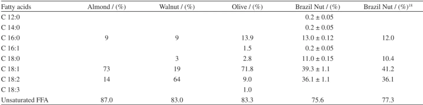
Fatty acid composition obtained by GLC shows a high unsaturation (75.6%) level in the oil, due essentially to C18:1 (39.3%) and C18:2 (36.1%) (Table 1). These values are very close to the one obtained by Assuncao.18 It is lower than olive (83.3%), almond (87.0%) and walnut (83.0%) oils. Linoleic acid percentage (18:2) is higher than in almond or olive oils. The high unsaturation of the fatty acids and the high amount of linoleic acid give to this oil some interesting properties for a healthy diet.
Tocopherol fractions
Total tocopherol was found to be 0.06 mg g-1 of the full nut. Three main tocopherols are found in Brazil nut oil (Table 2). Brazil nut oil is characterized by its high content in -tocopherol (88.3%). This composition is very close to the one obtained recently by Kornsteiner19 on Brazil nut purchased in a local market in Austria. It is richer then walnut oil and olive oil, which contained only 1.8% and 5.3%, respectively.20 Olive oil has a high content in -tocopherol (84.2%) against 11.3% for Brazil nut oil. Brazil nut oil is poor in -tocopherols (0.4%) in comparison with olive oil content (10.5%) and walnut oil (88.0%).21 -tocopherol is not found in Brazil nut oil whereas walnut contains 8.5%. -tocopherol is a characteristic component of Brazil nut oil and it could be used to discriminate this oil from others such as soy oil, which could be used as an adulterant.
Sterol fractions
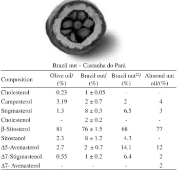
Brazil nut sterol total content in the oil was 0.19% ( 0.01) that is closed to the results obtained by Phillips22 with 0.09% on Brazil nuts consumed in US. Brazil nut oil has a similar sterol composition to olive oil. Its -sitosterol content (76%) is high and comparable to that of olive oil (81%) and Brazil nut oil consumed in US (68%). The two
Table 1. Fatty acid composition (%) of Brazil nut oil determined by GLC in comparison with other oil composition.18, 20, 21 Average value of the same
sample injected in triplicate
Fatty acids Almond / (%) Walnut / (%) Olive / (%) Brazil Nut / (%) Brazil Nut / (%)18
C 12:0 00.2 ± 0.05
C 14:0 00.2 ± 0.05
C 16:0 9 9 13.9 13.0 ± 0.12 12.0
C 16:1 1.5 00.2 ± 0.05
C 18:0 3 2.8 11.0 ± 0.15 10.4
C 18:1 73 19 71.8 39.3 ± 1.10 41.2
C 18:2 14 64 9.0 36.1 ± 1.10 36.1
C 18:3 1.0
Unsaturated FFA 87.0 83.0 83.3 75.6 77.3
Table 2. Composition (%) of Tocopherol fractions of Brazil nut oil determined by HPLC in comparison with Olive, Soybean and Walnut oil tocopherols.20, 21
Average value of the same sample injected in triplicate
Tocopherols Brazil nut oil / (%) Brazil nut oil19/ (%) Olive oil / (%) Soya bean oil / (%) Walnut oil / (%)
-tocopherol 11.3 ± 1.20 7 84.2 10 1.7
-tocopherol 88.3 ± 1.90 93 5.3 3 1.8
-tocopherol 00.4 ± 0.05 10.5 58 88.0
oils contain low amounts of stigmasterol but Brazil nut oil is richer than olive oil at 8% versus 1.3%.23 -sitosterol content in Brazil nut oil (76%) is also similar to almond oil (77%).24 Because of the similarity in sterol fractions among these three oils, this factor could not be use to differentiate Brazil nut oil from others oils (Table 3).
Phospholipid fractions
The concentration of phosphorous in Brazil nut oil was found in duplicate to be 377.3 mg L-1 ( 0.2) by flame spectrometry. The different classes of phospholipids, obtained by extraction, were analyzed by HPLC and by spectrophotometry. The percentage was recalculated based on the measured phosphorus content of 377.3 mg L-1 and compared to sunflower lecithin25 which is the most used phospholipids industrially (Table 4).
Brazil nut oil contains 31; 24; 21 and 24% of phosphatidylinositol (PI); phosphatidylcholine (PC); phosphatidylethanolamine (PE) and phosphatidic acid (PA) respectively, whereas sunflower oil contains 20 ; 47 ; 21 and 12% of the same components. PI content is higher (31%) then in sunflower oil (20%). PC content (24%) is half that of the sunflower oil content (47%) whereas phosphatidylserine (PS) is double the amount.25
Fatty acids extracted from phospholipids were analyzed by FAME/GLC (Table 5). The results are compared with the results obtained by Smiles et al.26 during the study of the effect of degumming reagents on the recovery and nature of lecithins from crude sunflower oil (Table 6).
Fatty acid composition of phospolipids of Brazil nut oil (Table 5) is different from the fatty acid composition of Brazil nut oil (Table 1). The main difference is that the oil contains more linoleic (36.1%) acid than phospholipids, which are present, on average, at 10%. PA and PI contain high level of lauric acid (10-12%) whereas the oil has a very low quantity (0.2%). PE contains 34% of linolenic acid while the oil does not contain any.
Brazil nut oil phospholipids contain less unsaturated fatty acids than those of sunflower oil
Linolenic acid (18:3) is not observed in sunflower phospholipids and Brazil nut oil while 34 and 11%, respectively of linolenic acid are found in PE and PA.
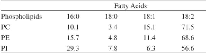
There was a very high content of linoleic acid (18:2) in PC, PE and PI (71.5; 68.6 and 56.6 % respectively) of sunflower oil while Brazil nut oil contained only 6; 8 and
Table 3. Composition (%) of Sterol fractions of Brazil nut oil determined
by GLC in comparison with Olive and Almond nut oil23, 24
Brazil nut – Castanha do Pará Composition Olive oil/
(%)
Brazil nut/ (%)
Brazil nut22/
(%)
Almond nut oil/(%)
Cholesterol 0.23 01 ± 0.05 -
-Campesterol 3.19 02 ± 0.70 2. 04
Stigmasterol 1.30 08 ± 0.30 06.5 03
Cholestenol - 02 ± 0.20 -
--Sitosterol 81.0 76 ± 1.50 680 77
Sitostanol 2.30 08 ± 1.20 04.3
-5-Avenasterol 2.70 02 ± 0.70 14.1 12 7-Stigmastenol 0.55 01 ± 0.20 06.4 02
7- Avenasterol - - - 02
Table 4. Composition (%) of Phospholipids fractions of Brazil nut oil
determined by spectrophotometry in comparison to Sunflower lecithin26
Phospholipids PI/(%) PC/(%) PE/(%) PA/(%) Total/(%) Sunflower
Lecithin 20 47 21 12 100
Brazil nut oil 31 24 21 24 100
Table 5. Composition (%) of Phospholipid fatty acids of Brazil nut oil
Fatty acids Spot 0 PI PC PE PA Unknown Unknown
12:0 - 12.0 01.0 - 10.0 04.0
-14:0 00.3 05.0 02.0 05.0 04.0 02.0 05.0
16:0 25.2 26.0 34.0 18.0 38.0 20.0 27.0
16:1 01.0 06.0 02.0 03.0 05.0 02.0 02.0
18:0 09.0 18.0 23.0 09.0 18.0 12.0 16.0
18:1 50.2 18.0 31.0 23.0 14.0 19.0 28.0
18:2 14.0 11.0 06.0 08.0 - 08.0 19.0
18:3 00.3 04.0 01.0 34.0 11.0 33.0 03.0
11% of PC, PE and PI. There were no traces of 18:2 in PA of Brazil nut oil.
Content of oleic acid (18:1) of Brazil nut oil phospholipids is high: 31; 23 and 18% respectively of PC, PE and PI compared to 15.1; 11.4 and 6.3 % in sunflower oil.
The stearic acid (18:0) is observed to contain 23; 9 and 18% of PC, PE and PI respectively in Brazil nut oil whereas sunflower oil contained lower contents with only 3.4; 4.8 and 7.8 % of PC, PE and PI, respectively.
Palmitic acid (16:0) content in Brazil nut oil is the highest with 34 and 18% respectively of PC and PE compared to those of sunflower oil 10.1 and 15.7%. PI of Brazil nut and sunflower oil contains nearly the same content in palmitic acid, 26 and 29.3%.
Discussion
Brazil nut oil has a good proportion of unsaturated fatty acids 75.6% that could be compared to that of walnut or olive oils (83%).20 The high content in linoleic acid (39.3%) and linolenic acid (36.1%) provides this oil with some interesting dietary characteristics.
The tocopherol fraction of Brazil nut oil is quite different from that of olive oil. The content of -tocopherols (88.3%) is much higher then that in other oils and could be a discriminator in case of falsification. Previous studies indicate that usually -tocopherol is more active against oxidation then -tocopherol. Mortensen and Skibsted27 established a hierarchy of the antioxidant properties of tocopherols and concluded that -tocopherol, -tocopherol, -tocopherol and -tocopherol have descending antioxidant properties. Nevertheless, Kaiser et al.28 showed that -tocopherol is as effective as -tocopherol in physical quenching of O2 but has a very low chemical reactivity. This tocopherol homologue might be particularly suitable for biological conditions in which an accumulation of oxidation products might weaken the antioxidant defense.
The phytosterol composition of Brazil nut oil is similar to olive and almond nut oils. Its high content in sitosterol could be useful as anti-cholesterol medicine.23, 24
Table 6. Composition (%) of Phospholipid fatty acids of sunflower oil
and soy bean oil27
Fatty Acids
Phospholipids 16:0 18:0 18:1 18:2
PC 10.1 03.4 15.1 71.5
PE 15.7 04.8 11.4 68.6
PI 29.3 07.8 06.3 56.6
PI: Phosphatidylinositol; PC: Phosphatidylcholine; PE: Phosphatidyl-ethanolamine.
Our experiment shows that Brazil nut oil contains an interesting composition of phospholipid fatty acids comparable to sunflower lecithin.25, 26
Based on these results, Brazil nut oil has the potential to have high commercial value in the health food industry with application in some areas of medical science.
Aknowledgments
Financial support was provided by the Project for Research and Industry (PRI) supported by the French Ministry of Foreign Affairs. Nuts were supplied by JBA Agroconcept.
References
1. Sun, S. S. M.; Leung, F. W.; Tomic, J. C.; J. Agric. Food Chem.
1987,35, 232
2. Ali, R.; Khan, M. N.; J. Am. Oil Chem. Soc. 1988,65, 1951. 3. Busson, V.; Journées Chevreul AFECG, Bordeaux-Pessac,
25-26 November 1999.
4. Schulz, V.; Hansel, R.; Tyler, V. E.;A Physician’s Guide to Herbal Medicine. 3rd ed., 168-173. Springer Berlin, 1998.
5. Ikeda, I.; Kawasaki, A.; Samzima, K.; Sugano, M.; J. Nutr. Sci. Vitaminol.1981,27, 243.
6. Klippel, K. F.; Hitl, D. M.; Schipp, B.; Br. J. Urol.1997,80, 427.
7. Dieber-Rotheneder, M.; Puhlwaeg, G. H.; Striegl, G., Esterbauer, H.;J. Lipid Res.1991,32, 1325.
8. Heinonen, O. P.; Albanes, D.; Virtamo, J.; Taylor, P. R.; Huttunen, J. K.; Hartman, A. M.; Haapakoski, J.; Malila, N.; Rautalahti, M.; Ripatti, S.; Mäenpää, H.; Teerenhovi, L.; Koss, L.; Virolainen, M.; Edwards, B.L. ; J. Natl. Cancer Inst.1998,
90, 440.
9. Kimmick, G. G.; Bell, R. A.; Bostick, R. M.; Nutrition and Cancer1997,27, 109.
10. Nesarethnam, K.; Palm Oil Developments1992,17, 5. 11. Yamada, K.; Terao, J.; Matsushita, S.; Lipids1987,22, 125. 12. Chunhieng, T.; Goli, T.; Piombo, G.; Pioch, D.; Brochier, J.;
Montet, D.; Bois et Forêts des tropiques 2004, 280, 91. 13. CIRAD-Amis-PAA Lab. Procedures. Preparation of Methylic
Esters, Montpellier, France, Code PC0919A, 1999, 1-6. 14. International Union of Pure and Applied Chemistry. Applied
Chemistry Division Oils and Fats Section. Standard Methods of the Oils and Fats Section of the IUPAC. London, 1965, 3-4.
15. CIRAD-Amis-PAA Plant LabProcedures.Solubilisation of Minerals from Fatty Products and Dosage by Plasma Emission
Spectrometry (ICP), Montpellier, France, Code PP0912A. 1997.
16. Ames, B. N.; Methods Enzymol. 1966,8, 115.
18. Assunção, F. P.; Bentes, M. H. S.; Serruya, H.; J. Am. Oil Chem. Soc.1984,61, 1031.
19. Kornsteiner, M.; Wagner, K. H.; Elmadfa, I.; Food Chem.2006.
98, 381.
20. Kamal-Eldin, A.; Andersson, R. ; J. Am. Oil Chem. Soc.1997,
74, 375.
21. Woodlandnut; http://woodlandnut.com/walnut.html, accessed in 2007.
22. Phillips, K. M.; Ruggio, D. M.; Ashraf-Khorassani, M.; J. Agric. Food Chem.2005,53, 9436.
23. Jimenez de Blas, O.; Gonzalez, A. D. V.;J. Am. Oil Chem. Soc.
1996,73, 1685.
24. Itoh, T.; Tamura, T.; Matsumoto, T.; Oléagineux 1974,5, 250. 25. Carelli, A. A.; Brevedan, M. I. V.; Crapiste, G. H.;J. Am. Oil
Chem. Soc.1997,74, 511.
26. Smiles, A.; Kakuda, Y.; Mac Donald, B. E.; J. Am. Oil Chem. Soc.1988,65, 1151.
27. Mortensen, A.; Skibsted, L. H.; FEBS Lett.1997,417, 261. 28. Kaiser, S.; Di Mascio, P.; Murphy, P.; Sies H. M. E.; Arch.
Biochem. Biophys.1990,277, 101.
Received: August 8, 2007