429
BRAZILIAN JOURNALOF OTORHINOLARYNGOLOGY 72 (3) MAY/JUNE 2006
http://www.rborl.org.br / e-mail: [email protected]
CASE REPORT
Rev Bras Otorrinolaringol 2006;72(3):429.
Automastoidectomy
João Alcides Miranda1, Fábio Akira Suzuki2, Marcello
Henrique de Carvalho Borges3, Andre Luis Sartini4
INTRODUCTION
Automastoidectomy is defined as extensive destruction of the middle ear cavity and the mastoid, appearing as an image reminiscent of a radical mastoidec-tomy cavity.1 This condition may arise as a complication of cholesteatomatous chronic otitis media2 or of keratosis obturans.3 Very little, however, has been described of this entity in literature.
CASE REPORT
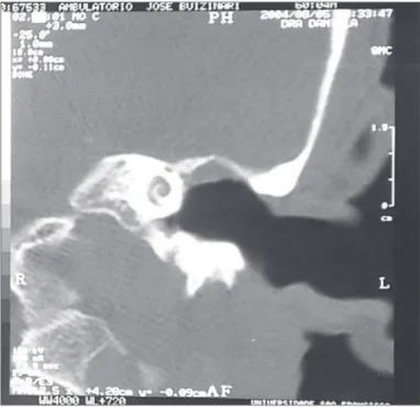
A male 60 year old patient pre-sented with left hypoacusia beginning 8 months ago. The patient complained of sporadic recurrent left otorrhea since in-fancy, the last episode occurring 18 months
ago. Otoscopy revealed an intact opacified left tympanic membrane; the manubrium of the malleus was not seen. Audiometry showed left severe mixed hearing loss and moderate right neurosensorial hearing loss. Computed tomography of the ear disclosed a wide left tympanic cavity and absence of the ossicular chain (Figure 1). The diagno-sis was left automastoidectomy. An explor-atory tympanotomy was discussed but the patient refused surgical treatment.
DISCUSSION
Automastoidectomy is a rare se-quela of diseases that affect the external and middle ear. Hawke and Shanker3 report a case of automastoidectomy resulting from keratosis obturans. This report describes a patient with left automastoidecto-my and a history suggesting chron-ic non-choleste-atomatous otitis media. Based on the patient’s his-tory and the im-age diagnosis, this appears to be a situation in which automastoidecto-my is unrelated to a cholesteatoma or simple chronic otitis media that progressed follow-ing closure of a possible tympanic perforation. These conditions would not convincingly explain the bone destruction found
in this case. Could this possibly be a case of cholesteatomatous chronic otitis media that hypothetically resolved spontaneously, leaving an automastoidectomy as a sequela? According to Hungria,4 this might be pos-sible, however no similar report has been published in medical literature.
CONCLUSION
Automastoidectomy is described as a rare complication of external and middle ear diseases; there has been no publication associating this condition with chronic otitis media other than the cholesteatomatous form of otitis media.
REFERENCES
1. The Encyclopaedia of Medical Imaging Volume VI 2. Disponível em http: // www. amershamhealth.com/medcyclopaedia/ medical/Volume20VI202/AUTOMASTOID-ECTOMY.ASP. Acessado em 13 de março de 2005.
2. Gaurano JL, Joharjy IA. Middle ear choles-teatoma: characteristic CT findings in 64 patients. Ann Saudi Med 2004;24(6):442-7. 3. Hawke M, Shanker L. Automastoidectomy
caused by Keratosis Obturans: a case report. J Otolaryngol 1986;15(6):348-50.
4. Hungria H. Otites Médias Crônicas Supu-rativas. Timpanoplastias. Em: Hungria H. Otorrinolaringologia. 8a ed. Rio de Janeiro: Guanabara Koogan; 2000. p. 371-91.
Keywords: automastoidectomy, cholesteatoma, chronic otitis media.
1 Resident of the Otorhinolaryngology Unit of the Sao Francisco University Hospital, Bragança Paulista, SP.
2 Doctor in Otorhinolaryngology graduated at UNIFESP - EPM, Head of the Otorhinolaryngology Unit of the Sao Francisco University Hospital, Bragança Paulista, SP, Vice-coordinator of the post-graduate course in Otorhinolaryngology of the Servidor Publico Estadual Hospital (HSPE).
3 Resident of the Otorhinolaryngology Unit of the Sao Francisco University Hospital, Bragança Paulista, SP.
4 Mestrando em Otorrinolaringologia pelo Hospital do Servidor Público Estadual, Médico Assistente do Serviço de Otorrinolaringologia do Hospital Universitário São Francisco, Bragança Paulista. This study was done in the Sao Francisco University Hospital, Bragança Paulista - SP.
Correspondence: João Alcides Miranda - Rua Bias Fortes 438 Boa Esperança MG 3717-000. E-mail: [email protected]
Paper submitted to the ABORL-CCF SGP (Management Publications System) on November 20th, 2005 and accepted for publication on April 27th, 2006.