From the Department of Dermatology and the Immunopathology Laboratory, Hospital das Clínicas, Faculty of Medicine, University of São Paulo – São Paulo/SP, Brazil.
Received for publication on March 10, 2003.
PREVALENCE OF ANTINUCLEAR AUTOANTIBODIES
IN THE SERUM OF NORMAL BLOOD DONORS
Solange Assuncion Villagra Fernandez, Alice Zoghbi Coelho Lobo, Zilda Najjar Prado de Oliveira, Ligia Maria Ichimura Fukumori, Alexandre Marques Périgo and Evandro A. Rivitti
FERNANDEZ SAV et al. - Prevalence of antinuclear autoantibodies in the serum of normal blood donors. Rev. Hosp. Clín. Fac. Med. S. Paulo 58(6):315-319, 2003.
OBJECTIVE: To examine the presence of serum antinuclear autoantibodies in a healthy population.
METHODS: Serum of 500 normal blood donors between 18 and 60 years of age were tested for the presence of autoantibodies. Antinuclear antibodies were detected by indirect immunofluorescence technique using HEp-2 epithelial cells as the substrate. The presence of dnaN was detected by indirect immunofluorescence technique using Critidia lucillae
as the substrate. Anti-SSA (RO), anti-SSB (LA), anti-Sm, and anti-RNP were determined by double radial immunodiffusion. RESULTS: In the evaluation of the presence of serum antibodies, antinuclear antibodies were detected in 22.6% of the sera. The presence of other antibodies was not significant. The majority of the titers were 1:40.
CONCLUSION: The presence of autoantibodies is not necessarily pathologic and has to be related to the age group, gender, and clinical condition of the patient.
DESCRIPTORS: Autoantibodies. Healthy population. Rheumatic diseases. Antinuclear antibodies. Autoimmunity.
Immune humoral response in the presence of autoantibodies against in-tracellular antigens characteristically occurs in a majority of connective tis-sue diseases. This phenomenon is found in systemic lupus erythematous, systemic sclerosis, Sjögren syndrome, mixed connective tissue disease, poly-myositis, and dermatopoly-myositis, among others1.
The detection of such autoanti-bodies is important not only for diag-nosis but also for the progdiag-nosis of the diseases, in addition to allowing clini-cal follow-up and treatment evaluation in many cases1.
It is relevant to point out that in healthy people and in patients with non-rheumatic conditions, such as chronic hepatic diseases, neoplasias, as well as active infections including
tu-berculosis, malaria, and subacute bac-terial endocarditis, these autoanti-bodies may be present1. In general,
they are found in lower titers than those detected in autoimmune dis-eases. It is believed that such titers may precede the appearance of dis-eases in normal and asymptomatic in-dividuals for many years2.
Previous studies have shown dif-ferent prevalences of autoantibodies in healthy populations. Baig and Shere (1989), who investigated the serum of blood donors and patients without autoimmune diseases in Saudi Arabia,
found antinuclear antibodies (ANA) present in 4.2%3. Vlam et al. (1993)
detected ANA present in 13% of nor-mal blood donors in a Belgian popu-lation2. Vazquez-Del Mercado et al.
(1995) reported that 4.7% of a Mexi-can population tested positive for ANA4.
The present study intends to evalu-ate the prevalence of autoantibodies in healthy people in relation to gen-der and age group. It is important to note that it is the first such study in Brazil, one that characterizes a specific population, that of blood donors of the Pro-Blood Foundation – Hemocenter of São Paulo, aiming to obtain param-eters for comparisons with other popu-lations.
immunofluorescence technique in HEp-2 cells. They were also tested for the identification of antibodies against antigens extracted from the nucleus such as anti-SSA/RO, anti-SSB/LA, anti-Sm, and anti-RNP by double ra-dial immunodiffusion. They were still tested for dnaN using indirect immun-ofluorescence (IIF) technique with Crithidia luciliae as the substrate.
In the blood bank, samples are ha-bitually tested for infectious diseases such as Chagas disease, syphilis, hepa-titis B and C, HTLV-1 and HTVL-2, and HIV-1 and HIV-2, that may contraindi-cate the utilization of the blood taken.
This study was approved by the In-stitutional Ethical Committee. The volunteers who had significant titers (ANA>1:160) for the studied param-eters were informed of that and were re-examined for any disease that could be clinically confirmed. They received follow-up evaluation and orientation when necessary.
PATIENTS AND METHODS
Blood samples collection
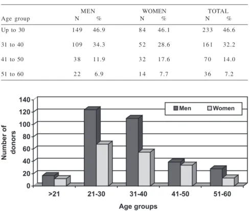
The blood samples were collected only after the volunteers had agreed to take part in the study and after they signed the consent form.. Five hundred consecutive blood samples of volun-teer donors (182 women and 318 men), between 18 and 60 years of age, with no clinical symptoms of any disease, were analyzed (Fig. 1). They were ob-tained from the Pro-Blood Foundation – Hemocenter of São Paulo.
Autoantibody detection
After collection in plain red-top tubes, the blood samples were centri-fuged (10 minutes at 2000 rpm), and the serum was separated into aliquots for performing the autoantibody-de-tection tests.
The indirect immunofluorescence (IIF) technique to detect antinuclear antibodies, described by Holbrow, Weir, and Jonhson in 1957, was used in slides covered with commercially obtained (Hemagen®) HEp-2 cells
(epi-thelial cells of human larynx carci-noma). The usage of human cells guar-antees the presence of nuclear antigens in such a concentration that enables the confirmation of the presence of antibodies in the serum.
At first, a screening test was carried out, in which serum samples were di-luted in phosphate-buffered saline, pH 7.4 (PBS) to a titer of 1:40 to detect positive reaction and characterization of the fluorescence pattern. The posi-tive samples were titered until testing negative.
The HEp-2 cells were incubated with the donor’s serum samples in a humid and dark camera for 30 minutes and were then washed in PBS. After-wards they were incubated with human anti-IgG antibody marked with fluo-rescein isothiocyanate. After being washed again, the slides were mounted with buffered glycerine pH 8. A Zeiss microscope equipped for epilumines-cence was used for reading the slides.
In order to detect native anti-DNA autoantibodies (DNAn), the indirect immunofluorescence (IIF) technique was used in slides covered with Crithidia lucillae (a protozoan that is rich in kinetoplast DNA) obtained from the Department of Rheumatology of our Institution.
The slides were incubated with the samples diluted 1:20 in Tris-buffered saline, pH 7.5 (TBS) and were then washed and incubated with human anti-IgG antibody (SIGMA brand) marked with fluorescein isothiocya-nate. A Zeiss microscope equipped for epiluminescence was used for reading the slides (immersion objective).
The double radial immunodiffu-sion technique, described by O. Ouchterlony in 1949, was used to
in-vestigate anti-SSA (RO), anti-SSB (LA), anti-Sm, and anti-RNP antibod-ies. The reaction was run on 0.6% agarose. The sources of antigens were extracted from the human spleen for SSA and rabbit thymus extract (Sigma brand) for SSB, Sm, and anti-RNP. The positive controls were com-mercially obtained (IMMCO brand).
Petri plates with agarose gel were perforated to restrain the samples (non-diluted serum), the controls, and the source of antigen. The plates were visually analyzed over a period of 24 to 48 hours for observation of the for-mation (positive) or non-forfor-mation (negative) of lines of immunopre-cipitation between the samples and the controls.
Infectious diseases serology
The Pro-Blood Foundation – Hemocenter of São Paulo uses the fol-lowing techniques to investigate infec-tious diseases in the blood of donors: ELISA and VDRL for syphilis; ELISA, and if positive, Western - Blot for HIV; ELISA for HTLV; ELISA, indirect hemaglutination and indirect immun-ofluorescence (IIF) for Chagas disease and ELISA for hepatitis B.
Statistical analysis
The statistical analysis was per-formed by the Department of Preven-tive Medicine of our Institution. The c2 test was used, adopting the value of P less than or equal to 5% as the indicator for statistical significance. The statistical analysis was carried out with the software STATATM (Statistical/
Data Analysis), version 7.0.
RESULTS
years (standard deviation = 9.7 years of age). No association was found be-tween gender and age of the donors (P = 0.27) (Table 1; Fig.1). Out of the 500 donors included in the sample, 113 were positive for ANA, representing a prevalence of 22.6% (CI95%:18.9% to 26.3%) as shown in figure 2. Among the donors who presented ANA+, 73 (64.6%) had a titer of 1:40, 23 (20.4%) a titer of 1:80, 10 (8.8%) a titer of 1:160, and 7 (6.2%) a titer equal or higher than 1:320. Out of this last group, 1 donor presented a titer of 1:320, 2 donors 1:1280, 2 others 1:2560, 1 a titer of 1:5120, and an-other of 1:10240. Titers of ANA equal to or higher than 1:320 are considered pathological in the literature, and 7 of the 500 donors included in the study presented titers in this category, which corresponds to a prevalence of 1.4% (CI95%: 0.3 to 2.4). The prevalence of ANA among the donors included in the study according to the different titers found is shown in table 2. The prevalence of other autoantibodies was very low: 1 donor presented anti-SM+ (and did not present ANA+), and 2 other donors presented anti-RO+ (one with ANA+ 1:40 and another with ANA+ 1:10240). The association be-tween gender and age group and the presence of ANA+ (in any titer) is shown in table 3. Female blood donors presented a higher risk of presenting ANA+ (PR =1.66; CI95%:1.20 to 2.28). As for the age group, donors under 40 years of age presented a tendency to a smaller prevalence of ANA+, with no statistical significance. The risk of pre-senting ANA+ according to gender did not present any significant alterations after the age adjustment (PR = 1.62; CI95%: 1.18 to 2.21), which means that age was not a confounding variable for this association. Only a few donors pre-sented ANA titers equal to or higher than 1:320, and this hindered the in-vestigation of the characteristics asso-ciated with such titers. There was a
Table 2 - Prevalence of ANA+ of the volunteer blood donors included in the study, according to different titers (n=500).
ANA (titer) N Prevalence (%) CI95% (%)
1:40 7 3 14.6 11.5 to 17.7
1:80 2 3 4.6 2.8 to 6.4
1:160 1 0 2.0 0.8 to 3.2
1:320 or more 7 1.4 0.3 to 2.4
Any titer 113 22.6 18.9 to 26.3
Table 1 - Distribution by gender and age group of the volunteer blood donors included in the study (n=500).
MEN WOMEN TOTAL
Age group N % N % N %
Up to 30 149 46.9 8 4 46.1 233 46.6
31 to 40 109 34.3 5 2 28.6 161 32.2
41 to 50 3 8 11.9 3 2 17.6 7 0 14.0
51 to 60 2 2 6.9 1 4 7.7 3 6 7.2
Figure 2 - Percentage of antinuclear antibodies positive in each age group, according to gender.
marginally significant tendency to-wards the presence of higher titers of ANA+ (PR = 4.4; CI95%: 0.9 to 22.3) among females, and no association was found between age group and titers of ANA higher than 1:320 (Table 4, Fig.3).
As for serologies for infectious dis-eases, only 2 donors presented positive serology for hepatitis B (anti-HBc) and were ANA positive with low titers (1:40).
DISCUSSION
Antinuclear antibodies (ANA) are present in some autoimmune diseases and in other non-autoimmune condi-tions, as mentioned before. In the autoimmune diseases, testing for ANA is considered a useful screening test due to its high sensitivity and low specificity. For instance, in case of sys-temic lupus erythematous, a positive test with titers higher than 1:40 and 1:80 presents a positive predictive value of 15% to 35%, indicating the need to complement the investigation with tests for other autoantibodies such as anti-DNAn, anti–RO, anti-Sm, and anti-LA2.In face of a positive test for
ANA, in addition to the patient clini-cal profile, some other conditions have to be taken into consideration, such as the presence of positive serologies for hepatitis B and C, HIV, HTLV, Chagas disease, and syphilis, since patients with such diseases may test positive for ANA although in low titers2. In the
present study, 2 out of the 112 ANA-positive samples presented ANA-positive serology for hepatitis B (ANA being found in low titers: 1:40), which may represent a false positive result due to the hepatitis. That is why it is always important to exclude the possibility of any infectious disease in face of a posi-tive result for ANA in low titers.
In the present study, the prevalence of ANA in the general population was
22.6%, higher than the prevalences found in the literature, which vary from 4% to 13%3,5,6,7. The majority had low
titers (83.9% with titers of 1:40 and 1:80), which is in accordance with the results obtained by other authors2,8. In
10 donors (8.9%), the ANA titer was 1:160, which is considered an interme-diate value2. Since there are few
stud-ies of this kind in the literature and most of them deal with the European population, which does not have the same ethnic characteristics as ours, it is difficult to determine whether this incompatible result is due to any
idi-osyncrasy of the sample studied. In or-der to clarify this point, it would be preferable if this study were applied in other populations. Taking into consid-eration that 8 of the observed donors presented ANA>1:160, a titer consid-ered significant for the diagnosis of collagenosis, it is advisable that such donors, although clinically healthy, should receive follow-up evaluation in order to detect any sign of the devel-opment of an autoimmune disease. Two of the observed donors presented positive anti-RO, one of them with an ANA titer of 1:160 and the other ANA
Table 3 - Prevalence ratios of ANA+ in any titer, according to age group and gender.
Characteristic Total ANA+ % PR CI95% P
Gender
Male 318 5 8 18.2 1.0
Female 182 5 5 30.2 1.66 1.20 to 2.28 0.002
Age group
41 or older 106 3 0 28.3 1.0
Up to 40 394 8 3 21.1 0.74 0.52 to 1.06 0.11
Table 4 - Prevalence ratios of antinuclear antibodies titers equal to or higher than 1:320, according to gender and age group.
Characteristic Total FAN+ % PR CI95% P>1:320
Gender
Male 318 2 0.6 1.0
Female 182 5 2.7 4.4 0.9 to 22.3 0.06
Age group
41 or older 106 1 0.9 1.0
Up to 41 394 6 1.5 1.6 0.2 to 13.2 0.65
titer of 1:40. The positivity of such an antibody associated with ANA signifi-cantly increases the possibility of an autoimmune disease that is not clini-cally detectable at the present moment or that has not yet developed. There-fore, these 2 donors should be segre-gated from the ANA+ assuredly healthy population, reducing the positivity of such an autoantibody in the healthy individuals to 20.6%. Concerning gender, there was a higher positivity of ANA among females, which agrees with the available data in the literature3,5,6,9. As for ANA
dis-tribution among age groups, there was no statistical difference, which
contra-dicts the observations reported in the literature of higher positivity of autoantibodies, including those of ANA, in the elderly as a result of the loss of autoregulation of the immune system due to senescence3,5,10.
CONCLUSION
This paper reports on a study of a specific population of blood donors between 18 and 60 years of age. Al-though it does not cover all the age groups, it allows the following conclu-sions based on the ones observed: 1. ANA may be positive in low titers
in the normal population, not nec-essarily being an indicator of col-lagenosis.
2. Infectious contagious diseases may lead to positivity for ANA although in low titers.
3. ANA positivity in healthy indi-viduals was higher among females. 4. Positivity for other autoantibodies was low and not always related to the positivity of ANA.
5. The present study leads the way to population studies in Brazil so that we may have a chart of ANA and of other autoantibody positivity in different population groups in our country.
RESUMO
FERNANDEZ SAV e col. - Prevalência de auto-anticorpos antinucleares no soro de doadores de sangue normais. Rev. Hosp. Clín. Fac. Med. S. Paulo 58(6):315-319, 2003.
OBJETIVO: O objetivo deste tra-balho foi detectar a presença de auto-anticorpos em pessoas sadias.
MÉTODOS: Foi estudado o soro de 500 doadores de sangue sadios, com idade entre 18 e 60 anos.
Anti-corpo antinuclear foi detectado por imunofluorescência indireta usando células Hep-2 como substrato. A pes-quisa de anti-DNA-nativo (DNA-n) foi feita com a técnica de imunofluo-rescência indireta usando Critidia lucillae como substrato. A pesquisa de SSA, SSB, Sm e anti-RNP foi feita utilizando a técnica de imunodifusão radial dupla.
REUSLTADOS: A presença de anticorpo antinuclear foi detectada em 22,6% das amostras estudadas. A
mai-oria apresentou títulos 1/40. A presen-ça de outros anticorpos não foi signi-ficativa.
CONCLUSÃO: A presença de auto-anticorpos não é necessariamente pa-tológica e deve ser correlacionada à idade, sexo e condição clínica do pa-ciente.
DESCRITORES: Autoanticorpos. População Sadia. Doenças Reumáti-cas. Anticorpos Antinucleares. Autoimunidade.
REFERENCES
1. Von Mühlen CA, Tan EM. Autoantibodies in the diagnosis of systemic rheumatic diseases. Semin Arthritis Rheum 1995; 24(5):323-58.
2. De Vlam K, De Keyser F, Verbruggen G, et al. Detection and identification of antinuclear autoantibodies in the serum of normal blood donors. Clin Exp Rheumatol 1993; 11(4):393-7. 3. Baig MM, Shere SJ. Prevalence of autoantibodies in Saudi
population. J Med 1989; 20(3,4):286-90.
4. Vazquez-Del Mercado M, Gonzalez-Bernaldez M, Garcia-De La Torre I. Prevalence of antinuclear antibodies in a Huichol population of Mexico. Lupus 1995; 4(2):164-5.
5. Manoussakis MN, Tzioufas AG, Silis MP, et al. High prevalence of anti –cardiolipin and other autoantibodies in a healthy elderly population. Clin Exp Immunol 1987; 69(3):557-65.
6. Goemaere S, Ackerman C, Ghoethals K, et al. Onset of symptoms of rheumatoid arthrits and relation to age, sex and menopausal transition. J Rheumatol 1990; 17(12):1620-2.
7. Azizah MR, Shahnaz M, Zulkifli MN, et al. Anti-nuclear, anti-mitochondrial, anti-smooth muscle and anti-parietal cell antibodies in the healthy Malaysian population. Malays J Pathol 1955; 17(2):83-6.
8. Tan EM, Feltkamp TE, Smolen JS, et al. Range of antinuclear antibodies in “healthy” individuals. Arthritis Rheum 1997; 40(9):1601-11.
9. Lahita RG. Sex steroids and the rheumatic diseases. Arthritis Rheum 1985; 28(2):121-6.