Predictors of abdominal injuries in blunt trauma
Predictors of abdominal injuries in blunt trauma
Predictors of abdominal injuries in blunt trauma
Predictors of abdominal injuries in blunt trauma
Predictors of abdominal injuries in blunt trauma
Fatores preditivos de lesões abdominais em vítimas de trauma fechado
Fatores preditivos de lesões abdominais em vítimas de trauma fechado
Fatores preditivos de lesões abdominais em vítimas de trauma fechado
Fatores preditivos de lesões abdominais em vítimas de trauma fechado
Fatores preditivos de lesões abdominais em vítimas de trauma fechado
SAMIRIS FARRATH1; JOSÉ GUSTAVO PARREIRA,TCBC-SP 1; JACQUELINE A. G. PERLINGEIRO,TCBC-SP 1; SILVIA C. SOLDA, TCBC-SP1;
JOSÉ CESAR ASSEF,TCBC-SP2
A B S T R A C T A B S T R A C T A B S T R A C T A B S T R A C T A B S T R A C T
Objective: Objective: Objective: Objective:
Objective: To identify predictors of abdominal injuries in victims of blunt trauma. Method:Method:Method:Method:Method: retrospective analysis of trauma protocols (collected prospectively) of adult victims of blunt trauma in a period of 15 months. Variables were compared between patients with abdominal injuries detected by computed tomography or/and laparotomy (Abbreviated Injury Scale abdome>0 - group I) and others (Abbreviated Injury Scale abdome= 0, group II). Results:Results:Results:Results:Results: A total of 3783 cases were included, with a mean age of 39.1 ± 17.7 years (14-99), 76.1% being male. Abdominal injuries were detected in 130 patients (3.4%). Patients sustaining abdominal injuries had significantly lower mean age (35.4 + 15.4 vs. 39.2 + 17.7), lower mean systolic blood pressure on admission (114.7 + 32.4 mmHg vs. 129.1 + 21.7 mmHg), lower mean Glasgow coma scale (12.9 + 3.9 vs. 14.3 + 2.0), as well as higher head AIS (0.95 + 1.5 vs. 0.67 + 1.1), higher thorax AIS (1.10 + 1.5 vs. 0.11 + 0.6) and higher extremities AIS (1.70 ± 1.8 vs. 1.03 ± 1.2). Patients sustaining abdominal injuries also presented higher frequency of severe injuries (AIS>3) in head (18.5% vs. 7.9%), thorax (29.2% vs. 2.4%) and extremities (40.0% vs. 13.7%). The highest odds ratios for the diagnosis of abdominal injuries were associated flail chest (21.8) and pelvic fractures (21.0). Conclusion:Conclusion:Conclusion:Conclusion: Abdominal injuriesConclusion: were more frequently observed in patients with hemodynamic instability, changes in Glasgow coma scale and severe lesions to the head, chest and extremities.
Key words: Key words: Key words: Key words:
Key words: Patients. Diagnosis. Wounds and injuries. Blunt injuries. Abdominal injuries.
Work performed in the Emergency Department of the Brotherhood of Holy Home of São Paulo, –Department of Surgery, Faculty of Medical Sciences of Holy Home of São Paulo-SP-BR.
1. Surgeon, Emergency Department, Brotherhood of Holy Home of São Paulo-SP-BR; 2. Director, Emergency Service, Brotherhood of Holy Home of São Paulo.
INTRODUCTION
INTRODUCTION
INTRODUCTION
INTRODUCTION
INTRODUCTION
I
n large cities, the most common mechanisms of blunt trauma include automobile accidents, pedestrian accidents and falls. The large energy dissipation may result in multiple injuries in different body segments and, among these, the abdomen presents some peculiarities. The liver and spleen are the organs most frequently injured. However, it is known that up to 40% of hemoperitoneums do not determine significant signs or symptoms at initial assessment. These false diagnoses result in deaths considered “preventable”, as they would not occur if the lesions had been initially recognized 1.There are several situations that complicate the diagnosis of abdominal injuries. Physical examination may be unreliable due to the presence of multiple trauma or change in the level of consciousness. The parameters of the clinical examination may be masked in patients with exogenous intoxication 2. Thus, one turns to complementary
tests, such as ultrasound and computed tomography. The Focused Assessment Sonography for Trau-ma (FAST) is the ultrasound performed in the emergency
room in order to detect free intraperitoneal fluid and pericardial effusion in trauma victims. This diagnostic method has limitations, mainly related to the volume of hemoperitoneum present at the examination, besides being dependent on the examiner 3. Even a complete ultrasound
exam, in which there is detailed evaluation of abdominal organs, can be false negative 4.
Computed tomography is currently the most accurate examination for this situation 5,6. However, it also
has its downsides. There is need for intravenous administration of iodinated contrast and radiation exposure. Depending on the protocol used, it may not be cost-effective compared to simpler tests. In addition, there are known limitations in the diagnosis of pancreatic and intestinal lesions 7.
as to the seriousness of the case and suggest a more detailed and directed diagnostic investigation. Additionally, one can determine a closer monitoring and influence the time of discharge.
The aim of our study is to identify predictors of abdominal injuries in victims of blunt trauma.
METHODS
METHODS
METHODS
METHODS
METHODS
In the Emergency Department of the Brotherhood of the Holy Home of São Paulo, we conducted a prospective data collection of all trauma patients admitted to the emergency room between 2008 and 2009. We collected data on identification, mechanism of injury, vital signs at admission, trauma indices, complementary exams, associated diseases, injuries diagnosed and treatment.
The evaluation protocol for abdominal imaging that is routinely used in our department uses the FAST, complete ultrasound (U.S.) and, selectively, computed tomography (CT), depending on the assessment of the risk of abdominal injury by the attending physician. In addition to the imaging investigation, we perform laboratory tests, such as leukocyte count, serum amylase and blood gas analysis for evaluation of possible abdominal injuries. Leukocytosis, increased amylase and metabolic acidosis may suggest lesions not identified by imaging methods.
This study was approved by the Ethics Committee on Human Research of the Brotherhood of the Holy Home of São Paulo under number 064/11. We conducted a retrospective analysis of protocols collected in the period from October, 10th 2008 and September, 1st
2009. We included all blunt trauma victims older than 13 years. The stratification of gravity of the sample was carried through the rates of trauma: Glasgow Coma Scale (GCS)
8, Revised Trauma Score (RTS) 9, Abbreviated Injury Scale
(AIS) 10, Injury Severity Score (ISS) 11 and Trauma Score
-Injury Severity Score (TRISS) 12. Variables were compared
between patients with abdominal injuries (AIS abdomen> 0, group I) diagnosed by computed tomography and/or laparotomy and the individuals without abdominal lesions (abdominal AIS = 0, group II) to identify predictors of such injuries. Injuries with AIS> 3 were considered severe. Patients with free intraperitoneal fluid and retroperitoneal hematomas, but no specific lesions in the viscera, were not considered as having abdominal injury and were included in group II.
For analysis, we considered only those variables about which information was present in more than 95% of charts. We used the chi-square or Fisher tests to evaluate categorical variables. Numerical variables are presented as mean ± standard deviation. We used the Student t test to compare means. We considered p <0.05 as statistically significant. We also calculated the odds ratio when appropriate.
RESULTS
RESULTS
RESULTS
RESULTS
RESULTS
We included 3783 blunt trauma victims, whose ages ranged between 14 and 99 years (mean 39.1 ± 17.7) and 2879 (76.1%) were male. The mean RTS, ISS and TRISS calculated for the sample were, respectively, 7.72 ± 0.6, 5.13 ± 8.3 and 0.97 ± 0.1. The mechanisms of trauma were accidents involving motorcyclists in 924 (24.4%), pedestrian accidents in 855 (22.6%), falls from own height in 644 (17.0%), falls from higher heights in 455 (12, 0%), assaults in 424 (11.2%), motor vehicle accidents in 337 (8.9%). The remaining 144 (3.9%) had associated trauma mechanisms, or did not fit the above groups.
The lesions found in the extremities were observed in 2233 (59.0%) patients, the cephalic segment in 1566 (41.4%), the thoracic in 216 (5.7%) and the abdo-minal segment in 130 (3.4%). In group I the most frequent abdominal injuries were of the liver, identified in 42 (32.3%) patients and the spleen, also in 42 (32.3%) (Table 1). The lesions of the stomach, small intestine and colon affected a total of 12 patients, corresponding to 0.3% of the total sample and 9.1% of group I. Severe abdominal injuries (AIS> 3) were identified in 84 patients, corresponding to 2.2% of the total sample and 64.6% of patients in group I. We found that 3424 patients had normal abdo-minal examination at admission. Of these, 54 (1.6%) had some abdominal injury. From the 359 victims who had an altered abdominal examination, 76 (21.2%) had nal injury. When we analyzed only patients with abdomi-nal injuries, we found that 54 (41.5%) had normal abdo-minal physical examination on admission. Only six patients had frank signs of peritonitis on physical examination and all these had abdominal injuries.
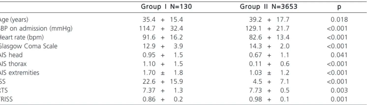
The comparison of numerical variables between the groups revealed that patients with abdominal injuries (group I) were characterized for having significantly (p <0.05) lower mean age (35.4 years ± 15.4 vs. 39.2 ± 17.7), lower mean systolic blood pressure (SBP) on admission (114.7 mmHg ± 32.4 vs. 129.1 ± 21.7), higher mean heart rate on admission (91.6 ± 16.2 bpm vs. 82.6 ± 13.4 bpm), lower mean Glasgow Coma Scale on admission (12.9 ± 3.9 vs. 14.3 ± 2.0), higher mean AIS for segments head (0.95 ± 1.5 vs. 0.67 ± 1.1), thorax (1.10 ± 1.5 vs. 0.11 ± 0.6) and extremities (1.70 ± 1.8 vs. 1.03 ± 1.2) (Table 2). Patients in group I presented significantly (p <0.05) lower mean RTS (7.37 ± 1.3 vs. 7.73 ± 0.5) and TRISS (0.86 ± 0.2 vs. 0.98 ± 0.1), as well as higher average ISS (22.6 ± 15.9 vs. 4.5 ± 7.1) when compared to group II.
We noted that there was a significant difference when comparing the frequency of trauma mechanisms between the groups (p <0.001). The frequency of pedestrian accidents was higher (33.8% vs. 22.2%), and the falls from own height, lower (1.5% vs. 17.6%) in the group of patients with abdominal injuries.
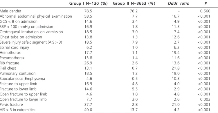
had a significantly higher frequency of abnormal abdominal examination (58.5% vs. 7.7%), GCS <8 at admission (14.6% vs. 6.4%), SBP <100 mmHg at admission (16.9% vs. 1.8%), chest drainage on admission (13.8% vs. 1.3%), severe injury to the cephalic segment (AIS > 3) (18.5% vs. 7.9%), spinal cord injury (6.2% vs. 1.0%), hemothorax (17.7% vs. 1.1%), pneumothorax (13.8 % vs. 1.4%), rib fractures (26.9% vs. 2.6%), flail chest (13.1% vs. 0.7%), pulmonary contusion (18.5% vs. 1 2%), subcutaneous emphysema (4.6% vs. 0.5%), severe injury to the thoracic segment (29.2% vs. 2.4%), fractures of the upper limbs (16.9% vs. 4.8%), fractures of the lower limbs (14.6% vs. 5.5%), open fractures of the upper limbs (4.6% vs. 1.0%), open fractures of the lower limbs (7.7 % vs. 3.0%), pelvic fractures (37.7% vs. 2.8%) and serious injuries (AIS> 3) in the extremities (40.0% vs. 13.7%) (Table 3). The highest odds ratios for diagnosis of abdominal injuries were the presence of flail chest (21.8) and pelvic fracture (21.0) (Table 3).
In group I, 39 laparotomies were performed, with 29 treatments. Sixty patients had nonoperative treatment of lesions in the liver, spleen and/or kidneys, ten
arteriographies with embolization having been required in this group. Mortality was significantly higher in group I (12.3% vs. 1.9%, p <0.001). No complications were identified by the delayed diagnosis of abdominal injuries.
DISCUSSION
DISCUSSION
DISCUSSION
DISCUSSION
DISCUSSION
VNo one knows for sure the percentage of patients with abdominal injuries secondary to blunt trau-ma. In studies with larger samples, such as the one from Mackersie et al. 13, about 3% of victims of blunt trauma
had some abdominal injury. In multiple trauma victims admitted with mild traumatic brain injury, their frequency increases to 10% 14. Studying only patients sustaining “high
energy” trauma, Deunk et al. 7 found approximately 30%
of abdominal injuries. In cases of trauma victims with fractures of the pelvis, the incidence of associated abdomi-nal injuries can achieve 40% 15.
The sample of this study has values of RTS, ISS and TRISS suggesting low-energy trauma. The frequency
Table 2 Table 2 Table 2 Table 2
-Table 2 - Characteristics of the numerical variables between groups I e II. Data presented as average + standard deviation.
Group I N=130 Group I N=130 Group I N=130 Group I N=130
Group I N=130 Group II N=3653Group II N=3653Group II N=3653Group II N=3653Group II N=3653 ppppp
Age (years) 35.4 + 15.4 39.2 + 17.7 0.018
SBP on admission (mmHg) 114.7 + 32.4 129.1 + 21.7 <0.001
Heart rate (bpm) 91.6 + 16.2 82.6 + 13.4 <0.001
Glasgow Coma Scale 12.9 + 3.9 14.3 + 2.0 <0.001
AIS head 0.95 + 1.5 0.67 + 1.1 0.041
AIS thorax 1.10 + 1.5 0.11 + 0.6 <0.001
AIS extremities 1.70 ± 1.8 1.03 ± 1.2 <0.001
ISS 22.6 + 15.9 4.5 + 7.1 <0.001
RTS 7.37 + 1.3 7.73 + 0.5 0.003
TRISS 0.86 + 0.2 0.98 + 0.1 0.001
SBP: Systolic Blood Pressure; mmHg: millimeters of mercury. Bpm: beatings per minute. AIS: Abbreviated Injury Scale. RTS: Revised trauma score. ISS: Injury Severity Score. TRISS: calculation of the survival probability.
Table 1 Table 1 Table 1 Table 1
-Table 1 - Abdominal injuries identified by laparotomy and/or computerized tomography in victims of blunt trauma.
Injured organ Injured organ Injured organ Injured organ
Injured organ N u m b e rN u m b e rN u m b e rN u m b e rN u m b e r Percentage of the patients with abdominal injuryPercentage of the patients with abdominal injuryPercentage of the patients with abdominal injuryPercentage of the patients with abdominal injuryPercentage of the patients with abdominal injury
Spleen 42 32.3%
Liver 42 32.3%
Kidney 18 13.8%
Small Intestine 8 6.1%
Mesentery 8 6.1%
Bladder/urethra 5 3.8%
Colon 3 2.3%
Pancreas 1 0.7%
Great vessels 3 2.3%
Stomach 1 0.7%
Ureter 1 0.7%
of abdominal injuries was 3.4%. Severe lesions were present in 2.2% and bowel injuries in 0.3%. We believe that this low incidence renders the diagnosis of abdominal injuries even more difficult.
It is also known that a normal physical examination does not rule out the possibility of abdominal injury. Both clinical history and physical examination and laboratory tests may show false negative results. In 2010, Michetti et al. 2 found that 10% of victims of blunt trauma
with normal physical examination on admission had abdo-minal injuries confirmed by imaging (computed tomography). In our study, we observed that from patients with normal abdominal examination, only 1.6% had abdo-minal injuries, but 41.5% of patients with some abdoabdo-minal injury had no change in abdominal examination. Therefore, additional tests should be employed to identify these potential injuries.
The FAST and full abdominal ultrasound, methods routinely used for the evaluation of blunt abdomi-nal trauma victims, have the problem false negativity 3,16-24.
Although there are groups that indicate the routine performance of computed tomography in blunt trauma victims, we know that there are limitations of its use. It requires the administration of intravenous and oral contrast material, resulting in anaphylactic reactions at a ratio of 1:1000. There is exposure to a dose of radiation, which can be associated with the onset of long-term neoplasia 25.
The risk of transfer should be considered. Depending on the distance between the emergency room and the CT
scanner, the patient must be hemodynamically normal for its performance. Another limitation is the availability of this test, which is not regular. Tomography of the abdomen may also show false negative results, especially in lesions of the pancreas, retroperitoneal duodenum and jejunum/ ileum 26,27. It is also known that the trauma care consumes
an important part of the health system’s budget 28. This
should also be considered in the systematic indication of computerized tomography in a sample of patients with low-energy trauma, in which the positivity of the test could hardly be more than 5%.
There is need to select patients at higher risk of injury to be submitted to computed tomography. It is with this objective that the idea to study variables that may be significantly associated with presence of abdominal injuri-es arisinjuri-es. Thinjuri-ese predictive factors can alert to a higher risk, allowing prioritization and targeting of diagnostic investigation.
In 1989, Mackersie et al. 13 studied the “indirect”
signs related to the presence of abdominal injuries in patients who suffered blunt trauma. These authors noted that the presence of base excess lower than -5mEq/L of arterial blood gases, arterial hypotension on admission or at the trauma scene, injuries to the chest and pelvic fractures were significantly associated with the presence of abdominal injuries.
In 2010, Deunk et al. 7 proposed a selective
indication for CT based on clinical, radiological, laboratory and ultrasound findings. In a study involving 1,040 victims of high-energy trauma, they identified nine independent
Table 3 Table 3 Table 3 Table 3
Table 3 - Distribution of the nominal variables between groups I e II according to gender. exams’ results and type of injury.
Group I N=130 (%) Group I N=130 (%)Group I N=130 (%)
Group I N=130 (%)Group I N=130 (%) Group II N=3653 (%)Group II N=3653 (%)Group II N=3653 (%)Group II N=3653 (%)Group II N=3653 (%) Odds ratioOdds ratioOdds ratioOdds ratioOdds ratio PPPPP
Male gender 78.5 76.2 - 0.560
Abnormal abdominal physical examination 58.5 7.7 16.7 <0.001
GCS < 8 on admission 14.6 3.4 4.9 <0.001
SBP < 100 mmHg on admission 16.9 1.8 11.3 <0.001
Orotraqueal Intubation on admission 18.5 3.0 7.4 <0.001
Chest tube on admission 13.8 1.3 12.6 <0.001
Severe injury cefaic segment (AIS > 3) 18.5 7.9 2.7 <0.001
Spinal cord injury 6.2 1.0 6.2 <0.001
Hemothorax 17.7 1.1 19.4 <0.001
Pneumothorax 13.8 1.4 11.6 <0.001
Rib fracture 26.9 2.6 13.6 <0.001
Flail chest 13.1 0.7 21.8 <0.001
Pulmonary contusion 18.5 1.2 19.0 <0.001
Subcutaneous Emphysema 4.6 0.5 10.3 <0.001
Fracture to upper limb 16.9 4.8 4.0 <0.001
Fracture to lower limb 14.6 5.5 2.9 <0.001
Open fracture to upper limb 4.6 1.0 4.8 <0.001
Open fracture to lower limb 7.7 3.0 2.6 0.003
Pelvis fracture 37.7 2.8 21.0 <0.001
AIS > 3 in extremities 40.0 13.7 4.2 <0.001
factors significantly associated with the presence of abdo-minal injuries: changes in plain chest, spine or pelvis radiography, positive FAST, positive abdominal examination, changes in the physical examination of the spine, base excess less than -3mEq/L in arterial blood gas, systolic blood pressure less than 90mmHg and the presence of fractures in long bones. Based on these data, CT is indicated in hemodynamically stable patients who concurrently presented: signs of neurological impairment (Glasgow Coma Scale less than 8, anisocoria, skull fracture), abnormal abdominal physical examination, pelvic, lumbar spine or extremities fractures, base excess less than -3mEq/ L on arterial blood gases, abnormalities on chest, pelvis or spine radiography, or positive FAST 7.
There is particular concern with the traumatized group with decreased level of consciousness, especially those with severe traumatic brain injury. Since there is no appropriate neurological level, the abdominal physical examination becomes impaired and serious injuries may go unnoticed, even in computerized tomography. The lesions most feared are those that occur in hollow viscera, because late diagnosis can have serious consequences 29. Precisely
in this group, the diagnosis by computed tomography is more difficult, which is a very dangerous combination.
Other studies have evaluated the presence of predictors of abdominal injuries in victims of blunt trauma. In 2004, Beck et al. 30 found a significant relationship
between abdominal injuries and abnormal radiographs of the pelvis and the need for endotracheal intubation. The data found in our sample clearly show the association of abdominal lesions with a few variables: hemodynamic instability on admission, decreased level of consciousness on admission, increased severity of lesions in segments head, chest and extremities, as well as fractures of the pelvis and of long bones (Table 3). Many of these data are consistent with the studies cited above 7,13,30. Interestingly, the presence
of pelvic fracture is the single factor that appears most often as a predictor of abdominal injuries. In the study of Deunk et al.7, in 2010, the odds ratio for the presence of
abdomi-nal injury in patients with pelvic fractures was 46.8. In our
study, the chance of a trauma patient with a fractured pelvis present an abdominal injury is 21 times higher when compared with patients without this type of fracture.
The association between chest and abdominal injuries is also already known 13. In our sample, the highest
odds ratio for the presence of abdominal lesions was observed in association with flail chest (OR = 21.8), a marker of severe chest trauma. Our data also confirmed the increased chance of injury in abdominal trauma with fractures of long bones and spine. However, we consider important to note that in our study the incidence of abdominal injuries was also higher in the presence of severe lesions of the cephalic segment and in patients with a decrease Glasgow Coma Scale. Clearly, for a proper diagnosis, patient should be viewed as a whole, as the signals from abdominal lesions can also be found in other body parts. Data from this study show a higher frequency of abdominal injuries in victims of pedestrian accidents, whereas for victims of falls from own height the incidence was lower.
Finally, we would draw attention to the sum of factors that complicate the diagnosis in clinical practice: abdominal injuries are more common precisely in situations of higher risk of going unnoticed, such as when there is lower level of consciousness, severe head trauma, need for intubation, need for analgesics (flail chest) or even in the operative treatment of fractures of the extremities, when an anesthetic is necessary.
The purpose of our study was precisely to widely assess which variables could be associated with abdominal injuries. Certainly, the use of these data can provide useful information to identify lesions that initially could go unnoticed, contributing to decreased morbidity and mortality associated with late diagnosis of abdominal injuries in victims of blunt trauma.
Data from this study allow us to conclude that the predictors of abdominal injuries in victims of blunt trau-ma are: mechanism of injury, hemodynamic instability, altered level of consciousness and presence of severe lesions in the skull, chest or extremities, especially flail chest and pelvic fractures.
R E S U M O R E S U M O R E S U M O R E S U M O R E S U M O
Objetivo: Objetivo: Objetivo: Objetivo:
Objetivo: Identificar fatores preditivos de lesões abdominais em vítimas de trauma fechado. Métodos:Métodos:Métodos:Métodos: Análise retrospectiva dosMétodos: dados das vítimas de trauma fechado com idade superior a 13 anos, em um período de 15 meses. Comparamos as variáveis entre os doentes com lesões abdominais diagnosticadas por tomografia computadorizada e/ou laparotomia – grupo I (Abbreviated Injury Scale abdome>0, grupo I) e os demais – grupo II (Abbreviated Injury Scale abdome=0,). Resultados:Resultados:Resultados:Resultados:Resultados: Foram incluídos 3783 casos, com média etária de 39,1 +17,7 anos (14 a 99 anos), sendo 76,1% do sexo masculino. Foram identificadas lesões abdominais em 130 doentes (3,4%). Os traumatizados com lesões abdominais apresentaram, significativamente, menor média etária (35,4 + 15,4 anos vs. 39,2 + 17,7 anos), menor média da pressão arterial sistólica à admissão (114,7 + 32,4mmHg vs. 129,1 + 21,7mmHg), menor média na escala de coma de Glasgow à admissão (12,9 + 3,9 vs. 14,3 + 2,0), maior média de AIS em segmento cefálico (0,95 + 1,5 vs. 0,67 + 1,1), maior média de AIS em segmento torácico (1,10 + 1,5 vs. 0,11 + 0,6) e maior média de AIS em extremidades (1,70 ± 1,8 vs. 1,03 ± 1,2). Os maiores Odds ratio foram presença de tórax flácido (21,8) e fraturas de pelve (21,0). Conclusão:Conclusão:Conclusão:Conclusão:Conclusão: As lesões abdominais foram mais frequentemente observadas nos doentes com instabilidade hemodinâmica, alteração na escala de coma de Glasgow, lesões graves em crânio, tórax ou extremidades.
Descritores: Descritores: Descritores: Descritores:
REFERENCES
REFERENCES
REFERENCES
REFERENCES
REFERENCES
1. Fabian TC, Croce, MA. Abdominal trauma, including indications for celiotomy. In: Mattox KL, Feliciano DV, Moore EE, editores. Trauma. 4a ed. New York: McGraw-Hill; 2000. p.1583-602.
2. Michetti CP, Sakran JV, Grabowski JG, Thompson EV, Bennett K, Fakhry SM. Physical examination is a poor screening test for abdo-minal-pelvic injury in adult blunt trauma patients. J Surg Res. 2010;159(1):456-61.
3. Shuster M, Abu-Laban RB, Boyd J, Gauthier C, Mergler S, Shepherd L, et al. Focused abdominal ultrasound for blunt trauma in an emergency department without advanced imaging or on-site surgical capability. CJEM. 2004;6(6):408-15.
4. Kornezos I, Chatziioannou A, Kokkonouzis I, Nebotakis P, Moschouris H, Yiarmenitis S, et al. Findings and limitations of focused ultrasound as a possible screening test in stable adult patients with blunt abdominal trauma: a Greek study. Eur Radiol. 2010;20(1):234-8.
5. Self ML, Blake AM, Whitley M, Nadalo L, Dunn E. The benefit of routine thoracic, abdominal, and pelvic computed tomography to evaluate trauma patients with closed head injuries. Am J Surg. 2003;186(6):609-13; discussion 613-4.
6. Salim A, Sangthong B, Martin M, Brown C, Plurad D, Demetriades D Whole body imaging in blunt multisystem trauma patients without obvious signs of injury: results of a prospective study. Arch Surg. 2006;141(5):468-73.
7. Deunk J, Brink M, Dekker HM, Kool DR, Blickman JG, van Vugt AB, et al. Predictors for the selection of patients for abdominal CT after blunt trauma: a proposal for a diagnostic algorithm. Ann Surg. 2010;251(3):512-20.
8. Teasdale G, Jennett B. Assessment of coma and impaired consciousness: a practical scale. Lancet. 1974;2(7872):81-84. 9. Champion HR, Sacco WJ, Copes WS, Gann DS, Gennarelli TA,
Flanagan ME. A revision of the Trauma Score. J Trauma. 1989;29(5):623-9.
10. Association for the Advancement of Automotive Medicine, Committee on Injury Scaling. The abbreviated injury scale-1990 Revision (AIS-90). Des Plaines, IL: Association for the Advancement of Automotive Medicine, 1990.
11. Baker SP, O’Neill B, Haddon W Jr, Long WB. The injury severity score: a method for describing patients with multiple injuries and evaluating emergency care. J Trauma. 1974;14(3):187-96. 12. Boyd CR, Tolson MA, Copes WS. Evaluating trauma care: the
TRISS method. Trauma Score and the Injury Severity Score. J Trauma. 1987;27(4):370-8.
13. Mackersie RC, Tiwary AD, Shackford SR, Hoyt DB. Intra-abdomi-nal injury following blunt trauma. Identifying the high-risk patient using objective risk factors. Arch Surg. 1989;124(7):809-13. 14. Wu SR, Shakibai S, McGahan JP, Richards JR. Combined head and
abdominal computed tomography for blunt trauma: which patients with minor head trauma benefit most? Emerg Radiol. 2006;13(2):61-7.
15. Parreira JG, Haddad L, Rasslan S. Lesões abdominais nos traumatizados com fraturas de bacia. Rev Col Bras Cir. 2002;29(3):153-60.
16. Miller MT, Pasquale MD, Bromberg WJ, Wasser TE, Cox J. Not so FAST. J Trauma. 2003;54(1):52-9; discussion 59-60.
17. Sirlin CB, Brown MA, Deutsch R, Andrade-Barreto OA, Fortlage DA, Hoyt DB, et al. Screening US for blunt abdominal trauma: objective predictors of false-negative findings and missed injuries. Radiology. 2003;229(3):766-74.
18. Hoffman L, Pierce D, Puumala S. Clinical predictors of injuries not identified by focused abdominal sonogram for trauma (FAST) examinations. J Emerg Med. 2009;36(3):271-9.
19. Ballard RB, Rozycki GS, Newman PG, Cubillos JE, Salomone JP, Ingram WL, et al. An algorithm to reduce the incidence of false-negative FAST examinations in patients at high risk for occult injury. Focused Assessment for the Sonographic Examination of the Trauma patient. J Am Coll Surg. 1999;189(2):145-50; discussion 150-1.
20. Poletti PA, Kinkel K, Vermeulen B, Irmay F, Unger PF, Terrier F. Blunt abdominal trauma: should US be used to detect both free fluid and organ injuries? Radiology. 2003;227(1):95-103. 21. Yoshii H, Sato M, Yamamoto S, Motegi M, Okusawa S, Kitano M,
et al. Usefulness and limitations of ultrasonography in the initial evaluation of blunt abdominal trauma. J Trauma. 1998;45(1):45-50; discussion 50-1.
22. Kendall JL, Faragher J, Hewitt GJ, Burcham G, Haukoos JS. Emergency Department Ultrasound is not a sensitive detector of solid organ injury. West J Emerg Med. 2009;10(1):1-5.
23. Tillou A, Gupta M, Baraff LJ, Schriger DL, Hoffman JR, Hiatt JR, et al. Is the use of pan-computed tomography for blunt trauma justified? A prospective evaluation. J Trauma. 2009;67(4):779-87. 24. Deunk J, Brink M, Dekker HM, Kool DR, van Kuijk C, Blickman JG, et al. Routine versus selective computed tomography of the abdomen, pelvis, and lumbar spine in blunt trauma: a prospective evaluation. J Trauma. 2009;66(4):1108-17.
25. Johnson DA, Helft PR, Rex DK. CT and radiation-related cancer risk-time for a paradigm shift? Nat Rev Gastroenterol Hepatol. 2009;6(12):738-40.
26. Atri M, Hanson JM, Grinblat L, Brofman N, Chughtai T, Tomlinson G. Surgically important bowel and/or mesenteric injury in blunt trauma: accuracy of multidetector CT for evaluation. Radiology. 2008;249(2):524-33.
27. Phelan HA, Velmahos GC, Jurkovich GJ, Friese RS, Minei JP, Menaker JA, et al. An evaluation of multidetector computed tomography in detecting pancreatic injury: results of a multicenter AAST study. J Trauma. 2009;66(3):641-6; discussion 646-7. 28. Lanzarotti S, Cook CS, Porter JM, Judkins DG, Williams MD. The
cost of trauma. Am Surg. 2003;69(9):766-70.
29. Sung CK, Kim KH. Missed Injuries in abdominal trauma. J Trauma. 1996;41(2):276-82.
30. Beck D, Marley R, Salvator A, Muakkassa F. Prospective study of the clinical predictors of a positive abdominal computed tomography in blunt trauma patients. J Trauma. 2004;57(2):296-300.
Received on 18/11/2011
Accepted for publication 19/01/2012 Conflict of interest: none
Source of funding: none
How to cite this article: How to cite this article:How to cite this article: How to cite this article:How to cite this article:
Farrath S, Parreira JG, Perlingeiro JAG, Solda SC, Assef JC. Predictors of abdominal injuries in blunt trauma. Rev Col Bras Cir. [periódico na Internet] 2012; 39(4). Disponível em URL: http://www.scielo.br/rcbc
Address correspondence to: Address correspondence to:Address correspondence to: Address correspondence to:Address correspondence to: Jose Gustavo Parreira