Effects of local pressure on cutaneous blood flow in pigs
Efeitos da pressão local no fluxo sanguíneo cutâneo de porcos
MiChel lUCiano holger toledano vaena, tCBC-rJ1; João paUlo sinneCKer2; BrUno Benedetti pinto1; Mario FritsCh toros neves1; Fernando serra-gUiMarães1; rUy garCia MarqUes, tCBC-rJ1.
INTRODUCTION
P
ressure ulcers are usually defined as localized le-sions on the skin, and may or may not include the underlying tissue. They usually occur on a prominent bone or arise related to a medical device or others. The lesion is the result of intense and/or prolonged pressure, or of shear pressure. Certain factors such as advanced age, the presence of multiple comorbidities, among others, may increase the risk of developing pressure ulcers during hospitalization1. The prevalenceof pressure ulcers (grades 1-4) in hospitalized patients in Europe is 18.1%2, and 13.5% in the United States3,
and their incidence when related to medical devices is 34.5%4. Considering that the prevalence in
deve-loping countries is equal that or greater, the overall economic impact is enormous.
Pressure relief is the most important aspect in the prevention of pressure ulcers, either by mobi-lization of the patient or by the use of specific
mat-tresses that increase the contact area, reducing the interface pressure5. The effects of increasing pressures
exerted on the surface of the skin in the microcircula-tory blood flow have been studied before, but without a more detailed quantitative analysis6. Understanding
these effects may help in the prevention of pressure ulcers by establishing more adequate parameters for the safety of support surfaces and medical devices that come in contact with the skin.
The aim of our study is to evaluate the ef-fects of increasing pressures on the microcirculatory blood flow of the skin of pigs.
METHODS
We used a system consisting of a subcuta-neous magnetic implant and external magnets of di-fferent intensities to generate an in vivo increasing compression of the skin. We introduced thirty
magne-1 - University of the State of Rio de Janeiro, Rio de Janeiro, RJ, Brazil. 2 - Brazilian Center for Physical Research, Department of Experimental Physics of Low Energies, Rio de Janeiro, RJ, Brazil.
A B S T R A C T
Objective: to evaluate the effects of increasing pressures on the cutaneous blood flow in the skin of pigs. Methods: we conducted an experimental study in pigs submitted to subcutaneous magnetic implants (n=30). After healing, were applied external magnets with vary-ing magnetic forces to the skin, generatvary-ing compression. We evaluated the cutaneous circulation of the skin under compression by the Laser Speckle Contrast Imaging (LSCI) technique. We measured the depth of the implants by ultrasonography, and applied computational simulations to the calculation of the different pressure values, considering the different distances between implants and external magnets.
Results: nineteen implants presented complications. The remaining 11 were submitted to different magnetic compression forces and perfusion analysis. Two linear regression models showed an inverse correlation between exerted pressure and cutaneous perfusion, with significant variation, mainly in the initial pressure increases, of up to 20mmHg. Conclusion: The main reduction in cutaneous blood flow resulted from initial increases of up to 20 mmHg. The results suggest that tissue ischemia can occur even in low-pressure regimes, which could contribute to the appearance of skin lesions, particularly ulcers related to medical devices.
tic implants (n=30) into two male pigs (Sus domesti-cus) weighing 17.2kg (pig #1) and 19kg (pig #2), with prior approval of the institution’s ethics committee for animal testing. After complete healing of the wound, we applied external magnets of four different intensi-ties to the skin, generating compression. We evalua-ted the cutaneous circulation of compressed skin with the Laser Speckle contrast Imaging (LSCI) technique, with the PeriCam PSI device.
Each magnetic implant consisted of a sili-cone capsule containing two N40 grade neodymium magnets (Nd2Fe14B). The capsule was made of silicone elastomer (medical grade) with shore 30A hardness, manufactured in an ellipsoid shape (50mm long and 22mm wide), with a flattened profile (4mm height), without edges or tips, to avoid tissue trauma. The two internal neodymium magnets were identical, disk-sha-ped, 6mm in diameter and 1.5mm thick, with field-s-trength intensities of 48 mT and axially magnetized, with nominal magnetic remanence of 1.25 T (Figure 1A).
We sedated the animals with intramuscular injection (acepromazine acetate 0.2mg/kg) and trans-ported them to the surgical center. After venous ca-theterization, we administered additional anesthetics (propofol and thiopental sodium) in dose-effect mode and performed tracheal intubation. Each pig was kept under general anesthesia during the introduction of the implants. The dorsal skin was trichotomized and prepared with antiseptic solution (chlorhexidine 2%). We marked the cutaneous incision sites with a der-mographic pen. We made all 2cm incisions over the dorsal skin, and introduced a straight cannula through each incision (Figure 1B). The cannula promoted blunt dissection and detachment of the sub-dermal plane, making a narrow tunnel parallel to the skin surface. We introduced each implant through the cutaneous incision (Figure 1C) and placed it in its final position under the dermis, and sutured the incisions. We used topical antibiotic spray on the sutured wounds. At the end of the procedure, each pig received a total of 15 implants under the dorsal skin, totaling 30 implants (Figure 1D). After the post-anesthetic recovery, the animals were released to feed and move freely during the wounds’ healing period.
Forty days after the procedure, were sedated the pigs again, transported them to the surgical center and anesthetized them. We kept the ambient tempera-ture constant to avoid changes in the cutaneous circula-tion. We evaluated the perfusion on the pigs’ dorsal skin over the implants using the LSCI technique. Skin sites that had apparent clinical changes (such as erythema, continuity solutions or fluctuation) were excluded from the analysis. The LSCI technique uses the “mottling” phenomenon to obtain a perfusion map of the tissues, capturing the changes in the “mottled” pattern, which correspond to the reflexes of erythrocytes in movement, with a camera located inside the projector of the Pe-ricam PSI device when illuminated by the Laser beam (Figure 2). We analyzed these changes mathematically and generated a tissue perfusion map.
Figure 1. A- Implant; B- Dissection; C- Insertion; D- final aspect.
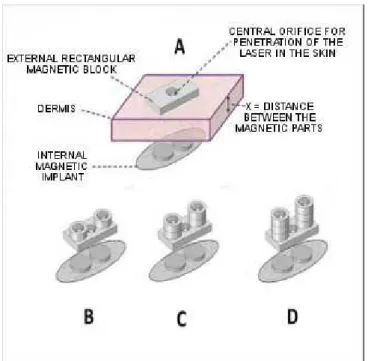
After this initial evaluation, we placed mag-nets of different forces on the dorsal skin on the same sites, above the implants. These external magnets con-sisted of a synthesized N48 grade rectangular magnetic neodymium block (25mm x 12mm x 3mm) with a 4mm center hole to allow penetration of the laser beam into the skin (Figure 3A). On this rectangular magnet, we added up to three pairs of cylindrical magnets (9mm X 3mm) of grade N42 neodymium. For each added pair, the total magnetization of the system increased, there-by increasing the magnetic attraction force between the implant under the skin and the external magnetic block, thus increasing the mechanical pressure on the skin sur-face (Figures 3B, 3C and 3D).
Figure 3. A- External block + implant; B, C, D- Magnets added.
After the procedure, we removed the outer magnets and subjected the dorsal skin to ultrasonogra-phic examination. Again, any implant sites containing subclinical collections not previously identified, such as seromas or abscesses, were excluded from the study. The depth of the implants relative to the skin surface was measured at 0.1mm intervals using a linear high fre-quency (12Mhz) transducer. This precise measurement of the implants’ depth allowed to establish the exact dis-tance (shown as the disdis-tance “x” in Figure 3A) between the magnetic components and, therefore, to determine
the pulling force exerted by the system. Thus, given the surface area (already known) of the outer magnetic blo-ck and defining the intensity of the magnetic force exer-ted by the system, it was possible to calculate the exact pressure the skin surface was submitted to. To calculate the pressure values exerted by the outer block on the skin surface, we performed computational simulations using the COMSOL Multiphysics® simulations software under license n. 2072699.
We tabulated the data obtained from the PeriCam cutaneous perfusion readings and compared them with the estimated blood pressure levels. We per-formed statistical analysis with the R software (The R Foundation for Statistical Computing, Vienna, Austria). After applying the Shapiro-Wilk test to the variables, we computed the Pearson correlation coefficient. We calcu-lated the linear regression models and the applied F test (p<0.05) for statistical significance.
RESULTS
DISCUSSION
Recent studies have questioned the safety of the standard limit of 32mmHg pressure used as a parameter for interface pressure on support surfaces7.
In hospitalized patients, a source of pressure on the skin surface may be the medical devices themselves employed to patient monitoring or treatment4. Ulcers
related to medical devices can be caused by nasal can-nulas, endotracheal tube attachments, pulse oximetry sensors, anti-embolism stockings, orthopedic splints etc. Whereas all of these devices are specially designed not to damage skin integrity, the high prevalence of ulcers related to medical devices suggests that current safety parameters should be questioned.
Over time, different animal models have been proposed in the literature to study the local circulatory effects of mechanical pressure exerted on the skin. Due to practical limitations, most studies are performed on rats. Although mouse skin circulation is provided by direct cutaneous arteries of the panniculus carnosus, which is absent in humans, several publications of di-fferent authors have employed and validated animal models of rats8. However, considering the anatomical
and histological similarities with the human skin, the pig is considered the best animal model for cutaneous healing9.
Pioneering studies on pressure ulcers deve-loped by Groth10, Kosiak11 and Dinsdale12, using
rab-bits, dogs and pigs, respectively, evaluated the effects of applying pressure to the skin of live animals,
basi-cally by macroscopibasi-cally inspecting the skin changes and subsequent histopathological analysis. This type of study was also employed by Daniel13, who in turn
demonstrated the greater vulnerability of deep tissues to pressure ischemia, while the dermis could withstand longer ischemic intervals without necrosis. These stu-dies, if taken as a whole, have not yet contemplated real-time dynamic changes in the dermal circulation due to increased pressure on the skin. Of course, the collection of information in live animals adds technical difficulties and ethical implications that do not occur in postmortem specimens14,15. Branemark16, however,
drew attention to this issue by using a vital microscopy camera study, demonstrating the influence of higher pressures on the skin and the ischemic changes in the microcirculation.
In the last two decades, in vivo cutaneous microcirculation research has been based more on non-invasive methods, such as optical microscopy and laser-Doppler techniques. Techniques derived from op-tical microscopy basically depend on transillumination, which tends to restrict anatomical areas that can be studied (such as video-capillaroscopy of the nail bed in humans), or may require vital microscopy camera te-chniques17. The limitation of such techniques is that
they basically provide morphological information about the microcirculation. Laser-Doppler techniques, on the other hand, provide quantitative information related to the skin blood flow. This latter method was validated by Salcido et al.18 in the research on the development
of pressure ulcers, but there are many technical issues
Figura 4. A) Gráfico de dispersão; B) Regressões lineares.
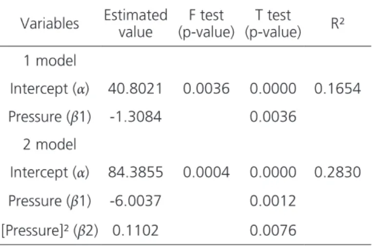
Table 1. Properties of the linear regression models applied to the scatter plot.
Variables Estimated value (p-value)F test (p-value)T test R²
1 model
Intercept (α) 40.8021 0.0036 0.0000 0.1654
Pressure (β1) -1.3084 0.0036
2 model
Intercept (α) 84.3855 0.0004 0.0000 0.2830
Pressure (β1) -6.0037 0.0012
that must be taken into account to guarantee the fide-lity and reproducibifide-lity of Laser-Doppler techniques19.
The LSCI technique has been used more re-cently and provides real-time, non-invasive skin cir-culation monitoring. It has been shown that the flow measurements between Laser Doppler and LSCI dis-play a linear relationship20, but one of the main
ad-vantages of LSCI is its high reproducibility21. In
addi-tion, the LSCI measurement depth is more superficial than the Laser Doppler techniques22. In our study, the
central orifice in the rectangular external magnetic blo-ck allowed the laser beam to penetrate the Region of Interest (ROI) while the pressure on the skin was gradu-ally increased with the additional magnets. The LSCI te-chnique allowed non-contact, non-invasive monitoring of skin blood flow. The magnetic system avoided the influence of external factors that could affect perfusion readings by the LSCI technique, such as variations in the animal position, variations in lighting conditions or changes in the ROI. The magnetic system also en-sured that the ROI remained within the tissue under compression. In addition, the long interval between the introduction of the implants and the evaluation of the dermal circulation allowed the complete healing of the surgical wounds, thus avoiding the influence of in-flammatory changes and the phenomenon of autono-mization on the evaluated skin segments.
Few studies have also used magnetic force to induce mechanical compression in animal models. Peir-ce23 developed an animal model of
ischemia-reperfu-sion injury by implanting a steel plate under the dorsal skin of rats. An external magnet was applied to the skin 24 hours after the implantation procedure, generating compression and local ischemia. Peirce’s experimental model advocates the initiation of compression cycles 24 hours after the surgical procedure. This model does not take into account the inflammatory changes inhe-rent in the surgical procedure, nor the local circulatory changes in the skin flap due to subcutaneous tissue dis-section. Despite these conceptual limitations, the Pier-ce study demonstrated that ischemia-reperfusion cycles were more damaging to the skin when compared with ischemia alone. These results were also demonstrated using non-magnetic compression models17.
Nguyen-Tu24 used Peirce’s animal model to
study the microvascular response of skin to pressure in obese rats. The results of that study suggest that obe-sity could play a protective role by reducing skin lesions induced by compression through changes in skin struc-ture. A clear limitation of the study, which the authors recognize, is related to the possible changes in the pressures applied to the skin due to the different skin thicknesses between groups. It is known that the mag-netic attraction force is inversely proportional to the distance applied. In fact, because of the non-linearity of the magnetic force, small variations in the implant’s depth under the skin can cause large variations in the pressure exerted on the surface of the skin. Although it should be noted that in the Nguyen-Tu study the same magnets were used in both groups of rats (obese and non-obese), different skin thicknesses may have gene-rated different pressures in each group. In our study, such thickness variations were not neglected, and com-putational simulations (using COMSOL Multiphysics® modeling software and ultrasound measurements) allowed the calculation of different pressure values, taking into account the different distances due to the different skin thicknesses.
Perfusion Units (PUs) are arbitrary units used by the LSCI technique and should be interpreted as re-al-time flow measurements, without absolute values. They serve to be compared to each other in real-time dynamic analysis. The results in our study suggest that dermal blood flow is extremely sensitive to the pres-sure exerted on the surface of the skin. Basically, the Model 2 curve shows a drop in blood flow to about half at pressures up to 10mmHg. Between 10 and 20 mmHg, the flow falls to one-fourth of normal physio-logical levels and continues to drop at 25mmHg. These results are in accordance with what would be expected within the knowledge of the mean value of cutaneous capillary pressure since the pioneering study of Landis25
and also of more recent publications26. However,
rea-dings of the LSCI technique also demonstrated blood flow measurements under higher pressure regimes. In fact, Shibata et al.6, using capillaroscopy based on a
not allow the quantification of changes in the flow due to increases in pressure. Based on the results of our study, it is likely that under pressures greater than 25-30mmHg, blood continues to circulate, but well below physiological levels in terms of flow.
These results corroborate the questioning about the safety of the standard pressure limit of 32mmHg used in the supportive surface definitions26.
In addition, they may help explain the onset of certain ulcers, particularly pressure ulcers related to medical devices. In clinical practice, mechanical pressure exer-ted on the skin surface generates locally increased in-terstitial pressures that may exceed capillary pressure. According to the anatomical topography of the com-pressed region, these pressures are transmitted
hete-rogeneously, resulting in partial blockage or complete collapse of the capillaries, generating tissue ischemia. When certain pathological conditions outweigh com-pensatory mechanisms (self-regulation of capillary cir-culation), pressure ulcers can arise even in the context of slight pressures exerted on the skin.
Our results suggest that the main reduction in cutaneous blood flow originates from the initial in-creases of pressure up to 20mmHg, reinforcing the im-portance of surveillance and early relief of the pressure exerted in the prevention of pressure ulcers. Even under mild pressure conditions, health professionals should be aware of pressure ulcers related to medical devices, in particular due to the specificities of the contact in-terface and to the frequent presence of comorbidities.
REFERENCES
1. Gardiner JC, Reed PL, Bonner JD, Haggerty DK, Hale DG. Incidence of hospital-acquired pressure ulcers - a population-based cohort study. Int Wound J. 2016;13(5):809-20.
2. Vanderwee K, Clark M, Dealey C, Gunningberg L, Defloor T. Pressure ulcer prevalence in Europe: a pilot study. J Eval Clin Pract. 2007;13(2):227-35.
3. VanGilder C, Amlung S, Harrison P, Meyer S. Results of the 2008-2009 International Pressure Ulcer Prevalence Survey and a 3-year, acute care, unit-specific analysis. Ostomy Wound Manage. 2009;55(11):39-45. 4. Black JM, Cuddigan JE, Walko MA, Didier LA, Lander
MJ, Kelpe MR. Medical device related pressure ulcers in hospitalized patients. Int Wound J. 2010;7(5):358-65.
5. Moysidis T, Niebel W, Bartsch K, Maier I, Lehmann N, Nonnemacher M, et al. Prevention of pressure ulcer: interaction of body characteristics and different mattresses. Int Wound J. 2011;8(6):578-84.
6. Shibata M, Yamakoshi T, Yamakoshi K, Komeda T. Observation of capillary flow in human skin during tissue compression using CCD video-microscopy. Conf Proc IEEE Eng Med Biol Soc. 2010;2010:5161-4.
7. Gefen A. The biomechanics of sitting-acquired pressure ulcers in patients with spinal cord injury or lesions. Int Wound J. 2007;4(3):222-31.
8. Salcido R, Popescu A, Ahn C. Animal models in pressure ulcer research. The J Spinal Cord Med. 2007;30(2):107-16.
9. Sullivan TP, Eaglstein WH, Davis SC, Mertz P. The pig as a model for human wound healing. Wound repair
Objetivo: avaliar os efeitos de pressões crescentes exercidas sobre a pele de porcos no fluxo sanguíneo cutâneo. Métodos: estudo experimental em porcos submetidos a implantes magnéticos subcutâneos (n=30). Após a cicatrização, foram aplicados sobre a pele, ímãs externos com forças magnéticas variadas, gerando compressão. A circulação cutânea da pele submetida à compressão foi avaliada pela técnica Laser Speckle Contrast Imaging (LSCI). A profundidade dos implantes foi medida por ultrassonografia, e simulações compu-tacionais foram aplicadas para o cálculo dos diferentes valores de pressão, considerando-se as variadas distâncias entre implantes e ímãs externos. Resultados: dezenove implantes apresentaram complicações. Os 11 restantes foram submetidos à diferentes compressões magnéticas e análise de perfusão. Dois modelos de regressão linear mostraram uma correlação inversa entre pressão exercida e perfusão cutânea com variação significativa principalmente nos acréscimos iniciais de pressão até 20mmHg. Conclusão: a principal redução do fluxo sanguíneo cutâneo resulta dos acréscimos iniciais de pressão de até 20mmHg. Os resultados sugerem que a isquemia tecidual pode ocorrer mesmo em regimes de baixa pressão, o que poderia contribuir para surgimento de lesões de pele, particularmente as úlceras relacionadas a dispositivos médicos.
Descritores: Lesão por Pressão. Pele. Microcirculação. Fluxo Sanguíneo Regional. Modelos Animais. Suínos.
Regen. 2001;9(2):66-76.
10. Groth KE. Klinische Beobachtungen und experimentelle Studien über die Entstehung des Dekubitus. Acta Chir Scand. 1942; Suppl 76:209. 11. Kosiak M. Etiology of decubitus ulcers. Arch Phys
Med Rehabil. 1961;42:19-29.
12. Dinsdale SM. Decubitus ulcers: role of pressure and friction in causation. Arch Phys Med Rehabil. 1974;55(4):147-52.
13. Daniel RK, Priest DL, Wheatley DC. Etiologic factors in pressure sores: an experimental model. Arch Phys Med Rehabil. 1981;62(10):492-8.
14. Marques RG, Morales MM, Petroianu A. Brazilian law for scientific use of animals. Acta Cir Bras. 2009;24(1):69-74.
15. Schanaider A, Silva PC. Uso de animais em cirurgia experimental. Acta Cir Bras. 2004;19(4):441-7. 16. Branemark PI. Microvascular function at reduced flow
rates. In: Kenedi RM, Coeden JM, editors. Bed sore biomechanics. London (UK): Macmillan Education; 1976. p. 63-8.
17. Tsuji S, Ichioka S, Sekiya N, Nakatsuka T. Analysis of ischemia-reperfusion injury in a microcirculatory model of pressure ulcers. Wound Repair Regen. 2005;13(2):209-15.
18. Salcido R, Fisher SB, Donofrio JC, Bieschke M, Knapp C, Liang R, et al. An animal model and computer-controlled surface pressure delivery system for the production of pressure ulcers. J Rehabil Res Dev. 1995;32(2):149-61.
19. Roustit M, Cracowski JL. Non-invasive assessment of skin microvascular function in humans: an insight into methods. Microcirculation. 2012;19(1):47-64. 20. Millet C, Roustit M, Blaise S, Cracowski JL.
Comparison between laser speckle contrast imaging and laser Doppler imaging to assess skin blood flow
in humans. Microvasc Res. 2011;82(2):147-51. 21. Mahé G, Humeau-Heurtier A, Durand S, Leftheriotis
G, Abraham P. Assessment of skin microvascular function and dysfunction with laser speckle contrast imaging. Cir Cardiovasc Imaging. 2012;5(1):155-63. 22. O’Doherty J, McNamara P, Clancy NT, Enfield JG,
Leahy MJ. Comparison of instruments for investigation of microcirculatory blood flow and red blood cell concentration. J Biomed Opt. 2009;14(3):034025-. 23. Peirce SM, Skalak TC, Rodeheaver GT.
Ischemia-reperfusion injury in chronic pressure ulcer formation: a skin model in the rat. Wound Repair Regen. 2000;8(1):68-76.
24. Nguyen-Tu MS, Begey AL, Decorps J, Boizot J, Sommer P, Fromy B, et al. Skin microvascular response to pressure load in obese mice. Microvasc Res. 2013;90:138-43.
25. Landis EM. Micro-injection studies of capillary blood pressure in human skin. Heart. 1930;15(15):209-28. 26. de Graaff JC, Ubbink DT, Lagarde SM, Jacobs MJ. The
feasibility and reliability of capillary blood pressure measurements in the fingernail fold. Microvasc Res. 2002;63(3):270-8.
Received in: 28/04/2017
Accepted for publication: 01/06/2017 Conflict of interest: none.
Source of funding: FAPERJ – Carlos Chagas Filho Founda-tion for Research Support of the State of Rio de Janeiro.
Mailing address:
Michel Luciano Holger Toledano Vaena