Influence of the colon in liver regeneration of rats submitted to
hepatectomy and colectomy
Influência do cólon na regeneração do fígado de ratos submetidos à hepatectomia
e colectomia
Marília Carvalho Moreira1; ítalo Medeiros azevedo1; CláUdia nUnes oliveira1; aldo da CUnha Medeiros, eCBC-rn1.
INTRODUCTION
T
he liver is one of the most complex organs in the human body. Its mass is measured in proportion to the individuals’ body weight1, and this ratio is restoredafter hepatic resection2. Half of all patients with
colo-rectal cancer develop hepatic metastases in the course of this disease3. Patients with metastases may benefit
from hepatic resection, as it provides an opportunity for healing4, with isolated segmentectomy and lobectomy
being the most common surgical interventions. The re-sults have been relatively good if the resection safety margins and the liver functional reserve are adequate5.
Long-term survival after liver resection for colorectal me-tastases has improved significantly in recent years6.
The-se facts justify the study of hepatic regeneration in the presence of simultaneous colectomy, due to the high incidence of colorectal disease with metastases and to the frequency with which these procedures are perfor-med at the same operative time.
Liver regeneration has been the subject of
studies over the years. However, the mechanisms by which the organ is stimulated to replicate and the re-lationship between cells and cytokines have not yet been fully elucidated. Nutritional and other factors have been evaluated, all demonstrating some influen-ce on the regeneration proinfluen-cess7-9. New knowledge has
emerged on liver regeneration, emphasizing the per-formance of growth factors and other cytokines10,11.
In animal models, hepatic regeneration mechanisms have been investigated in detail. Hepatocytes early ex-press tumor necrosis factor-α (TNF-α) and interleukin-6 (IL-6), mainly produced by Kupffer cells, and the pro-liferation and growth of hepatocytes are induced pri-marily in response to the transforming growth factor-α (TGF-α) and hepatocyte growth factor (HGF), among others10.
One of the first studies to investigate the co-lon role in hepatic regeneration examined the effect of ileocolectomy associated with 50% hepatectomy on the regenerative response, evaluating thymidine kina-se activity and mitotic figures as regeneration markers.
1 - Federal University of Rio Grande do Norte, Post-graduation Program in Health Sciences, Natal, RN, Brazil.
A B S T R A C T
Objective: to evaluate whether colectomy, associated with 70% hepatectomy, influences liver regeneration in rats. Methods: we dis-tributed 18 Wistar rats in three groups of six animals each. In group I (sham), we performed laparotomy; In group II, colectomy + 70% hepatectomy; In group III, only 70% hepatectomy. On the 6th postoperative day, we collected blood by cardiac puncture under anesthesia, followed by euthanasia. We performed serum dosages of aspartate aminotransferase (AST), alanine aminotransferase (ALT), albumin and alkaline phosphatase (AF), hepatocyte growth factor (HGF) and transforming growth factor-α (TGF-α). We calculated liver regeneration by the formula: liver weight ratio per 100g body weight at the time of euthanasia / liver weight preoperatively projected for 100g body weight × 100. Results: ALT and AST levels were significantly lower in group II when compared with group III (p<0.001). Albuminemia
showed significantly higher levels in group II. Levels of HGF and TGF- in group II were significantly higher than in group III. The percentage
of hepatic regeneration was significantly higher in group II than in group III. Conclusion: Colectomy performed simultaneously with 70% hepatectomy had a positive influence on liver regeneration in rats. Further research is needed to reveal the molecular mechanisms of this effect and to characterize the colon influence in liver physiology.
When compared, this surgical procedure generated a significantly greater regenerative response than sole hepatectomy or hepatectomy with ileum resection12.
Moser et al.13 studied the participation of genetic
fac-tors in hepatic regeneration after colectomy in 2006. However, a study by Hachiya et al.14 in 2008
conclu-ded that the process of liver regeneration after syn-chronous resection of the liver and colon in rats was reduced.
Faced with the controversy, we seek to con-tribute to the theme. The objective of the present stu-dy was to examine the influence of extensive colon resection on liver function and regeneration in an ex-perimental rat model.
METHODS
The Institutional Committee on Ethics in the Use of Animals approved the research project under protocol number 054-10. The animal care followed the standards of the Brazilian legislation for the scientific use of animals (Law 11.794/2008, CONCEA).
We used 18 male, adult Wistar rats (Rattus norvegicus), weighing 294 ± 13g, supplied by the He-alth Center of the Federal University of Rio Grande do Norte (UFRN). The animals were housed in individual polypropylene cages with 12-hour light-dark cycles, controlled humidity and temperature, with ad libitum access to water and chow for rodents. For seven days prior to the experiment, they remained in the labora-tory for acclimatization. One day before the surgical interventions, they took only water, and were then randomly divided into three groups with six rats each: in group I (sham), we performed laparotomy; in group II, colectomy + 70% hepatectomy; in group III, only 70% hepatectomy. All animals were anesthetized with intraperitoneal injection of ketamine (70mg/kg) and xylazine (10mg/kg), and operated with aseptic tech-nique after abdominal wall trichotomy and antisepsis with 70% ethyl alcohol.
The animals of group II, 70% hepatectomy + colectomy, underwent median laparotomy, through which we resected the whole cecum and 5cm of the proximal colon, proceeding with an end-to-end,
sin-gle-plane ileocolic anastomosis, with simple, separated stitches of 6 0 polypropylene, with the aid of a DFV sur-gical microscope (São Paulo, Brazil), 10x magnification. Concomitantly, we resected the left and middle lobes of the liver (70% hepatectomy). In group III, hepatec-tomy, the animals were submitted to resection of the left and middle lobes of the liver (70% hepatectomy). In the sham group, we carried out a median laparo-tomy and mild manipulation of the cecum and liver un-der the same conditions of anesthesia and antisepsis. In all animals, after checking hemostasis, we sutured the abdominal incision in two planes with 4-0 nylon sutures. After the intervention, postoperative pain con-trol was done with intramuscular meperidine at a dose of 10mg/kg once daily for the first three days. We kept the animals under observation for six days, during whi-ch we observed weight loss parameters through digital weighing, with sensitivity to variation of one gram.
The animals received only water in the first 24 postoperative hours, followed by a solid diet until euthanasia and, in the observation period, were kept in a postoperative control room. On the sixth postope-rative day, we weighed and anesthetized the animals with the same technique described above, and collec-ted 5ml blood samples by cardiac puncture for labo-ratory tests. We then submitted them to euthanasia with an anesthetic overdose (100mg/Kg intraperitone-al thiopentintraperitone-al sodium). We resected the remaining liver (right lobe), washed it with 0.9% saline solution and weighed it on a precision scale. In the sham group, we weighed the whole liver.
Serum dosages
Calculation of hepatic regeneration
Initially, we calculated the hepatic mass / body mass ratio of sham animals (HMBMR). After the observation period, we weighed the rats (B) in a preci-sion scale, removed the entire liver and weighed it too (A). We expressed the acquired data as a percentage of the ratio of A to B, multiplied by 100, calculated by the formula: HMBMR= (Liver mass / Body mass) x 100.
This ratio established the percentage that the liver represents over the body mass of each animal. We evaluated the changes in the HMBMR of the animals of the studied groups as degree of hepatic regeneration. Hepatic Regeneration (HR) was defined as: HR=([HMB-MReuta - HMBMRpos] / HMBMRpos) x 100. Where: HR is the percentage of hepatic regeneration; HMBMReu-ta is the Hepatic Mass-Body Mass Ratio in euthanasia (after the observation period); HMBMRpos is the Hepa-tic Mass-Body Mass Ratio in the immediate postopera-tive period (shortly after hepatectomy).
Statistical analysis
We used the ANOVA test followed by the Tukey test to compare the laboratory parameters
be-tween groups. To evaluate the difference bebe-tween the means of liver regeneration between groups, we applied the Student’s t-test. For all tests, we set the significance level at 5%, using the statistical package SPSS®21.
RESULTS
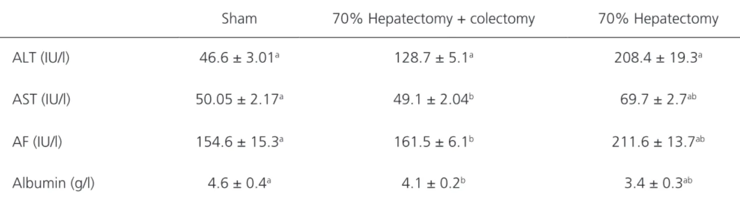
All animals survived the experiments and there was no significant difference in the evolution of their body weights between groups. On the 6th posto-perative day, biochemical measurements showed sig-nificantly higher levels of ALT in animals submitted to 70% hepatectomy + colectomy when compared with the sham group (p<0.01). However, ALT, AST and AF levels in the group of animals submitted solely to patectomy were significantly higher than in the he-patectomy + colectomy group (p<0.01). Albuminemia was significantly higher in the rats of the sham and hepatectomy + colectomy groups than in the hepa-tectomy group (p<0.01). There was no significant di-fference of albuminlevels between the sham and the hepatectomy + colectomy groups (p>0.05). Table 1 summarized the values of the biochemical data.
Table 1. Values of biochemical data and their statistical interpretation.
Sham 70% Hepatectomy + colectomy 70% Hepatectomy
ALT (IU/l) 46.6 ± 3.01a 128.7 ± 5.1a 208.4 ± 19.3a
AST (IU/l) 50.05 ± 2.17a 49.1 ± 2.04b 69.7 ± 2.7ab
AF (IU/l) 154.6 ± 15.3a 161.5 ± 6.1b 211.6 ± 13.7ab
Albumin (g/l) 4.6 ± 0.4a 4.1 ± 0.2b 3.4 ± 0.3ab
Tukey test: mean ± SD values followed by the same letter are statistically significant, with p < 0.01. AST, Aspartate aminotransferase; ALT, Alanine aminotransferase; AF, Alkaline Phosphatase.
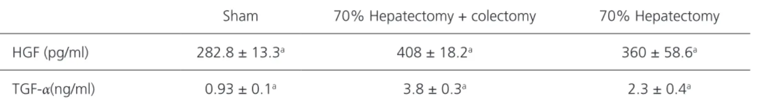
Table 2 shows that the values of the HGF and TGF α of the animals submitted to hepatectomy + co-lectomy were significantly higher than in the sham and hepatectomy groups (p<0.01).
The calculation of the percentage of
DISCUSSION
Hepatic regeneration is a very complex topic that arouses great interest due to the way it happens through cellular interactions, humoral and molecular me-chanisms, and influence of portal system organs, which have not yet been fully elucidated. In a previous study, we demonstrated that the ileus acts positively on the pa-rameters of hepatic regeneration in rats15.
The present study showed that animals submit-ted to 70% hepatectomy simultaneous to a resection of the cecum and part of the colon had significantly bet-ter hepatic regeneration during the observation period than animals submitted to 70% hepatectomy alone. The hepatic resection simultaneous to colectomy did not increase the risk of postoperative complications and all rats survived until the end of the experiments. Our re-sults suggest that simultaneous colon and liver resection contributed to improve hepatic regeneration parameters assessed on the sixth postoperative day and, at the same time, liver function and injury tests had more favorable levels than in animals with isolated hepatectomy. Ha-chiya et al.14 performed an ileocolectomy simultaneously
with hepatectomy in rats and concluded that there was a reduction in regeneration and impairment of endothelial cell function in the remaining liver. One criticism to their model is that they added ileus resection to the animals.
It is known that ileus is essential to the process of liver regeneration15. A study in rats submitted to hepatectomy
and simultaneous resection of a segment of only 1cm of the colon concluded that there was a higher degree of liver regeneration than in the animals submitted to isolated hepatectomy16. The theme is controversial, the
studies are scarce in the literature and the methodology is much varied.
We indirectly analyzed the degree of hepatic impairment due to injury caused by interventions in the liver and colon, through ALT, AST, AF and albumin. Being a cytoplasmic and mitochondrial enzyme, AST is found in many organs besides the liver, including heart, skeletal muscle, kidneys and brain tissues. However, ALT is cyto-plasmic, is mainly found in the liver and is more specific than AST17. Serum transaminases are sensitive in the
de-monstration of hepatocyte damage and, independent of etiological factors, their values remain high while hepatic lesions persist17. Table 1 show that ALT levels were higher
in the hepatectomy + colectomy group compared with sham, and this serum level was significantly lower than in the isolated hepatectomy group. As regards to AST, AF and albumin, their serum levels did not differ signifi-cantly between the hepatectomy + colectomy and sham groups. These data are relevant because they may mean that the absence of the colon should have exerted a pro-tective effect on the liver and had a positive influence on
Table 2. Values of growth factors and their statistical interpretation.
Sham 70% Hepatectomy + colectomy 70% Hepatectomy
HGF (pg/ml) 282.8 ± 13.3a 408 ± 18.2a 360 ± 58.6a
TGF-α(ng/ml) 0.93 ± 0.1a 3.8 ± 0.3a 2.3 ± 0.4a
Tukey test: mean ± SD values followed by the same letter are statistically significant, with p < 0.01.
Table 3. Descriptive data and inferential test of liver regeneration.
Groups
70%
Hepatectomy 70% Hepatectomy + colectomy p-value
Regeneration (%) 18.8 ± 8.90 52.7 ± 16.32 0.003
liver regeneration.
In order to calculate the percentage of hepatic regeneration, we chose to compare only the two groups with hepatectomy, since in the sham group, there was no intervention in the liver and we considered liver rege-neration null.
There are many growth factors produced by he-patocytes during regeneration18. TGF-α has been shown
to be mitogenic for hepatocytes in cultures, being more active than other growth factors, which are mitogenic for various types of non-parenchymal cells, especially endo-thelial cells. TGF-α-deficient mice have a normal hepatic regeneration after hepatectomy19. HGF is a potent
hepa-tocyte proliferation factor10. In the present study, the
as-sociation of colectomy with hepatectomy had a positive relationship with serum levels of HGF and TGF-α on the sixth postoperative day, coinciding with a higher percen-tage of hepatic regeneration than in the group of ani-mals submitted to isolated hepatectomy. These findings are consistent with findings of other authors20.
Our study demonstrated that colectomy posi-tively influenced liver regeneration after 70% hepatec-tomy in rats. Further research is needed to reveal the molecular mechanisms of this effect and to characterize the influence of the colon on other parameters of liver physiology.
REFERENCES
1. Tarlás MR, Ramalho FS, Ramalho LNZ, Castro-e-Silva T, Brandão DF, Ferreira J, et al. Cellular aspects of liver regeneration. Acta Cir Bras. 2006;21(Suppl. 1):63-6.
2. Fausto N. Liver regeneration. J Hepatol. 2000;32(Suppl. 1):19-31.
3. Faivre J, Manfredi S, Bouvier AM. [Epidemiology of colorectal cancer liver metastases]. Bull Acad Natl Med. 2003;187(5):815-22. French.
4. Wicherts DA, Miller R, de Haas RJ, Bitsakou G, Vibert E, Veilhan LA, et al. Long-term results of two-stage hepatectomy for irresectable colorectal cancer liver metastases. Ann Surg. 2008;248(6):994-1005. 5. Inoue Y, Hayashi M, Komeda K, Masubuchi S,
Yamamoto M, Yamana H, et al. Resection margin with anatomic or nonanatomic hepatectomy for
liver metastasis from colorectal cancer. J Gastrointest Surg. 2012;16(6):1171-80.
6. Tomlinson JS, Jarnajin WR, DeMatteo RP, Fong Y, Kornprat P, Gonen M, et al. Actual 10-year survival after resection of colorectal liver metastases defines cure. J Clin Oncol. 2007;25(29):4575-80.
7. Silva RM, Malafaia O, Torres OJ, Czeczko NG, Marinho Jr CH, Kozlowski RK. Evaluation of liver regeneration diet supplemented with omega-3 fatty acids: experimental study in rats. Rev Col Bras Cir. 2015;42(6):393-7.
8. Toderke EL, Baretta GAP, Gama Filho OP, Matias JEF. Sirolimus influence on hepatectomy-induced liver regeneration in rats. Rev Col Bras Cir. 2014;41(3):203-7.
9. Salomão LS, Young SB, Galhardo MA, Pereira LA, Pires AR, Boaventura GT, et al. Evaluation of liver regeneration by modulation with ischemic Objetivo: avaliar se a colectomia, associada à hepatectomia 70%, influencia a regeneração do fígado em ratos. Métodos: foram utiliza-dos 18 ratos Wistar distribuíutiliza-dos em três grupos de seis animais cada. No grupo I (sham) foi realizada laparotomia; no grupo II colectomia + hepatectomia 70%; no grupo III apenas hepatectomia 70%. No sexto dia pós-operatório foi colhido sangue por punção cardíaca, sob anestesia, seguido de eutanásia. Foram realizadas dosagens séricas de aspartato aminotransferase (AST), alanina aminotransferase (ALT), albumina e fosfatase alcalina (FA), fator de crescimento de hepatócitos (HGF) e fator de crescimento transformador-α (TGF-α). A regene-ração do fígado foi calculada pela fórmula: razão peso do fígado por 100g do peso corporal no momento da eutanásia/peso do fígado no pré-operatório projetado por 100g de peso corporal ×100. Resultados: Os níveis de ALT e AST foram significativamente menores no grupo II quando comparados com o grupo III (p<0,001). A albuminemia mostrou níveis significativamente mais elevados no grupo II. Os níveis de HGF e TGF-α no grupo II foram significativamente mais elevados que no grupo III. O percentual de regeneração hepática foi significativamente mais elevado no grupo II do que no grupo III. Conclusão: o estudo demonstrou que a colectomia realizada simulta-neamente à hepatectomia 70% influenciou positivamente na regeneração do fígado em ratos. Pesquisas adicionais são necessárias para revelar os mecanismos moleculares deste efeito e para caracterizar a influência do cólon na fisiologia do fígado.
Descritores: Regeneração Hepática. Colectomia. Hepatectomia. Ratos.
preconditioning after ischemia and reperfusion and partial hepatectomy. Rev Col Bras Cir. 2012;39(3):211-5.
10. Fausto N, Campbell JS, Riehle KJ. Liver regeneration. Hepatology. 2006;43(2 Suppl 1):S45-53.
11. Jesus RP, Waitzberg DL, Campos FG. Regeneração hepática: papel dos fatores de crescimento e nutrientes. Rev Assoc Med Bras. 2000;46(3):242-54. 12. Kahn D, Von Sommoggy S, Hickman R, Terblanche
J. Ileocolectomy enhances the regenerative response after partial hepatectomy in the pig. S Afr J Surg. 1990;28(1):11-3.
13. Moser MJ, Gong Y, Zhang MN, Lipschitz J, Cohen A, Minuk GY. The effects of colectomy on immediate-early proto-oncogene expression and hepatic regeneration in the rat. Dig Dis Sci. 2006;51(7):1179-82.
14. Hachiya Y, Chijiiwa K, Noshiro H, Tanaka M. Impaired liver regeneration after synchronous liver and colon resection in rats. Hepatogastroenterology. 2008;55(82-83):641-6.
15. Medeiros AC, Azevedo AC, Oséas JM, Gomes MD, Oliveira FG, Rocha KB, et al. The ileum positively regulates hepatic regeneration in rats. Acta Cir Bras. 2014;29(2):93-8.
16. Sasanuma H, Mortensen FV, Knudsen AR, Funch-Jensen P, Okada M, Nagai H, et al. Increased liver regeneration rate and decreased liver function after synchronous liver and colon resection in rats. Ann
Surg Innov Res. 2009;3(1):1-7.
17. McGill MR. The past and present of serum aminotransferases and the future of liver injury biomarkers. EXCLI J. 2016;15(6):817-28.
18. Matsumoto K, Miyake Y, Umeda Y, Matsushita H, Matsuda H, Takaki A, et al. Serial changes of serum growth fator levels and liver regeneration after partial hepatectomy in healthy humans. Int J Mol Sci. 2013;14(10):20877-89.
19. Russell WE, Kaufmann WK, Sitaric S, Luetteke NC, Lee DC. Liver regeneration and hepatocarcinogenesis in transforming growth factor-alpha-targeted mice. Mol Carcinog. 1996;15(3):183-9.
20. Efimova EA, Glanemann M, Nussler AK, Schumacher G, Settmacher U, Jonas S, et al. Changes in serum levels of growth factors in healthy individuals after living related liver donation. Transplant Proc. 2005;37(2):1074-5.
Received in: 24/04/2017
Accepted for publication: 01/06/2017 Conflict of interest: none.
Source of funding: National Council for Scientific and Te-chnological Development, Protocol No. 4449083 / 2014-4.
Mailing address:
Marília Carvalho Moreira