Rev Odontol UNESP. 2016 July-Aug; 45(4): 183-188 © 2016 - ISSN 1807-2577 ORIGINAL ARTICLE
Doi: http://dx.doi.org/10.1590/1807-2577.27815
Evaluation of bone loss due to primary occlusal trauma
in two experimental models of occlusal overload
Avaliação da reabsorção óssea promovida pelo trauma oclusal primário em dois modelos
experimentais de sobrecarga oclusal
Ana Cristina Távora de Albuquerque LOPES
a, Mirela Anne Quartaroli TÉO
a,
Mônica Grazieli CORRÊA
b, Bella Luna Colombini ISHIKIRIAMA
c,
Mirella Lindoso Gomes CAMPOS
a*
aUSC – Universidade do Sagrado Coração, Bauru, SP, Brasil
bUNIP – Universidade Paulista, São Paulo, SP, Brasil
cFaculdade de Odontologia de Bauru, USP – Universidade de São Paulo, Bauru, SP, Brasil
Resumo
Introdução: Trauma oclusal primário (TO) é a injúria causada no periodonto de inserção de dentes com periodonto de altura normal devido a forças oclusais que excedem sua capacidade adaptativa. Objetivo: Avaliar histometricamente a reabsorção óssea alveolar na região da furca do 1º molar inferior em ratos submetidos experimentalmente a 2 modelos de sobrecarga oclusal. Material e método: 45 animais aleatoriamente divididos em 3 Grupos: Interferência Oclusal (GI, n = 15) - fixação de um segmento de fio ortodôntico na face oclusal do 1º molar inferior; Sobrecarga Oclusal (GS, n = 15) - desgaste das cúspides dos molares inferiores contralaterais e dos 2º e 3º molares do lado do 1º molar que teve suas dimensões mantidas; Grupo Controle Negativo (CN, n = 15) - avaliação das dimensões iniciais do ligamento periodontal. Cinco animais/grupo foram submetidos à eutanásia após 14, 21 e 28 dias. Resultado: A avaliação intergrupos mostrou que houve reabsorção óssea significativa em GI (p<0,001) e GS (p<0,01) quando comparados com CN. O grupo GI teve reabsorção óssea significativamente maior quando comparado ao grupo GS em 14 (p<0,01), 21 (p<0,01) e 28 dias (p<0,01). A avaliação intragrupos não mostrou influência significativa do tempo na reabsorção óssea em GI e GS, independente da técnica utilizada (p>0,05). A espessura do ligamento periodontal manteve-se estável no grupo CN (p>0,05). Conclusão: GI e GS foram eficazes na reprodução experimental do TO e GI promoveu maior reabsorção óssea alveolar quando comparado a GS, mostrando que o impacto causado pela sobrecarga oclusal no grupo GI aumentou a lesão por TO.
Descritores: Reabsorção óssea; oclusão dental; ferimentos e lesões; ratos.
Abstract
Introduction: Primary occlusal trauma (OT) is an injury of the periodontium with normal height as a result of occlusal forces which exceed their adaptive capacity. Objective: To evaluate, histometrically, the alveolar bone loss in the furcation region of rats experimentally submitted to 2 models of occlusal overload. Material and method: 45 animals randomly divided into 3 groups: Occlusal Interference (OI, n = 15) - fixing an orthodontic wire segment on the occlusal surface of the first lower molar; Occlusal Overload (OO, n = 15) - wearing of the cusps of the lower contralateral molars, the second and third molars next to the first molar that had its dimensions maintained; Negative Control (NC, n = 15) - evaluation of the initial dimensions of the periodontal ligament (PL). Five animals / group were sacrificed after 14, 21 and 28 days. Result: Intergroup evaluation showed significant bone loss in OI (p<0.001) and OO (p<0.01) compared to NC. OI had significantly higher bone loss compared to OO at 14 (p<0.01), 21 (p <0.01) and 28 days (p<0.01). The intragroup evaluation showed no significant influence of time on bone loss in OI and OO, regardless of the technique used (p>0.05). The thickness of the PL remained stable in NC (p>0.05).
Conclusion: OI and OO were effective in the experimental reproduction of OT, and OI promoted greater alveolar bone loss compared to OO, showing that the impact of occlusal overload in OI increased the extent of the OT injury.
INTRODUCTION
he periodontium comprises root cement, alveolar bone and periodontal ligament itself. he main functions of this sustaining apparatus are to connect the tooth physically, more speciically the root, to the dental socket and mechanically to adapt to the functional demands arising from occlusion1,2. his permits physiological
micro-adaptations without rupturing the homeostasis of this system and consequent appearance of injuries.
Ideal functional occlusion is characterized by an appropriate intercuspal tooth, to preserve all physiological components of the stomatognathic system, namely the dental occlusion, the temporomandibular joint, the tooth attachment and the entire neuromuscular mechanism involved in the process of biomechanics of occlusion3. Harmonious occlusion occurs when there is a mandibular
relationship in centric and eccentric relation in which there are no interceptive or delector contacts of the occlusal surfaces4. Changes
in the occlusal contacts imply an adaptation of both the periodontal tissues as well as the entire stomatognathic system.
Premature occlusal, deined as any contact between opposite teeth which occurs primarily at the planned closing4; occlusal
interference, deined as contact between the teeth which prevents the remaining occlusal surfaces from making stable and harmonious contact, enabling deviation of the mandible from its physiological closing4; and, occlusal overload, deined as a mechanical load that
exceeds the adaptive capacity of the support structures5,6 and which
can occur when the mechanical loads are guided in an eccentric axis can lead the periodontium to collapse. his is because the tissues which compose the support apparatus for the teeth have a limited capacity of adaptation to occlusal forces.
When the functional or parafunctional occlusal forces exceed the capacity for adaptation and repair of the teeth attachment, an injury, limited or progressive, occurs in the supporting tissues. his is called occlusal trauma. When the damage caused by the excessive occlusal forces exceeds the adaptive capacity of the attachment structure of one tooth or a group of teeth with normal height of the periodontium, primary occlusal trauma occurs. Secondary occlusal trauma, in turn, is the injury caused by excessive occlusal forces on a tooth or a group of teeth with reduced height of the periodontium1.
Several studies have been conducted with the aim of understanding the cellular, molecular and genetic mechanisms involved in occlusal trauma7-11. Clinically, however, there is an ethical implication that
limits the conduct of prospective studies on occlusal trauma in humans, making it diicult to measure occlusal overload and its implications on bone, cementum and the periodontal ligament12,13.
For this reason, experimental studies are frequently conducted using animal models to evaluate the efect of occlusal forces on the periodontium. here are studies of occlusal trauma that have been developed satisfactorily by constructing occlusal apparatus that led to occlusal interference8,14 or occlusal overload, experimentally
mimicking partially edentulous patients15-17.
he distribution of occlusal forces in the periodontium, due to the type of traumatogenic occlusion, may cause distinct bone injuries according to the force vectors, intensity and frequency of
their application. hus, the aim of the present study was to evaluate, histometrically, the efectiveness of two models of experimental reproduction of occlusal trauma. One model mimicked partial edentulism and the other model by creating occlusal interference and its impact on the occurrence of bone loss in 1st lower molars
in rats, subjected to occlusal overload.
MATERIAL AND METHOD
Characteristics of the Sample
Forty-ive adult, male Wistar rats weighing between 300 and 400g were used. he animals were housed in plastic cages with access to food and water ad libitum. his study was accepted by the Committee for Ethics in Experiments with Animals (Approval # 1245-1).
Induction of Occlusal Trauma and Experimental Design
he 45 animals were randomly divided into 3 groups: Occlusal Interference Group (OI, N=15): he animals were anesthetized by intramuscular administration of ketamine (1mL/kg) (Dopalen; Vetbrands LTDA, Jacareí, SP, Brazil) and xylazine
hydrochloride (0.3 mL/kg) (Virbaxil; Virbac do Brasil Indústria
e Comércio LTDA, Roseira, SP, Brazil). he maxilla were opened using the Doku apparatus (1966) to insert a segment of orthodontic wire (0.5mm diameter and approximately 1mm length) in the occlusal surface of the irst bottom molar randomly chosen by lottery, in order to create occlusal interference, using increments of light-curing resin to attach it (Z100; 3M, Sumaré, SP, Brazil).
he occlusal surface of the selected molar was previously cleaned using a microbrush, followed by conditioning of the occlusal surface with 37% phosphoric acid (Villevie Dentalville do Brazil, Joinville,
SC, Brazil), and adhesive was applied (Single Bond; 3M, Sumaré,
SP, Brazil) according to manufacturer’s speciications. he diameter of the orthodontic wire standardized the height of the occlusal interference, with the resin inserted up to the limit of the height of the wire and not going beyond it. he animals were euthanized ater 14, 21 and 28 days..
Occlusal overload group (OO, N=15): he animals were anesthetized and had their maxilla opened, as described above. Occlusal wear was performed on the occlusal surface of all cuspids of the lower 2nd and 3rd molars next to the 1st molar that had its
vertical dimensions kept and of the 1st, 2nd and 3rd contralateral
molars. hus, a no 1016HL diamond drill-bit (KG Sorensen,
Cotia, SP, Brasil), attached to a cooled, high-speed motor (MRS 400 PB, Dabiatlante, Ribeirão Preto, SP, Brasil), was used to create experimentally a situation of occlusal overload. he animals were euthanized ater 14, 21 and 28 days.
Euthanasia of the Animals and Histological Processing
he animals underwent general anesthesia, as described in the period of OT induction, and euthanized using transcardiac perfusion with 10% formol in 0.1M phosphate bufer (pH 7.0) for approximately 10 minutes for each animal. his technique was selected to provide better setting of the pieces and, consequently, a better result of the histological processing for histometric purposes. Following the setting, the mandible was removed and hemi-sectioned at the symphysis. he material obtained was immersed in 10% formol with phosphate bufer (pH 7.0) for 24 hours. he hemi-mandibles were decalciied using 10% EDTA for 16 weeks at room temperature; the solution was renewed daily. Ater demineralization, the specimens were dehydrated in increasing concentrations of ethanol, diaphanized in xylene and embedded in parain. Longitudinal, mesio-distal sections of 6µm thickness were obtained using a microtome (Leica RM2155, Germany). he slices were stained using hematoxylin and eosin.
Histometric Analysis
Interradicular bone loss
Serial slices of 6μm thickness were obtained from the vestibular bone plate, and the slices in which the bifurcated region was identiied were selected. To estimate the volume, the irst slice was discarded and the remaining equidistant slices were selected according to the total number of histological slices obtained for each tooth. hus, 10 equidistant, histological slices were selected for each tooth for histometric evaluation, and scanned at 50X magniication (5X objective and 10X ocular, Axioskop 2 plus, Zeiss, Jena, Germany). he Computer Eye (Digital Vision, Dedham, MD, USA) program was used to capture the images of the cuts. Using the point system of a checkerboard reticulum, with the aid of the Image-Pro (Media Cybernetics, Silver Spring, MD, USA) sotware,
the area of conjunctive tissue coming from the loss of bone tissue from the interradicular region, or the thickness of the periodontal ligament of the CN group, was measured. he reticulum comprised squares of 0.08mm edges and 0.0064mm2 areas. he reticulum was
positioned such that it always included coronary and radicular dentin and bone tissue. he points that coincided with the intersections of the edges of the adjacent squares, inside the area of conjunctive tissue present in the interradicular region as a consequence of bone loss, were computed. he area of bone loss in the furcation region was determined from the average of the readings of the 10 equidistant slices for each tooth, expressed in mm2.
Calibration of the examiner
To evaluate the calibration of the examiner, an intra-class correlation was conducted prior to the inal readings. Twenty slices were selected randomly, and their measurements were made and repeated 3 weeks ater the irst measurement. he high value of the correlation coeicient (r=0.989; Conidence Interval 95%: 0.972-0.996) shows consistency in the reproducibility of the reading, showing good calibration of the examiner.
RESULT
Normality of the sample was demonstrated by the Shapiro-Wilk test. hus, the ANOVA test (α = 0.05) with the Tukey test as the post-test were used for the detection of diferences (α = 0.05).
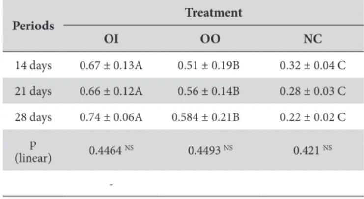
Intragroup analyses of the OI and OO groups showed no statistically signiicant diferences (p>0.05). his demonstrates that the bone loss observed at 14 (Figures 1A and 1D) days was similar to that observed at 21 (Figures 1B and 1E) and 28 (Figures 1C and 1F) days for both groups. hat is, there was no inluence of time on the increase of bone loss coming from OT ater 14 days of experiment for the OI and OO groups (Table 1). he NC group (Figures 1G, 1H and 1I) showed no change in the dimensions of the PL at 14, 21 and 28 days (p>0.05), demonstrating that time did not inluence the initial dimensions of the PL.
Intergroup analysis showed signiicant diference (p<0.05) when comparing groups OI x OO at 14 (0.67 ± 0.13 x 0.51 ± 0.19) (Figures 1A and 1D), 21 (0.66 ± 0.12 x 0.56 ± 0.14) (Figures 1B and 1E) and 28 days (0.74 ± 0.06 x 0.584 ± 0.21) (Figures 1C and 1F). his shows greater bone loss in Group OI for all periods of evaluation. When comparing groups OI x NC at 14 days (0.67 ± 0.13 x 0.32 ± 0.04) (Figures 1A and 1G), 21 days (0.66 ± 0.12 x 0.28 ± 0.03) (Figures 1B and 1H) and 28 days (0.74 ± 0.06 x 0.22 ± 0.02) (1C and 1I) and OO x NC at 14 days (0.51 ± 0.19 x 0.32 ± 0.04) (Figures 1D and 1G), 21 days (0.56 ± 0.14 x 0.28 ± 0.03) (Figures 1E and 1H) and 28 days (0.584 ± 0.21 x 0.22 ± 0.02) (Figures 1F and 1I), signiicant diferences were observed, showing greater bone loss for OI and OO compared with the NC group in all periods (p<0.01). his shows that both proposed experimental models were efective in reproducing primary occlusal trauma. Results are shown in Table 1.
DISCUSSION
It is known that there is alveolar bone loss in occlusal trauma1,8,15,
because all periodontal support tissues are involved. However, there is no information in the literature about the type of occlusal condition and its pathogenic intensity in the periodontium. Due to
Table 1. Mean ± Standard Deviation of bone loss (mm2) in the furcation region of the 1st lower molars, based on the experimental model and on time
Periods Treatment
OI OO NC
14 days 0.67 ± 0.13A 0.51 ± 0.19B 0.32 ± 0.04 C
21 days 0.66 ± 0.12A 0.56 ± 0.14B 0.28 ± 0.03 C
28 days 0.74 ± 0.06A 0.584 ± 0.21B 0.22 ± 0.02 C
p
(linear) 0.4464 NS 0.4493 NS 0.421 NS
-Mean ± SD of detectable alveolar bone loss area on furcation region of irst lower molars (mm2) based on treatment and time. Means followed by diferent letters in
ethical limitations on prospective evaluation of OT in humans, 2 experimental models in rats were developed for the present study, with the goal of evaluating the consequence of the type of occlusion on the development of bone loss in primary OT. In one of the models, occlusal interference7,8,11 was created to imitate experimentally the
occlusal interferences and premature occlusal contacts that lead to the increase in the vertical dimension of occlusion, leading to occlusal instability by altering the force vectors and changing the magnitude of the applied forces to one tooth. In the other model, the unilateral vertical dimension was reduced, creating occlusion on only one of the irst molars, with the goal of reproducing experimentally the cases of partial edentulism15, also creating
occlusal overload experimentally.
Intragroup analysis showed that there is no signiicant diference in the extent of bone loss veriied in the alveolar bone in the interradicular region of the irst lower molar of the animals at 14, 21 and 28 days (p>0.05). hese data lead to the belief that the periodontium underwent functional adaptation, having adapted
to a pathological state due to signiicant changes in the magnitude, frequency of event and change in direction of the force vectors of the occlusal demand18,19. hus, it may be hypothesized that the 1st lower
molar of the animals may have undergone functional adaptation due to possible tooth migration or inclination involving force vectors20,
permitting control of the damage during the applied experimental period (14, 21 and 28 days). Consequently, no histometrically signiicant intragroup diferences were seen regarding bone loss in OI and OO during the evaluation periods of primary OT.
Intergroup analysis showed the efectiveness of both models in the development of OT, showing signiicant bone loss in the furcated region of the 1st lower molar when comparing the OI and
OO groups with the NC group (p<0.01). he OI x OO intergroup analysis showed that the OI group lost signiicantly more bone when compared to the OO group at days 14, 21 and 28 (p<0.05). his shows that the occlusal interference model seems to be more harmful to the alveolar bone when compared to the overload model applied to OO. his may have occurred due to the intensity and
direction of the forces in the furcation region of these teeth and the consequent proportional activation of chemical mediators facing the traumatological demand9,10. It also may be hypothesized that
there was greater activation of osteoclasts by the over-regulation of the binding of the receptor of the activator of the nuclear factor Kappa B (RANK/RANKL) and/or by the reduction in the production of osteoprotegerin (OPG). his would allow greater diferentiation of the pre-osteoclasts in osteoclasts21, resulting in
greater bone loss in the OI group. here may likely have occurred greater magnitude of the forces, greater frequency of the occlusal contact events and alteration in the direction of the force vectors on the periodontium of the rats.
he scientiic literature that uses animal models for experimental reproduction of occlusal trauma are limited in the comparisons of the results of the present study with previous studies, since
most of them use the secondary or the combined occlusal trauma model15,22-24. he data should be evaluated with the constraints that
the animal model provides, because none of the existing animal models duplicate human physiology. However, they are of great value when a prospective clinical evaluation of the development of an injury is impossible. More histological, histochemical and immunological studies should be conducted, using both shorter and longer times, for greater clariication of the damage caused by OT to the alveolar bone and to the other tissues of the periodontium.
hus, within the limits of the present study, it may be concluded that the models of occlusal interference (OI) and occlusal overload (OO) efectively reproduced primary OT. It also may be seen that there was greater alveolar bone loss in the group in which occlusal interference was used.
REFERENCES
1. Hallmon WW. Occlusal trauma: effect and impact on the periodontium. Ann Periodontol. 1999 Dec;4(1):102-8. http://dx.doi.org/10.1902/ annals.1999.4.1.102. PMid:10863382.
2. Suzuki R, Nemoto E, Shimauchi H. Cyclic tensile force up-regulates BMP-2 expression through MAP kinase and COX-2/PGE2 signaling pathways in human periodontal ligament cells. Exp Cell Res. 2014 Apr;323(1):232-41. http://dx.doi.org/10.1016/j.yexcr.2014.02.013. PMid:24561081.
3. Liu H, Jiang H, Wang Y. The biological effects of occlusal trauma on the stomatognathic system - a focus on animal studies. J Oral Rehabil. 2013 Feb;40(2):130-8. http://dx.doi.org/10.1111/joor.12017. PMid:23211044.
4. Academy of Prosthodontics. The glossary of prosthodontic terms. J Prosthet Dent. 2005 Jul;94(1):10-92. http://dx.doi.org/10.1016/j. prosdent.2005.03.013. PMid:16080238.
5. Isidor F. Influence of forces on peri-implant bone. Clin Oral Implants Res. 2006 Oct;17(Suppl 2):8-18. http://dx.doi.org/10.1111/j.1600-0501.2006.01360.x. PMid:16968378.
6. Kan JP, Judge RB, Palamara JE. In vitro bone strain analysis of implant following occlusal overload. Clin Oral Implants Res. 2014 Feb;25(2):e73-82. http://dx.doi.org/10.1111/clr.12059. PMid:23067316.
7. Kaku M, Uoshima K, Yamashita Y, Miura H. Investigation of periodontal ligament reaction upon excessive occlusal load--osteopontin induction among periodontal ligament cells. J Periodontal Res. 2005 Feb;40(1):59-66. http://dx.doi.org/10.1111/j.1600-0765.2004.00773.x. PMid:15613081.
8. Campos ML, Corrêa MG, Júnior FH, Casati MZ, Sallum EA, Sallum AW. Cigarette smoke inhalation increases the alveolar bone loss caused by primary occlusal trauma in a rat model. J Periodontal Res. 2014 Apr;49(2):179-85. http://dx.doi.org/10.1111/jre.12091. PMid:23679047.
9. Onishi T, Ooshima T, Sobue S, El-Sharaby A, Kurisu K, Wakisaka S. Altered expression level of calbindin D28k in the periodontal ligament of rat molar in response to changes in occlusal force. J Periodontal Res. 2000 Oct;35(5):301-9. http://dx.doi.org/10.1034/j.1600-0765.2000.035005301.x. PMid:11005158.
10. Caviedes-Bucheli J, Azuero-Holguin MM, Correa-Ortiz JA, Aguilar-Mora MV, Pedroza-Flores JD, Ulate E, et al. Effect of experimentally induced occlusal trauma on substance p expression in human dental pulp and periodontal ligament. J Endod. 2011 May;37(5):627-30. http:// dx.doi.org/10.1016/j.joen.2011.02.013. PMid:21496661.
11. Kawamoto S, Nagaoka E. The effect of oestrogen deficiency on the alveolar bone resorption caused by traumatic occlusion. J Oral Rehabil. 2000 Jul;27(7):587-94. http://dx.doi.org/10.1046/j.1365-2842.2000.00542.x. PMid:10931251.
12. Chambrone L, Chambrone LA, Lima LA. Effects of occlusal overload on peri-implant tissue health: a systematic review of animal-model studies. J Periodontol. 2010 Oct;81(10):1367-78. http://dx.doi.org/10.1902/jop.2010.100176. PMid:20507230.
13. Naert I, Duyck J, Vandamme K. Occlusal overload and bone/implant loss. Clin Oral Implants Res. 2012 Oct;23(Suppl 6):95-107. http:// dx.doi.org/10.1111/j.1600-0501.2012.02550.x. PMid:23062133.
14. Oliveira Diniz CK, Corrêa MG, Casati MZ, Nociti FH Jr, Ruiz KG, Bovi Ambrosano GM, et al. Diabetes mellitus may increase bone loss after occlusal trauma and experimental periodontitis. J Periodontol. 2012 Oct;83(10):1297-303. http://dx.doi.org/10.1902/jop.2012.110514. PMid:22309176.
15. Nogueira-Filho GR, Fróes EB No, Casati MZ, Reis SR, Tunes RS, Tunes UR, et al. Nicotine effects on alveolar bone changes induced by occlusal trauma: a histometric study in rats. J Periodontol. 2004 Mar;75(3):348-52. http://dx.doi.org/10.1902/jop.2004.75.3.348. PMid:15088871.
17. Wan HY, Sun HQ, Sun GX, Li X, Shang ZZ. The early phase response of rat alveolar bone to traumatic occlusion. Arch Oral Biol. 2012 Jun;57(6):737-43. http://dx.doi.org/10.1016/j.archoralbio.2012.01.002. PMid:22297033.
18. Roesler H. The history of some fundamental concepts in bone biomechanics. J Biomech. 1987;20(11-12):1025-34. http://dx.doi.org/10.1016/0021-9290(87)90020-0. PMid:3323196.
19. Ruff C, Holt B, Trinkaus E. Who’s afraid of the big bad Wolff?: “Wolff ’s law” and bone functional adaptation. Am J Phys Anthropol. 2006 Apr;129(4):484-98. http://dx.doi.org/10.1002/ajpa.20371. PMid:16425178.
20. Poiate IA, de Vasconcellos AB, Santana RB, Poiate E Jr. Three-dimensional stress distribution in the human periodontal ligament in masticatory, parafunctional, and trauma loads: finite element analysis. J Periodontol. 2009 Nov;80(11):1859-67. http://dx.doi.org/10.1902/ jop.2009.090220. PMid:19905956.
21. Haugeberg G, Orstavik RE, Kvien TK. Effects of rheumatoid arthritis on bone. Curr Opin Rheumatol. 2003 Jul;15(4):469-75. http://dx.doi. org/10.1097/00002281-200307000-00016. PMid:12819477.
22. Budtz-Jorgensen E. Bruxism and trauma from occlusion: an experimental model in Macaca monkeys. J Clin Periodontol. 1980 Apr;7(2):149-62. http://dx.doi.org/10.1111/j.1600-051X.1980.tb01958.x. PMid:6769975.
23. Lindhe J, Ericsson I. The influence of trauma from occlusion on reduced but healthy periodontal tissues in dogs. J Clin Periodontol. 1976 May;3(2):110-22. http://dx.doi.org/10.1111/j.1600-051X.1976.tb01857.x. PMid:1064595.
24. Ericsson I, Lindhe J. Lack of effect of trauma from occlusion on the recurrence of experimental periodontitis. J Clin Periodontol. 1977 May;4(2):115-27. http://dx.doi.org/10.1111/j.1600-051X.1977.tb01891.x. PMid:266504.
CONFLICTS OF INTERESTS
he authors declare no conlicts of interest.
*CORRESPONDING AUTHOR
Mirella Lindoso Gomes Campos, USC – Universidade do Sagrado Coração, Rua Irmã Arminda, 10-50, Jardim Brasil, 17011-160 Bauru - SP, Brasil, e-mail: [email protected]