UNIVERSIDADE FEDERAL DE SÃO CARLOS
CENTRO DE CIÊNCIAS BIOLÓGICAS E DA SAÚDE
PROGRAMA INTERINSTITUCIONAL DE PÓS-GRADUAÇÃO EM
CIÊNCIAS FISIOLÓGICAS - UFSCar-UNESP
Vinicius Guzzoni
Remodelamento da matriz extracelular e respostas
cardíacas funcionais em ratos idosos submetidos a
treinamento resistido.
SÃO CARLOS - SP
FEDERAL UNIVERSITY OF SÃO CARLOS
BIOLOGICAL SCIENCE AND HEALTH CENTER
INTERINSTITUTIONAL POST-GRADUATION PROGRAM IN
PHYSIOLOGICAL SCIENCES - UFSCar-UNESP
Vinicius Guzzoni
Effects of high-intensity resistance training in the
extracellular matrix (ECM) and diastolic function in
left ventricle of old male rats.
SÃO CARLOS - SP
UNIVERSIDADE FEDERAL DE SÃO CARLOS
CENTRO DE CIÊNCIAS BIOLÓGICAS E DA SAÚDE
PROGRAMA INTERINSTITUCIONAL DE PÓS-GRADUAÇÃO EM
CIÊNCIAS FISIOLÓGICAS - UFSCar-UNESP
VINICIUS GUZZONI
REMODELAMENTO DA MATRIZ EXTRACELULAR E RESPOSTAS CARDÍACAS FUNCIONAIS EM RATOS IDOSOS SUBMETIDOS A TREINAMENTO RESISTIDO.
Tese apresentado ao Programa de Pós Graduação em Ciências Fisiológicas, para a obtencção do título de Doutor em Ciências Fisiológicas.
Orientadora: Profa. Dra. Heloísa S. Selistre de Araujo Co-orientadora: Profa. Dra. Ana Paula Davel
Ficha catalográfica elaborada pelo DePT da Biblioteca Comunitária UFSCar Processamento Técnico
com os dados fornecidos pelo(a) autor(a)
G993r
Guzzoni, Vinicius
Remodelamento da matriz extracelular e respostas cardíacas funcionais em ratos idosos submetidos a treinamento resistido / Vinicius Guzzoni. -- São Carlos : UFSCar, 2016.
67 p.
Tese (Doutorado) -- Universidade Federal de São Carlos, 2016.
TESE DE DOUTORADO
Vinicius Guzzoni
Remodelamento da matriz extracelular e
respostas cardíacas funcionais em ratos idosos
submetidos a treinamento resistido
Universidade Federal de São Carlos
Área de Concentração: Ciências Fisiológicas
Orientadora: Profa. Dra. Heloísa S. Selistre de Araujo2 Co-orientadora: Profa. Dra. Ana Paula Davel3
Pesquisadores Colaboradores:
Profa. Dra. Rita de Cássia Marqueti Durigan4 Prof. Dr. João Luiz Quagliotti Durigan4 Prof. Dr. Hernandes Faustino de Carvalho3
Prof. Dr. Carlos Lenz César5
1
Programa Interinstitucional de Pós-Graduação em Ciências Fisiológicas UFSCar-UNESP,
2
Departamento de Ciências Fisiológicas, Universidade Federal de São Carlos, UFSCar
3
Departamento de Biologia Estrutural e Funcional, Universidade Estadual de Campinas, UNICAMP.
4
Universidade de Brasília, Campus de Ceilândia,
AGRADECIMENTOS
A DEUS sobre tudo e todos!
À Nossa Senhora e todos os meus Santos Intercessores.
À minha família, meus amados pais, Vitor e Zaira pelo incondicional apoio e amor.
Às minhas irmãs Christina e Viviane Guzzoni pelo respaldo e carinho.
Em especial, agradeço do fundo do meu coração ao meus tios José Pedro Parise e Marcia
Gavioli Parise, por me conceder um ambiente muito agradável para residir e extremamente
propício para estudar na cidade de São Carlos.
À minha avó Ana Florinda (dona Florinha), mulher indescritível, sábia, que me auxiliou em
todos os aspectos. Devo muito a esta mulher a concretização deste trabalho.
Aos meus avós falecidos Leopoldina Motta Guzzoni, Vito Guzzoni e Nelson Parise pela
proteção diária.
À todos os membros da minha família em geral, maior graça que Deus me concedeu, o pilar
da minha vida. Um eterno obrigado! Tudo foi feito por e para cada um de vocês. Em
destaque, ao meu tio Nelson Parise, meu exemplo de vida e profissão e Fran, minha tia, com
sua ternura, humildade e inteligência que cativam as pessoas.
À minha afilhada Vitória de França Parise que me trouxe uma alegria indescritível e
responsabilidade para as lutas cotidianas.
À minha orientadora Profa. Dra. Heloísa Sobreiro Selistre de Araújo pelo total auxílio,
supervisão e competência profissional na condução deste estudo. Eternamente grato por seus
ensinamentos. Obrigado por me proporcionar um ambiente rico e vasto para execução deste
estudo.
À Rita de Cássia Marqueti Durigan e João Luiz Quagliotti Durigan, precursores deste
trabalho e pessoas imprescindíveis na minha formação acadêmica e pessoal. Sem a confiança
deles, nada do que foi escrito neste trabalho aconteceria.
Aos estudantes de Iniciação Científica Marcelo Mekaro e Tayná Costa pelo total
comprometimento em me ajudar em experimentos e atividades relacionadas com este estudo.
À co-orientadora deste estudo, Ana Paula Davel pelo amparo e orientação. Pela dedicação e
disponibilidade em vir até São Carlos para conduzir os experimentos.
Ao Prof. Dr. Orlando Moreira Filho pela concessão do microscópio para as análises
histológicas no Departamento de Genética e Evolução desta Universidade.
Ao Prof. Dr. José Antunes Rodrigues e aluno André de Souza Mecawi pela colaboração nos
ensaios e calorosa recepção em seu Laboratório no Departamento de Fisiologia da Faculdade
de Medicina da Universidade de São Paulo (USP), Ribeirão Preto – SP.
Aos Prof. Dr. Hernandes Faustino de Carvalho e Prof. Dr. Carlos Lenz César e ao técnico
Vitor pela orientação e solidário oferecimento do microscópio para os experimentos
histológicos no Instituto Nacional de Ciência e Tecnologia de Fotônica Aplicada à Biologia
Celular (INFABIC) localizado na UNICAMP, Campinas - SP.
Aos amigos de experimento Fabio Bogni, Fernando Fabrizzi e Giselle Lopes. Vocês foram
À “família LBBM”: Carol, Carmem, Cintia, Kelli, Camila, Uliana, Patty, Araceli, Livia,
Patricia Manzini, Sabrina, Monica, Léo, Taís, Milene, Bruna, Wanessa, Grazi, Rafael,
Antonio, Tamires e Victoria. Nosso sadio convívio diário foi fundamental para, acima a
construção dessa linda amizade. Levo todos, com carinho, para sempre!
Às grandes companheiras, incentivadoras, amigas, confidentes, Cynthia e Karina, mineiras,
que sempre estiveram ao meu lado.
Aos meus amigos de Taquaritinga, muitos para nomear, nenhum para esquecer.
À cada professor das disciplinas que frequentei, da banca de qualificação e do estágio
PESCD: Sandra Lia do Amaral, Carlos Cressani, Azair de Souza, Carina Andrade, Gerson J.
Rodrigues e Gilberto E. Shiguemoto. Docentes que contribuíram fortemente para a
concretização deste estudo.
Ao secretário Alexandre pelo pronto auxílio com documentos, dúvidas e burocracia dos
trâmites.
À Bety e Márcia, pelo auxílio em diversas atividades.
Em especial e, com eterna gratidão, à John Lawler grande pesquisador e meu supervisor no
Texas (EUA) (Redox Biology & Cell Signaling Laboratory) na Texas A&M University
(TAMU), onde realizei meu Doutorado Sanduíche. Inesquecível, indescritível! Obrigado por
abrir as portas do seu Lab onde pude saborear e vivenciar a ciência em um país-referência no
segmento.
À James Matthew Kuczmarski, Jeffrey M. Hord e Curran Reddy, companheiros e “irmãos”
de laboratório na TAMU. Vocês foram meus maiores professores. Muito obrigado, sempre!
Aos meus eternos e grandes amigos americanos Andrew Stivers (e família), Enoc Medina,
Barbara Castanheira Ferrara Barbosa. Vocês são parte determinante deste trabalho e da
minha vida.
À minha professora de inglês nos EUA Geraldine Wick (Geri). Vc foi/é especial.
Infelizmente partiu sem que eu pudesse me despedir. Muito disso é pra você, minha querida
amiga.
Às minhas escolas de inglês de São Carlos, The FIELDS e The Four.
À minha comunidade católica de São Carlos (São Nicolau de Flue) na pessoa do Pároco
Eduardo Malaspina, grande discípulo, e a cada membro da minha Célula Católica Santa
Izabel pelas constantes orações, em especial a líder e eterna amiga Maria Clara Santos.
Obrigado por sempre estar comigo Clara.
À Paróquia de São Sebastião de São Carlos, lugar maravilhoso para professar a fé. E ao meu
querido pai, pároco de minha cidade, padre João.
Aos meus grandes e eternos amigos João e Fabiana Zepon. Sou extremamente grato pelos
ensinamentos e carinho para comigo. Conhecer vocês foi uma benção de Deus!
Aos meus amigos de futebol da USP. Obrigado por me proporcionarem um momento de
lazer e amizade às terças-feiras. Espero que nossa amizade sempre perdure.
À Igreja A&M Church, em College Station (TX), cidade que residi no período de Doutorado
Sanduíche. Pessoas maravilhosas que me acolheram e certamente continuam a orar por mim.
À cada pessoa que trabalhou indiretamente, mas com exímio valor. Ao restaurante
universitário, biblioteca, equipe de limpeza, guardas, entre muitos outros que fazem parte
Agradeço às dificuldades, medos e inseguranças que deparei aos longos desses anos. No
enfrentamento destes, guiado pelas bênçãos Divinas, certamente vieram os mais belos
aprendizados.
À CAPES, por me conceder as bolsas de estudo (nacional e internacional).
LISTA DE ILUSTRAÇÕES
LIST OF ABREVIATION
ECM – extracellular matrix;
RT – resistance training;
LV – left ventricle;
BW – body weight;
SBP – systolic blood pressure;
DBP – diastolic blood pressure;
MBP – mean blood pressure;
LVSP – left ventricle systolic pressure;
LVEDP – left ventricle end-diastolic pressure;
ANG-II – angiotensin II-type;
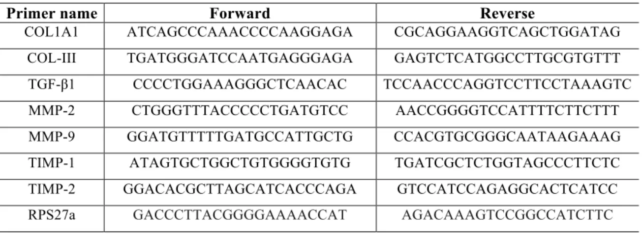
Table 1 - List of oligonucleotides primers: COL1A1: type-IA1 collagen; COL3A1: type-III collagen; TGF-β1, transforming growth factor beta-1; MMP-2: metalloproteinase 2; MMP-9: metalloproteinase 9; TIMP-1: tissue inhibitor of metalloproteinases-1; TIMP-2: tissue inhibitor metalloproteinases-2; RPS27a: ribosomal protein S27a.
Primer name Forward Reverse
COL1A1 ATCAGCCCAAACCCCAAGGAGA CGCAGGAAGGTCAGCTGGATAG
COL-III TGATGGGATCCAATGAGGGAGA GAGTCTCATGGCCTTGCGTGTTT
TGF-β1 CCCCTGGAAAGGGCTCAACAC TCCAACCCAGGTCCTTCCTAAAGTC
MMP-2 CTGGGTTTACCCCCTGATGTCC AACCGGGGTCCATTTTCTTCTTT
MMP-9 GGATGTTTTTGATGCCATTGCTG CCACGTGCGGGCAATAAGAAAG
TIMP-1 ATAGTGCTGGCTGTGGGGTGTG TGATCGCTCTGGTAGCCCTTCTC
TIMP-2 GGACACGCTTAGCATCACCCAGA GTCCATCCAGAGGCACTCATCC
Table 2 - Body weight (BW) and cardiac hypertrophy rates of the experimental groups. BWi: body weight at the beginning of RT; BWf: body weight in the end of RT; LV: left ventricle weight; Whole heart: whole heart weight; LV/BW: LV weight/body weight ratio; LV/tibia: LV weight/tibia length; Values are expressed by means ± SEM. Groups: YS: young sedentary; YT: young trained; OS: old sedentary; OT: old trained. Different characters mean statistical difference (ANOVA two-way, Tukey post-hoc test p ≤ 0.05). +YT vs. YS; *OS vs. YS; #OT vs. YT. n = 10/group.
YS YT OS OT
BWi, g 313 ± 4 304 ± 6 510 ± 13 519 ± 9
BWf, g 486a ± 10 426a + ± 9 467 a ± 15 480a ± 9
LV, mg 1058 ± 21 902+ ± 19 1061 ± 39 1053# ± 26
Whole heart, g 1.34 ± 0.03 1.16+ ± 0.03 1.50* ± 0.06 1.47# ± 0.03
Tibia, cm 4.46 ± 0.03 4.43 ± 0.05 4.58 ± 0.04 4.64 ± 0.03
LV/BWf, mg/g 2.18 ± 0.02 2.27 ± 0.03 2.20 ± 0.08 2.20 ± 0.04
LV/tibia, mg/cm 244 ± 5.7 208+ ± 9.7 223 ± 11 236 ± 7.8
Figure 1 - Overload (g) carried by old and young trained rats along the 12 weeks of RT. Blue and red lines correspond to the young trained rats (YT), and old trained rats (OT) respectively. n=31; a significant difference in comparison to the previous week (paired t Student test; p ≤ 0.05); b significant difference between YT x OT (unpaired t Student test; p ≤ 0.05). Values are expressed as means ± SEM.
Source: Author
0 1 2 3 4 5 6 7 8 9 10 11 12 13 700
850 1000 1150 1300 1450 1600
weeks
ov
e
rloa
d of
R
T (
g)
OT YT
a ab
ab
ab
ab
a a a a
ab
ab
ab
ab
b
Figure 2 - LV images of hematoxilin-eosin staining (H&E), 20X (A), Masson´s trichrome imaging, 20X (B) and Second harmonic generation (SHG), 40X (C). Connective tissue is staining in blue (yellow arrows, B) and in green collagen (white arrows, C); Values are expressed as means ± SEM. LV myocyte width (µm) (A), % of LV connective tissue (B) and %collagen deposition/area (C). Groups: YS: young sedentary; YT: young trained; OS: old sedentary; OT: old trained. Different characters mean statistical difference (ANOVA two-way, Tukey post-hoc
test p ≤ 0.05). +YT vs. YS; *OS vs. YS; &OS vs. OT. n = 7/group.
A B
C D
E F
20μm 20μm
YS YT
OS OT
20μm 20μm
YS YT OS OT
0 5 10 15 20 LV m y oc y te widt h ( µ m) * &
YS YT OS OT
Source: author
20μm
20μm
20μm
20μm
YS YT OS OT
0.0 0.5 1.0 1.5
Se
c
o
n
d
H
a
rm
o
n
ic
G
e
n
e
ra
ti
o
n
(A
U
) +
&
YS YT
Figure 3 -Bands representing the catalytic activity of MMP-2 in the gel (A); Densitometry of pro-MMP-2 (B) and active-MMP-2 (C). Values are expressed as means ± SEM. Groups; YS: young sedentary; YT: young trained; OS: old sedentary; OT: old trained.Different characters mean statistical difference (ANOVA two-way, Tukey post-hoc test p ≤ 0.05). +YT vs. YS; *OS vs. YS; #OT vs. YT; &OS vs. OT. n = 10/group.
A
B C
Source: author
YS YS YS YS
YT YT YT YT
OS OS OS OS
OT OT OT OT
YS YT OS OT
0 1 2 3 4 pr o-M M P -2 ( A U ) # + & *
YS YT OS OT
Figure 4 -Gene expression of collagen I (COL-I) (A), collagen III (COL-III) (B), transforming growth factor-β1 (TGF-β1) (C), tissue inhibitor of metalloproteinase 1 (TIMP-1) (D), metalloproteinase-9 (MMP-9 (E) and metalloproteinase-2 (MMP-2) (F) in the LV tissues measured by real time PCR. Values are expressed as means ± SEM and normalized by the reference gene (RPS27a). Groups; YS: young sedentary; YT: young trained; OS: old sedentary; OT: old trained. Different characters mean statistical difference (ANOVA two-way, Tukey post-hoc test p ≤ 0.05). +YT vs. YS; *OS vs. YS; #OT vs. YT; &OS vs. OT. n = 8/group.
Source: author
YS YT OS OT
0.0 0.5 1.0 1.5 mRNA CO L - II I expres s ion / RPS 27 a ( AU ) * #
A
B
C
D
YS YT OS OT
0.0 0.5 1.0 1.5 mRNA M M P -2 expres s ion / RPS 27 a ( AU ) # *
YS YT OS OT
Figure 5 - Protein expression of metalloproteinase-2 (MMP-2) (A), metalloproteinase-9 (MMP-9) (B); tissue inhibitor of metalloproteinase 1 (TIMP-1) (C), tissue inhibitor of metalloproteinase 2 (TIMP-2) (D) and transforming growth factor-β1 (TGF-β1) (E) in LV tissues, measured by Western immunoblot. Values are expressed as means ± SEM. Groups; YS: young sedentary; YT: young trained; OS: old sedentary; OT: old trained. No statistical differences were observed in all proteins. n=9/group.
Source: author
YS YT OS OT
0.0 0.5 1.0 1.5 MMP -2 p ro te in e x p re s s io n (A U )
YS YT OS OT 0.0 0.5 1.0 1.5 MMP -9 p ro te in e x p re s s io n (A U ) A B
YS YT OS OT
0.00 0.05 0.10 0.15 0.20 0.25 TIM P -1 pr ot e in e x pr e s s ion (A U )
YS YT OS OT
0.00 0.05 0.10 0.15 0.20 0.25 TIM P -2 pr ot e in e x pr e s s ion (A U )
YS YT OS OT
Table 3 - Arterial and intra-ventricular measurements.
YS YT OS OT
SBP, mmHg 122.8 ± 2.7 129.9 ± 4.8 100.5* ± 6.4 109# ± 4.1
DBP, mmHg 87.6 ± 3.1 92.6 ± 2.7 70* ± 4.9 78# ± 2.8
MBP, mmHg 104.3 ± 3.2 108.9 ± 3.2 84.8* ± 5.5 92.8# ± 3.2
HR bpm 204 ± 8 195 ± 6 200 ± 18 189 ± 6
LVSP, mmHg 135.8 ± 4 137.7 ± 3 107.6* ± 7.7 99.8# ± 5.4
+dP/dt, mmHg/s 5586.7 ± 347 5613 ± 365 6263.6 ± 590 5298.3 ± 448
LVEDP, mmHg 7.3 ± 1 7.3 ± 0.8 10.9* ± 0.4 8.4& ± 0.9
-dP/dt, mmHg/s -4052.6 ± 308 -4167.5 ± 185 -3232 ± 424 -3335.3# ± 230
Tau, s 0.033 ± 0 0.028 ± 0 0.016* ± 0 0.017# ± 0
Source: Author
TABLE OF CONTENTS – SUMÁRIO
INTRODUÇÃO ... 12 JUSTIFICATIVA ... 15 OBJETIVO GERAL ... 16 OBJETIVOS ESPECÍFICOS ... 16 INTRODUCTION ... 17 MATERIAL AND METHODS ... 22
Experimental Group ... 22 Resistance Training ... 22 Measurements of hemodynamic parameters ... 23 Histological analysis and Second Harmonic Generation (SHG) ... 24 Gelatin zymography ... 24 RNA isolation and Analysis ... 25 Reverse Transcription ... 26 Oligonucleotide primers ... 26 Real-‐time polymerase chain reactions ... 26 Western immunoblot ... 27 Determination of cardiac peptides concentration ... 28 STATISTICS ... 29 RESULTS AND DISCUSSION ... 30
RESUMO
INTRODUÇÃO: O remodelamento da matriz extracelular (MEC) cardíaca é um evento dentre
estas mudanças estruturais no ventrículo esquerdo (VE) que é orquestrado pelos níveis de
metaloproteinases (MMP) e seus inibidores endógenos (TIMPs). Na tentativa de prevenir tais
efeitos decorrentes da idade avançada, o exercício aeróbico tem sido sugerido por melhorar a
fibrose e a função cardíaca. Entretanto, os efeitos do treinamento resistido (TR) nestas variáveis
necessitam de melhor compreensão. OBJETIVO: investigar os efeitos crônicos do TR de alta
intensidade na MEC do VE e a função cardíaca em ratos idosos. METODOLOGIA: ratos de 3 e
21 meses de idade foram designados como grupos: jovem sedentário (YS), jovem treinado (YT),
idoso sedentário (OS) e idoso treinado (OT). Os grupos treinados foram submetidos à um
protocolo de 12 semanas de escalada sob alta intensidade, 3 vezes por semana. Decorridos 48h
pós última sessão de treino, medidas hemodinâmicas foram registradas: pressão arterial sistólica
(SAP), pressão arterial diastólica (DAP), pressão arterial média (MAP), frequência cardíaca
(HR), constante de decaímento da pressão ventricular (Tau), derivada temporal positiva da
pressão intraventricular (+dP/dt), razão de decaímento da pressão ventricular (-dP/dt), pressão
arterial sistólica máxima do VE (LVSP) e pressão arterial diastólica final do VE (LVEDP).
Largura dos cardiomiócitos, % de tecido conectivo e de colágeno intersticial foram analisados no
VE. A atividade da MMP-2 foi detectada por zimografia, assim como a expressao gênica e
proteica de alguns constituintes da MEC. Moduladores da hipertrofia e fibrose cardíaca,
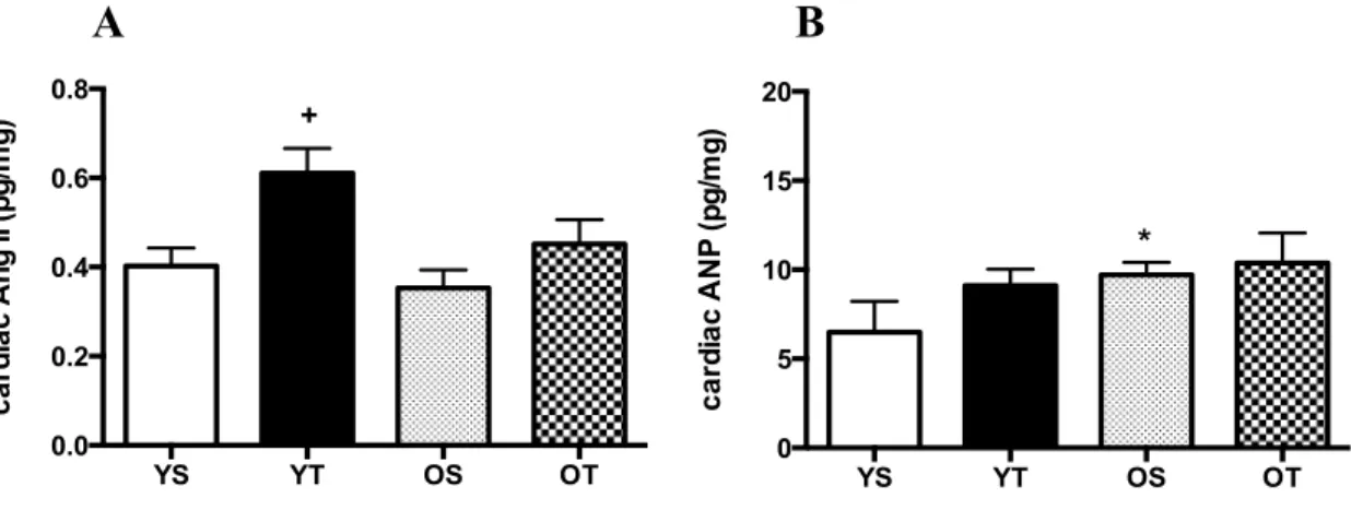
angiotensina II (Ang-II) e peptídeo natriurético atrial (ANP) foram avaliados. RESULTADOS: a
largura do cardiomiócito e a concentração de colágeno diminuíram em ratos OT comparados ao
grupo OS. TR aumentou a atividade da MMP-2 e atenuou os aumentos na LVEDP de ratos
idosos. Ratos OT não apresentaram alterações significativas na expressão dos elementos da MEC
e nos peptídeos cardíacos Ang-II e ANP. O TR diminuiu significativamente a expressão gênica
elevada de TIMP-1, TGF-β e COL-1, observados no grupo OS. CONCLUSÃO: o TR foi eficaz
em diminuir o colágeno cardíaco o que pode ser associado com a melhora na função diastólica, o
que pode estar relacionado com o aumento na atividade da MMP-2 em VE de ratos idosos. O TR
atenuou a via de sinalização TGF-β–TIMP-1–COL-1, a nível transcricional. Portanto este estudo
revela a importância do treinamento resistido na homesostase da MEC e melhora da função
Palavras-chave: Envelhecimento. Treinamento Resistido. Ventrículo Esquerdo. Matriz
ABSTRACT
INTRODUCTION: It is well documented that aging causes morphological and functional
alterations in the heart. Cardiac ECM remodeling is one event of structural changes in left
ventricle (LV), which is modulated by MMPs/TIMPs balance and may lead to cardiac fibrosis.
To prevent such effects inherent of aging, aerobic exercise training has been suggested to
improve the cardiac fibrosis and function. However the effects of resistance training (RT)
remains unclear. Whether that RT could alter cardiac function following cardiac ECM
remodeling is uncertain. PURPOSE: to investigate the chronic effects of high intensity
resistance training (RT) in the extracellular matrix (ECM) remodeling of left ventricle (LV) and
cardiac function in old rats. PROCEDURES: Rats with 3 and 21 months-age were assigned as
young sedentary (YS), young trained (YT), old sedentary (OS) and old trained (OT). The trained
groups (YT and OT) were submitted to high-intensity RT protocol (3 times a week during 12
weeks). After 48h post-training, hemodynamic and intra-ventricular pressures were recorded. LV
myocyte width, LV connective tissue and collagen fibrils were analyzed. MMP-2 activity, gene
and protein expression from ECM components as well as angiotensin II (Ang-II) and atrial
natriuretic peptide (ANP) were evaluated FINDINGS: LV myocyte width and connective tissue
were reduced in OT rats. RT increased the MMP-2 activity in OT rats and improved the
age-related increase in the left ventricle end diastolic pressure (LVEDP). The RT unchanged Ang-II
and ANP in LV of old rats. CONCLUSION: RT was effective to decrease LV connective tissue,
which was associated with increased ECM remodeling by MMP-2 activity in LV tissue and
improvement of LVEDP in aging rats. Our results point out the importance of RT in ECM
homeostasis and diastolic function in experimental aging model.
Key-words: Aging. Resistance Training, Left Ventricle, Extracellular Matrix, Diastolic
INTRODUÇÃO
O envelhecimento é um processo inerente a todos os seres vivos que se estabelece
como um fator de risco independente para a manifestação de doenças cardiovasculares
(LAKATTA; LEVY, 2003; NICCOLI; PARTRIDGE, 2012; STERN; BEHAR; GOTTLIEB,
2003). Entretanto, é difícil determinar precisamente as adaptações cardíacas frente ao processo
natural do envelhecimento e àquelas ocasionadas por doenças cardiovasculares decorrentes.
A fibrose cardíaca é uma característica do processo natural de envelhecimento que
vem sendo documentado ao longo dos anos (BOLUYT et al., 1994; FEINBERG;
VOGELSTEIN, 1984; GOLDSMITH; BRADSHAW; SPINALE, 2013; HORN, 2015;
(OLIVETTI et al., 1991ab; QUEREJETA et al., 2004) por favorecer uma maior rigidez da
câmara cardíaca asssociado com piora do relaxamento ventricular, o que contribui para o
desenvolvimento da disfunção diastólica (HORN, 2015; LAKATTA et al., 1987; REED et al.,
2011). A fibrose intersticial constitui um dos eventos do remodelamento cardíaco (HORN, 2015)
cuja definição é a resposta compensatória decorrente do aumento da sobrecarga de pressão
imposta ao ventrículo esquerdo (VE) - devido ao enrijecimento arterial decorrente da idade - o
que leva à alguns eventos celulares, tais como: apoptose e necrose, diminuição do número de
cardiomiócitos com consequente hipertrofia das células remanescentes, deposição de gordura
visceral epicárdica e acúmulo de proteínas da MEC – a fibrose intersticial (ANVERSA et al.,
1990; HORN, 2015; OLIVETTI et al., 1991ab). O acúmulo de tecido fibroso estabelece-se por 2
processos: fibrose reativa ou reparativa. A fibrose reativa ocorre em resposta à um aumento do
estresse da parede miocárdica com consequente espessamento das fibras de colágeno já
existentes dentro do espaço intramuscular. Por outro lado, fibrose reparativa é a síntese de novas
redes de colágeno nos espaços criados por células necróticas e apoptóticas (BURSTEIN;
NATTEL, 2008; TARONE et al., 2014; WEBER, 1989). Entretanto, é difícil estabelecer se estes
processos contribuem de forma conjunta ou isolada na manifestação da fibrose cardíaca em
decorrência do envelhecimento.
De uma forma geral, o acúmulo de colágeno configura a fibrose cardíaca (HORN
et al., 2012; JUGDUTT, 2003; LAKATTA; LEVY, 2003). Entretanto o colágeno está inserido
dentro de uma uma vasta rede de macromoléculas extracelulares, constituída de moléculas
principal função da MEC é fornecer suporte para as células contráteis – os cardiomiócitos
(JOURDAN-LESAUX et al., 2010). Juntamente com a proteína contrátil titina, a MEC também
possui papel fundamental na elasticidade e complacência do músculo cardiaco e transmissão do
estresse mecânico durante o ciclo cardíaco (GIELEN et al., 2005; NAGUEH et al., 2004; PUGH;
WEI, 2001; STEWART et al., 2003). Além do mais, a MEC é um ambiente rico de moléculas
sinalizadoras, tais como citocinas, fatores de crescimento e um reservatório de proteases
(GOLDSMITH; BORG, 2002; KASSIRI; KHOKHA, 2005). Dessa forma, ela está em constante
processo de síntese, degradação e resíntese de proteínas (turnover protéico). Por sua vez, o termo remodelamento da MEC é estabelecido como uma sequência de processos de degradação,
remoção dos subprodutos, reposição celular e consequente mudança estrutural do tecido (APTE;
PARKS, 2015). Frente às mudanças estruturais que ocorrem no coração envelhecido,
principalmente hipertrofia e fibrose cardíacas, utilizaremos aqui o termo remodelamento da
MEC.
O balanço entre a síntese e degradação das proteínas da MEC é orquestrado por
proteases dependentes de zinco, denominadas metaloproteases de matriz (MMPs) cuja atividade
é modulada principalmente por seus inibidores endógenos, os chamados inibidores teciduais de
metaloproteases, os TIMPs (CREEMERS et al., 2003; JAYASANKAR et al., 2004; MOTT;
WERB, 2004; SPINALE et al., 1998). As gelatinases (MMP-2 e MMP-9) são enzimas que
degradam gelatinas, os subprodutos da degradação de colágeno devido à ação da colagenase
MMP-1 (KASSIRI; KHOKHA, 2005). Entretanto, Foi demonstrada diminuição e aumento na
expressão protéica das MMPs e TIMPs respectivamente, em coração de ratos idosos (KWAK et
al., 2011). Além do mais, o TGF-β1 é um importante regulador do TIMP-1 e está relacionado
com a fibrose no coração envelhecido (BONNEMA et al., 2007; CHEN et al., 2000; TSUTSUI et
al., 2007). Angiotensina II (Ang-II) e peptídeo natriurético atrial (ANP) são importantes
sinalizadores da fibrose cardíaca e hipertrofia cardíaca, respectivamente (ASAKURA et al.,
2002; CHEN et al., 2000; GONZÁLEZ; LÓPEZ; Dı́EZ, 2004; KNOWLES et al., 2001; LIJNEN;
PETROV; FAGARD, 2001; OLIVER et al., 1997; TAKAHASHI et al., 1994; TSUTSUI et al.,
2007). A Ang-II atua como uma peptídeo de sinalização upstream ao TGF-β1, mediando as respostas relacionadas à fibrose cardíaca (BAKER; ACETO, 1990; SADOSHIMA; IZUMO,
1993; SCHUNKERT et al., 1990; THANNICKAL et al., 2003) assim como da produção de
função cardíaca (KJAER; HESSE, 2001). O ANP é um peptídeo cardíaco associado com as
adaptações à sobrecarga cardíaca de pressão (JIN et al., 2000) sendo, portanto, considerado um
marcador de hipertrofia patológica (DIETZ, 2005). Embora sua função esteja relacionada com
limitação da fibrose cardíaca (LEVIN; GARDNER; SAMSON, 1998; VARDENY; TACHENY;
SOLOMON, 2013), concentrações de ANP aumentam no VE de ratos envelhecidos (WU;
KWAN; TANG, 1997).
Na tentativa de amenizar a excessiva fibrose cardíaca relacionada à idade
avançada, o treinamento aeróbico tem sido amplamente estudado (KWAK et al., 2011;
THOMAS et al., 2001; WRIGHT et al., 2014). Os trabalhos, em geral, demonstraram seus efeitos
benéficos por promover redução da fibrose cardíaca relacionada com a idade avançada (KWAK
et al., 2011; THOMAS et al., 2000), associado com o aumento na expressão MMP-2 e redução
na expressão de TGF-β1 e TIMP-1 em VE de ratos idosos (KWAK et al., 2011). Outros estudos
sugerem que este efeito possa promover melhora nos parâmetros funcionais cardíacos (CHOI et
al., 2009; KWAK; SONG; LAWLER, 2006; LAKATTA; LEVY, 2003; RAYA et al., 1997;
TSURUDA et al., 2004). Entretanto, os efeitos do treinamento resistido nesse parâmetros ainda
necessitam ser melhor compreendidos. Ainda, se essas alterações estruturais da MEC cardíaca
em idoso poderiam ser moduladas pelo treinamento físico resistido e as consequentes respostas
cardíacas funcionais são ainda pouco conhecidas. Embora haja poucos estudos sobre os efeitos
do treinamento resistido nos parâmetros de remodelamento cardíaco em modelo de idoso, este
tipo de treinamento físico tem sido amplamente recomendado para este segmento da população
por sua eficácia em mimizar as perdas de força, massa muscular e óssea inerentes ao avanço da
idade (EVANS, 1999; HÄKKINEN et al., 1998). Considerando que o treinamento resistido
impõe uma sobrecarga de pressão – e de volume – sobre o VE (BERNARDO et al., 2010;
GROSSMAN; JONES; MCLAURIN, 1975), nós hipotetizamos que o treinamento resistido
aumenta o remodelamento da MEC do VE e melhora a função cardíaca em ratos idosos.
JUSTIFICATIVA
Diante das previsões de um ascendente aumento da população idosa, devido à
melhora da expectativa de vida por influência de diversos fatores, estudos contemplando temas
sobre o envelhecimento mostram-se relevantes, principalmente no que tange à envelhecer com
qualidade de vida.
Clinicamente, a prática de exercício físico com características aeróbicas tem se
mostrado eficaz nos aspectos morfológicos e funcionais cardíacos na população idosa. Embora o
treinamento físico resistido seja extremamente recomendado para este tipo de população, visto
seus benefícios músculo-esqueléticos, ainda se faz necessário uma melhor compreensão dos
efeitos deste nos parâmetros cardíacos tanto no aspecto morfológico quanto no funcionamento da
bomba cardíaca. Portanto este estudo possui relevância experimental com a intenção de elucidar
alguns pontos importantes dentro do tema remodelamento cardíaco na idade avançada, especificamente da MEC, de modo que possa contribuir com novas pesquisas na área. Visto a
dificuldade em se adequar protolocos de treinamento resistido para modelos experimentais e a
escassez de trabalhos científicos até o momento, acreditamos que esta pesquisa venha acrescentar
à literatura científica na tentativa de avançar as fronteiras do conhecimento na área em questão.
OBJETIVO GERAL
Testar os efeitos crônicos do treinamento físico resistido de alta intensidade sobre
as adaptações morfológicas cardíacas e o remodelamento da MEC em VE de ratos idosos, bem
como a função cardíaca.
OBJETIVOS ESPECÍFICOS
Avaliar os aspectos morfológicos do VE de ratos idosos; avaliar a atividade
enzimática da MMP-2 do VE em ratos idosos; investigar a expressão genética e protéica dos
elementos envolvidos no remodelamento da MEC de VE de ratos idosos e identificar as vias de
sinalização upstream do remodelamento da MEC em VE de ratos idosos.
INTRODUCTION
Progressive fibrosis is a hallmark of the aging heart, as confirmed in animal and human studies (NEILAN et al., 2013). Left ventricular (LV) hypertrophy and myocardial fibrosis are compensatory responses to changing load conditions in the aged heart which are caused by diminished vascular compliance and elevated cardiac pressure overload (GRAHAM et al., 2011; O’ROURKE; HASHIMOTO, 2007). These changes elevate the ventricular work required to eject blood during systole, resulting in cardiac remodeling (CHEN et al., 1998). In general, cardiac remodeling stands for morphological, cellular, molecular and functional changes in the myocardium caused by alterations in overload placed into the heart (COHN et al., 2000; HORN, 2015; QUARLES et al., 2015). Pathological cardiac remodeling is associated with myocardial infarction, inflammatory myocardial disease, hypertension (pressure overload), aortic regurgitation (pressure overload) and cardiomyopathies. The increased pressure overload elicits compensatory responses to the myocardium, including reduced cardiomyocyte numbers, lengthening of the remaining cardiomyocytes and proliferation of cardiac fibroblasts followed by collagen accumulation in the ventricular chamber (COHN et al., 2000; JANSSENS; LIJNEN, 2006; LAKATTA; LEVY, 2003; TORELLA et al., 2004). Myocardial collagen accumulation, or fibrosis, is managed by balanced equilibrium between extracellular matrix (ECM) synthesis, maturation, processing and degradation – the ECM turnover (ANVERSA et al., 1990; CAPASSO; FITZPATRICK; ANVERSA, 1992; CAPASSO et al., 1990; CENTURIONE et al., 2003; EGHBALI et al., 1989; FRATICELLI et al., 1989; HORN, 2015; SUSSMAN; ANVERSA, 2004; KWAK et al., 2011).
Matrix metalloproteinases (MMPs) are endopeptidases involved in ECM turnover and cardiac remodeling for degrading ECM proteins (AHMED et al., 2006; CHAKRABORTI et al., 2003; TSURUDA et al., 2004). Paradoxically, increased MMP levels mediate LV hypertrophy by enhancing ECM constituents, including collagen, fibronectin, elastin and laminin, leading to fibrosis with subsequent LV chamber dilatation and impaired LV function (JANSSENS; LIJNEN, 2006; OPIE et al., 2006). MMPs are synthesized by myofibroblasts, inflammatory cells, and myocytes (CLEUTJENS et al., 1995; COKER et al., 2001; HORN, 2015;
or pro-MMP) (TURNER; PORTER, 2012). MMP-2 and MMP-9 (gelatinases) are the main enzymes of cardiac remodeling because they break denatured collagen (gelatin) (COKER et al., 1998; HADLER-OLSEN et al., 2011; SPINALE et al., 1998), fibronectin, elastin and laminin in the rat myocardium (CHEUNG et al., 2000; TYAGI; RATAJSKA; WEBER, 1993; SPINALE, 2007). MMP activity is regulated by endogenous inhibitors, the tissue inhibitor metalloproteinases (TIMPs), which interact with MMPs at a 1-to-1 stoichiometric ratio (BONNEMA et al., 2007). Whereas MMPs have been associated with collagen degradation, there is recent evidence showing fibrosis following MMP activity (APTE; PARKS, 2015; GOLDSMITH; BRADSHAW; SPINALE, 2013). Expression of MMPs is regulated transcriptionally by growth factors, hormones and inflammatory cytokines (DESCHAMPS; SPINALE, 2006; FANJUL-FERNÁNDEZ et al., 2010). Angiotensin (Ang-II), atrial natriuretic peptide (ANP) and transforming growth factor beta (TGF-β) have been reported as one of the key upstream signaling pathways of cardiac hypertrophy and fibrosis (ASAKURA et al., 2002; CHEN et al., 2000; GONZÁLEZ; LÓPEZ; DÍEZ, 2004; LIJNEN; PETROV; FAGARD, 2001; KWAK, et al., 2011; ROSENKRANZ, 2004; TSUTSUI et al., 2007).
The ECM provides scaffolding for myocyte alignment, crucial for systolic
LAKATTA; LEVY, 2003; NICCOLI; PARTRIDGE, 2012; STERN; BEHAR; GOTTLIEB,
2003).
Exercise training has been postulated for its beneficial cardiovascular effects (ELLISON et al., 2012) and the reduced risk of cardiac events (NACI; IOANNIDIS, 2013) especially in the elderly (CONONIE et al., 1991). As a whole, exercise training provides enhancements in maximal cardiac output, increase in stroke volume and decrease in resting heart rate (MIHL; DASSEN; KUIPERS, 2008). For that reasons, unlike pathological remodeling, exercise training is a potent stimuli for physiological cardiac remodeling (MIHL; DASSEN; KUIPERS, 2008). Endurance training or aerobic exercises (e.g., walking, running and swimming) involves isotonic contractions of large skeletal muscle mass and are performed for extended periods (e.g., 30-60min) using oxygen as the main energy supply for sustaining repetitive high-intensity, low-resistance exercise (MORGANROTH et al., 1975; NADER, 2006). It triggers substantial skeletal muscle vasodilatation and cardiac volume overload (pre-load) caused by increasing venous return (PLUIM et al., 2000). The volume overload lead to increased stretching force on the myocardium (ANVERSA; OLIVETTI, CAPASSO, 1991), which stimulates the ventricular dilatation (VINEREANU et al., 2000), characterized by increasing left ventricular internal width and left ventricular wall thickness (PLUIM et al., 2000). The endurance exercise results in eccentric cardiac hypertrophy with normal or improve of ventricular function (HOSSACK, 1987; MELO et al., 2009). Such adaptations have been reported both in aging humans (ARBAB-ZADEH et al., 2004; TAKEMOTO et al., 1992) and experimental animal models (BRENNER; APSTEIN; SAUPE, 2001; JIN et al., 2000).
However, studies addressing the effects of the resistance training on cardiac remodeling, ECM turnover and cardiac functional are scarce. Although endurance exercise training has been shown to improve the aged-associated LV remodeling, cardiac fibrosis and cardiac function, the effects of high-intensity resistance training in cardiac ECM remodeling and its implications in the cardiac function are not well understood.
2006; WHARBURTON; GLEDHILL; QUINNEY, 2001), reduction of muscle mass (sarcopenia) and loss of strength inherent of aging (KRAEMER, 2002; HURLEY; ROTH, 2000; ROTH et al., 2002; WINETT; CARPINELLI, 2001). It involves smaller muscle mass with few repetitions of muscle contractions (usually <20) until exhaustion at high or maximal exercise intensities during short-duration periods (NADER, 2006). The American Heart Association (AHA) recommends strength-training exercises at least 60% of 1 RM (one Repetition Maximum) of intensity (FIATARONE et al., 1990; HAGERMAN et al., 2000). Recommendations by the majority of studies and health organizations state that the RT must be progressive, executed at low repetitions and moderate volume, with overload against the concentric phase of movement (KRAEMER, 2002). Pollock et al (1994) recommends resistance training for elderly persons performing 1-8 sets, 12 repetitions and 8-10 exercises twice a week. Unlike the endurance exercise training, RT induces pathological cardiac remodeling, which could potentiate, to some extent, the compensatory responses of natural aging process. Resistance-type exercise imposes high pressure overload to the heart due to increased myocardial wall stress (FAGARD, 1996; MELO et al., 2015) inducing cardiac concentric hypertrophy (MACDOUGALL et al., 1985; MIHL; DASSEN; KUIPERS, 2008). However, Pluim et al (2000) concluded in a meta-analysis that ventricular hypertrophy, eccentric or concentric, is not dependent upon type-specific exercise training (endurance or resistance). Elevations in blood pressures (systolic and diastolic), cardiac output and heart rate have been documented during RT execution (FISMAN et al., 1997; HOOGSTEEN et al., 2004), although Fleck and Kraemer (2004) reported reduces in rate-pressure product (product of heart rate and blood arterial rate-pressure) following RT programmes, which may reduce cardiac demands during daily activities (MCCARTNEY et al., 1993). Furthermore, heavy RT may lead dramatic acute increases in both systolic and diastolic blood pressure when Valsava manoeuvre is evoked (MACDOUGALL et al., 1985 MCCARTNEY, 1999) as well as increases in systemic vascular resistance due to isometric contraction imposed by heavier loads (FERNANDES et al., 2015).
MATERIAL AND METHODS
Experimental Group
Twenty male Wistar rats Rattus norvegicus albins with 3 (young) and 21 (old)
months’ age were used at the beginning of resistance training (RT) protocol. The animals were
housed at a constant room temperature (22±2°C) and light cycle (12:12-h light-dark cycle) with
free access to standard rat chow and tap water. All procedures were performed in accordance
with the USA Guide for care and use of laboratory animals. The study received approval from
the Ethics Committee on Animal Experimentation of Federal University of Sao Carlos, SP,
Brazil (protocol 015/2012) according to the Ethics Committee on Animal Use (Federal Law
11.794) and Protection Code of Animals (State Law 11.977). Rats were randomly divided into
four groups: YS (young-sedentary), YT (young trained), OS (old sedentary) and OT (old trained).
Animal studies may provide similar benefits to cardiovascular health of the growing aged
population (HACKER et al., 2006). Animals of the 6-month-olds represent a group of mature
rats, as indicated by the fact that their rapid growth phase, and rats with 24 months of age show
low mortality rate. Thus, the effects of aging can be investigated independently of confounding
factors, such as growth or overt pathology (TURTURRO et al., 1999). Basically, one day of
human’s life corresponds to thirty days of a rat, which imply rat with 24 months of age (720 days
of life) is similar to a elderly from 58-60 years-old (BENEDICT; SHERMAN, 1937).
Resistance Training
The RT was adapted to Hornberger and Farrar (2004) study. Rats were adapted to
climbing a ladder during 3 days with a load apparatus attached to their tails. They were initially
placed at the bottom of the ladder and familiarized with climbing. We eventually stimulate the
animals to initiate climbing in order to learn the upward movement. On the last familiarization
day, the maximal load carried through entire length of the ladder was considered the start
overload to start the resistance training protocol. This RT was performed in alternative days,
three times per week during 12 weeks. The length of the ladder allowed the animals to make 8–
progressive load of 65, 85, 95 and 100% of the maximal carrying capacity of each animal. At the
end of these 4 climbs, an additional 30-g weight was added to the load apparatus until animal
were not able to climb the entire ladder successfully. At the top of the ladder the rats arrived in a
housing chamber where they rest for 120 seconds. RT session consisted of 5-8 climbs per climb
over 6-8 seconds. At the end of the ET, the young and old animals achieved 6-mo and 24 mo-age
respectively.
Measurements of hemodynamic parameters
Forty-eighty hours after the last exercise training session, rats were anesthetized
with a ketamine-xylazine mixture (95 and 12mg/kg respectively, ip) as described for mice and
adapted here for rats (KOCH et al., 1995; ROCKMAN et al., 1996) and a polyethylene catheter
(PE-50, 8cm, filled with heparinized saline) was introduced through the right carotid artery into
the left ventricle (LV). Arterial blood pressure – systolic, diastolic and mean arterial (SBP, DBP
and MBP respectively) – heart rate (HR), maximal left ventricular systolic pressure (LVSP), LV
end-diastolic pressure (LVEDP) and time constant of isovolumic relaxation (Tau) were obtained
by a pressure transducer (Transpac IV-Abbbot, Critical Care Systems, Nashua, NH) and recorded
continuously in unconscious rats. The positive and negative first derivatives of LV pressure vs.
time (+dP/dt and -dP/dt, respectively) were also recorded (Mc Lab 8E, AD Instruments). In
general, LVSP is related to the pressure load (or afterload) (DOUGHTY et al., 1997) and +dP/dt
(a cardiac contractile index) with systolic performance (MASON, 1969). LVEDP, -dP/dt and Tau
are used to measure the diastolic function (BOUDEWIJN et al., 2002). The LV catheter was then
pulled out, and arterial blood pressure was measured – systolic and diastolic arterial pressure
(SBP and DBP respectively). The maintenance of diastolic blood pressure at proper values was
the guarantee that the aortic valve was not damaged during the procedure (DAVEL et al., 2008).
After arterial and LV pressure recordings, animals were euthanized and the heart was isolated
and weighed. The LV weight was normalized to body weight. This ratio was used as an index of
LV hypertrophy. Of note, we were not able to measure the arterial pressure in conscious animals
Histological analysis and Second Harmonic Generation (SHG)
The LVs were fixed with 4% paraformaldehyde in phosphate-buffered saline for
24h. After dehydration in ethanol, the material was embedded in paraffin for histological
determinations. Serial 6µm sections were cut, stained with hematoxylin-eosin. Different cuts
were stain with Masson’s trichrome to visualize collagen and myocytes. Colorimetric staining for
connective tissue was an adaptation of the Masson’s trichrome technique (KIM et al., 2008).
Afterwards, the cuts were analyzed under a light microscope. Histological sections were
visualized with an Olympus Upright light Microscope (DP2-BSW 2.2 software). ImageJ 1.45S
software (Media Cybernetics, Silver Spring, MD) was used to evaluate LV myocyte width. Five
images of the myocardium were randomly acquired. Seven cells of each image, for a total of 45
LV myocytes per animal, were acquired and measured at 40x magnification. To quantify the
width of myocytes, longitudinal myocytes with discernable lateral borders were visualized from
microscopic images of H&E-stained histological sections. The distance spanning the lateral
borders and perpendicular to the longitudinal axis of each identified myocyte was measured
recorded as the LV myocyte width. Although measurements of myocyte width have
methodological limitations we used such parameter to determine myocyte hypertrophy (KELLY
et al., 2009).
SHG was identified using an Olympus confocal system (IX-81 inverted
microscope, the FV300 scan head, the FV-5 COMB2 laser combiner, and two Hamamatsu model
3896 PMTs). SHG was examined by excitation with a Titanium sapphire laser (Tsunami, Spectra
Physics) at 800 nm and 80 MHz repetition rate, coupled to the scan head by an external port, and
collected by the condenser in the forward direction with a bandpass filter at 400 nm (Oriel
Corporation, Stratford, CT) and a blue shortpass filter to reject any fluorescence signal. The
Fluoview software was used to reconstruct the images (BRUNI-CARDOSO et al., 2010).
Gelatin zymography
Gelatin zymography of MMP-2 was performed as previously described for cardiac
incubated in extraction buffer (10 mM cacodylic acid, pH 5.0; 150mM NaCl; 1 µM ZnCl2; 20
mM CaCl2; 1.5 mM NaN3 and 0.01% Triton X-100 [v/v]) overnight (18h) at 4°C with
continuous mixing. The solution was centrifuged for 10 min (13000 g at 4°C) and total protein
was determined with the BCA Protein Assay Kit (Pierce, Rockford, IL, USA) in a
spectrophotometer at 562 nm, according to manufacture’s instruction. Twenty micrograms
(20µg/mL) of the solution was loaded into a sodium dodecyl sulfate (SDS)-10% polyacrylamide
gel prepared with 1 mg/mL gelatin. After electrophoresis, the gels were washed twice for 20 min
in 2.5% Triton X-100 to remove the SDS. Gels were rinsed and incubated in buffer substrate [50
mM Tris-HCl (pH 8.0); 5 mM CaCl2; 0.02% NaN3] at 37°C for 20 h. Afterward, gels were
stained with Coomassie brilliant blue for 1.5 h and destained with acetic acid: methanol: water
(1:4:5) for visualization of the activity bands. Gelatin-degrading enzymes were visualized as
clear white bands against a blue background, indicating proteolysis of the substrate protein. The
samples were also performed in presence of 15 mM EDTA that inhibited the MMPs activity
confirming their existence. The molecular mass of gelatinolytic activities was determined by
comparison to reference protein molecular mass marker PageRulerTM Prestained Protein Ladder
(Fermentas Life Sciences, Burlington, ON). Activity bands were identified according their
molecular weights (72 kDa: pro-MMP-2 and 57 kDa: cleaved or active-MMP-2). The gels were
then photographed with a Canon G6 Power Shot 7.1 mega pixels camera. Densitometric
quantitative analysis of the MMP-2 protein bands seen in the zymography gels was performed
using the Gene Tools version 3.06 software (Syngene, Cambridge, UK).
RNA isolation and Analysis
One frozen fragment of each muscle was homogenized and the total RNA isolated
using the Trizol reagent (Invitrogen, Carlsbad, CA) according to the manufacturer’s instructions.
The extracted RNA was dissolved in tris-HCl and ethylene-diaminetetracetic acid (TE) pH 7.6
and quantified spectrophotometrically. The purity was assessed by determining the ratio of the
absorbance at 260 nm to that at 280 nm. All samples had ratios above 2.2. The integrity of the
RNA was then confirmed by inspection of the ethidium bromide (Invitrogen, Carlsbad, CA)
Reverse Transcription
We reverse transcribed 1 mg of RNA to synthesize cDNA. A reverse transcription
(RT) reaction mixture containing 1 mg of cellular RNA, 5x reverse transcription buffer, a dNTP
(Promega, Madison, WI) mixture containing 0.2 mmol·L-1 each of dATP, dCTP, dGTP and 0.1
mol·L-1 of dTTP, 1 ml of oligo (dT) primer (Invitrogen, Carlsbad, CA) and 200U of M-MLV RT
enzyme (Promega, Madison, WI) was incubated at 70ºC for 10 min, 42ºC for 60 min and finally
heated at 95ºC for 10 min before quick chilling on ice.
Oligonucleotide primers
Oligonucleotide primers were designed using the Primer Express Software 2.0
(Applied Biosystems, Foster City, CA) (MENON; SINGH; SINGH, 2005). All of sequences
were synthesized using Imprint. The sequences primes used are depicted in the Table 1. RPS27a
works as the best reference gene due to constant amplification among the groups. The list of
oligonucleotide Primers is described in Table 1.
Real-time polymerase chain reactions
The RNA transcript levels for the different experimental and control muscles were
analysed simultaneously and the reactions carried out in duplicate in the C1000 Thermal Cycler
(CFX 96TM Real Time System, Bio-Rad) using fluorescent dye SYBR green detection (Applied
Biosystems, Foster City, CA) and 180 nM of each primer in a final volume of 50 µl. Thermal
cycling conditions to MMP-2 and RPS27a included 10 min at 95°C, and then 40 cycles each of
15 s at 94°C, 30 s at 48°C to MMP-2 and at 56°C to RPS27a, respectively, and 1 min at 72°C,
then 10 min at 72°C. For each gene, all samples were amplified simultaneously in duplicate in
one assay run. Data were analysed using the comparative cycle threshold (Ct) method. The target
gene expression was normalized to RPS27A (ribosomal protein S27a). Different normalization
tests were applied in order to choose RPS27A as a reference gene. This gene encodes a fusion
protein found in every eukaryotic cell, which plays an important role in degradation of proteins.
It is induced by cell stress such as hypoxia to help in protecting cells from damage (EL-KASHEF
et al., 2015).
Western immunoblot
Protein abundance in the whole extract of ventricular muscle was determined with
western immunoblotting. Samples were homogenized using tissue homogenizer for small tissues
in ice-cold RIPA buffer (10 µl/mL of proteinase inhibitor). Samples were then centrifuged at
1000g for 10 minutes. Protein concentration was determined using a BCA assay (Thermo-Fisher,
Waltham, MA) and tissue lysates were stored at -80°C.
For gel electrophoresis, all homogenates were suspended in sample buffer (Tris
HCl, pH 6.8 with 2% SDS, 60 mM DTT, and 25% glycerol) and heated for 5 minutes at 95°C.
Protein (30 µg/ml) was loaded onto 10% polyacrylamide gels and separated in a Bio-Rad Protein
III gel-box (Hercules, CA; 60V for 1.3-1.5 hours). Gels were transferred (100V for 1.5 hours) to
nitrocellulose membrane and stained with a 0.1% Ponceau red solution in 5% acetic acid to
confirm equal loading. Ponceau red stained membranes were digitized and then washed in Tris
buffer saline + 0.1% Tween-20 (TTBS) for further antibody detection. Membranes were blocked
in 5% non-fat dry milk in TTBS and then probed for various proteins using primary antibody
solutions in blocking buffers (KWAK et al., 2011). Primary antibodies used included: mouse
monoclonal anti- glyceraldehyde 3-phosphate dehydrogenase (GAPDH) (1:30.000; ABCAM);
MMP-2 (1:2500; ABCAM); MMP-9 (1:3000; ABCAM); TIMP-1 (1:2500; ABCAM), TIMP-2
(1:2500; ABCAM) and TGF-β (1:3000; ABCAM). Anti-Mouse specie-specific secondary
antibody (1:15000; ABCAM) was incubated with membranes following primary antibodies.
Membranes were then stained with chemilluminenscent peroxidase substrate (#CPS160-Kit
Sigma-Aldrich) and imaged using a Molecular Imager ChemiDocTM XRS+ Imaging System
(Bio-Rad).
ImageJ (1.48 version, National Institutes of Health, USA) and Image Lab (Version
5.2.1 Bio-Rad Laboratories, 2014) softwares were used for densitometry analysis. Protein
abundance was expressed as relative units normalized to GAPDH. Ponceau red was a staining
Determination of cardiac peptides concentration
For measurements of cardiac angiotensin II-type (Ang-II) and atrial natriuretic
peptide (ANP) concentrations LVs were manually dissected and homogenized with 0.045N HCl
in ethanol (10ml/g of tissue) containing peptidase inhibitors (1mm p hydroxymercury benzoate,
30 mm 10-phenanthroline, 1mm phenylmethanesulphonylfluoride, 1mm pepstatin A and 7.5%
ethylenediaminetetraacetic acid 50µl/ml of blood) and 0.0043% protease-free bovine serum
albumin. Protein concentrations in the plasma and in crude homogenates were determined by the
Bradford method (Bio-Rad). These homogenates were centrifuged (1600g for 40min at 4°C), and
the supernatants were evaporated, followed by dilution of the pellet in 2ml of 0.1%
trifluoroacetic acid (TFA). Next, tissue peptides were extracted onto a Bond Elut SPE-Column
(Peninsula Laboratories Inc., Belmont, CA, USA). The columns were pre-activated by sequential
washes with 4ml of 60% acetonitrile/0.1% TFA and 20ml of 0.1% TFA. After sample
application, the columns were washed with 20 ml of 0.1% TFA. The adsorbed peptides were
eluted with 3ml of 60% acetonitrile/0.1% TFA into polypropylene tubes. After evaporation,
angiotensin peptide levels were measured by radioimmunoassay (MECAWI et al., 2013). The
antibodies for Ang-I and Ang-II were obtained from Peninsula Laboratories (Ang I: T4166 and
Ang II: T4007) and the ANP antibody was kindly donated by Jolanta Gutkowska (Hotel Dieu,
University of Montreal, Canada). The sensitivity of the radioimmunoassay and the coefficients of
intra-and inter-assay variability were 1.2pg/mL, 9.85 and 7.4% for Ang-I, 0.39 pg/mL, 7.9% and
STATISTICS
Results were expressed as means ± SEM. The RT overload and rat’s body weight
were analyzed by nonparametric-paired t Student test in order to compare the differences among
the weeks. To verify the differences in the RT overload between the groups (YT x OT), we used
one-way ANOVA. Shapiro-Wilk’s W and Levene’s tests were applied to evaluate the normality
of means and homogeneity of the variances, respectively. All the other results were analyzed by
RESULTS AND DISCUSSION
RT performance and body weight
In order to investigate the chronic effects of RT, rats were euthanized 48h-post
exercise training period. The heart was excised and dissected into LV for cardiac hypertrophy
analysis.
The RT was described as a progressive resistance training (Figure 1) whose
intensity was controlled by increasing overloads. The American College of Sports Medicine
(ACSM) states that resistance training should target major muscle groups for 2 and/or 3 days a
week at intensity upper to 65% of subject’s one-repetition maximum (DONNELLY et al., 2009).
Thus, we used the Hornenberger and Farrar (2004) exercise training protocol in an attempt to
mimic such progressive resistance training with some modifications (intensity and frequency of
exercise training) for healthy elderly). The overload of RT corresponds the sum of rat’s weight
and the absolute load of carrying. For that reason, old rats initiate the RT with higher overload
than that of young ones in the first week of training (b, p ≤0.05). In the second week, OT rats
maintained the higher overload of training compared to young-matched group (b, p ≤0.05) and
this difference was observed at week 6 again. Moreover, in the second week, the overload
significantly increased for both groups when compared to the respective week 1 (a, p ≤0.05). The
overload continued increasing over the weeks, which means the RT was performed at maximal
intensity. Also, this RT might be a strength exercise training protocol with muscular hypertrophic
adaptations, since we found hindlimb hypertrophy of both trained animals groups (data not
shown). In the eighth week, the overload was higher in young rats than that for old animals
meaning that old rats’ performance remained constant in the following weeks, supposing lower
tolerance to a high intensity exercise training of old rats compared to young rats at a specific time
course.
The body weight of young animals (YS and YT) increased after 12 weeks.
However, old rats (OS and OT) showed lower body weight in relation to their initial body weight
(p<0.05) (Table 2). The RT did not affect the BWf of old rats whereas young trained rats showed
decreasing adipocyte mass and total triglycerides (SWIFT et al., 2014). However, RT appears to
contribute minimally for reduction of body fat (DONNELLY et al., 2009). The weight loss
observed in YT rats could be associated with increased energy expenditure once intramuscular
triglycerides utilization and fat oxidation are increased due to exercise energetic demand at the
rest periods after maximal-intensity exercise training regimens (KANG et al., 2009; KIENS;
RICHTER, 1998). Goto et al (2007) shown the resistance exercise training prior to an aerobic
workout has increased lipolysis during subsequent aerobic exercise, suggesting its pivotal role in
energetic metabolism. Here, the maintenance of body weight following 12 weeks in old rats
could be related to the lower tolerance showed in OT rats (Figure 1) or due to decreased food
ingestion of old rats, even though we did not control such variable. It has been reported that
energy expenditure declines with aging (WEINSIER et al., 2000; WESTERTERP, 2000) which
not explain our findings in these aging rats.
Aging and RT effects on cardiac weight rates
Given that the cardiac hypertrophy is a compensatory response to the overload
placed on aged LV, we sought to investigate the cardiomyocyte width, heart weight and the left
ventricle-to-body weight ratio, as an index of LV hypertrophy (CHOI et al., 2009). In OS rats,
whole heart weight was greater than that for YS rats, suggesting aged-related cardiac hypertrophy
(p≤0.05, Table 2). However, LV/BWf ratio revealed no changes among all experimental groups,
which is in accordance with Hacker et al (2006). We would expect to find increased LV mass in
old groups such as in other studies using rats in sedentary (CHOI et al., 2009; RINALDI et al.,
2006; THOMAS et al., 2001) or training conditions (KWAK; SONG; LAWLER, 2006).
However, Raya et al (1997) detected decreases in LV-to-body weight ratio with aging. These
divergences could be related to the differences in the age and strain of the rats. The RT reduced
significantly the absolute LV weight in YT rats when compared with YS group (Table 2),
whereas in other studies there was no significant differences on absolute LV mass between young
and old rats (KWAK; SONG; LAWLER, 2006) and after exercise training (CHOI et al., 2009).
We also analyzed the left ventricle-to-length ratio (LV/tibia) to investigate the
changes in weight (ROSSONI et al., 2011). Nevertheless we did not find differences among the
experimental groups (Table 2). Early studies had not find increases in heart weight/tibia length
ratio, even after exercise training (BRENNER; APSTEIN; SAUPE, 2001; ROSSONI et al.,
2011).
Many investigators have assumed that an increase in the heart weight-to-body
weight ratio is the main evidence of hypertrophy. Since the exercise training programs cause a
decrease in the body weight in male rats, this change in body weight is frequently responsible for
increasing the heart weight-to-body weight ratio. In attempt to clear the divergences here and
among the other studies, it makes necessary histological studies that define the normal
myocardial cell size for a given age of the animal.
Effects of RT and aging on LV structure
A relevant consequence of age-induced remodeling of the heart is the
accumulation of connective tissue (CENTURIONE et al., 2003; GAZOTI et al., 2001), with
increases in myocardial collagen concentration in the tissue (NGUYEN et al., 2001). Some
microscopy experiments have proposed in order to examine tissues morphology. We sought to
detect insoluble proteins including collagen by SHG microscopy which identifies the
morphology and arrangement of collagen fibers (TSAI et al., 2014) as well as LV myocyte width
and connective tissue were quantified.
LV myocyte width was larger in the OS than that in YS rats. Furthermore, we
observed that RT significantly reduced the age-related LV myocyte width (Figure 2A and 2B).
Contrary to LV/BWf and LV/tibia ratios, we were able to observe evidences of cardiac
hypertrophy by microscopic analysis. Studies of Kwak (2006, 2011) also have demonstrated
larger cardiac myocyte as a function of aging and exercise training.
Moreover, we noted marked increase of LV collagen concentration in OS vs. YS group (Figure 2B and 2E), which is in accordance with others (CHOI et al., 2009; KWAK et al.,
2011; THOMAS et al., 2001; WRIGHT et al., 2014). Such findings suggest remarkable LV