A R T I C L E
Preliminary comparisons between in vivo ultrasonographic
virtual histology and histopathological findings of
endarterectomized carotid plaque
Comparações preliminares entre a histologia virtual ultrassonográfica in vivo e os
achados histopatológicos da placa carotídea produto de endarterectomia
Fábio Hüsemann Menezes1, hiago do Carmo Silveira2, Sandra Aparecida Ferreira Silveira3, Sérgio Xavier Salles-Cunha4, Konradin Metze5, Ana Silvia Carvalho de Menezes6
Abstract
Background: Extracranial carotid artery atherosclerosis is a major preventable cause of strokes, the second most common cause of death in developed countries. he degree of arterial lumen stenosis is the basis for surgical indications, but does not provide information about other plaque aspects. Studies in the literature suggest that the morphological characteristics of the plaque and its composition should also be included in the assessment of this disease. Objective:
Investigate the correlation between atherosclerotic plaque composition deined by computer-assisted analysis of ultrasound images (virtual histology - USVH) and conventional histology. Method: he images of twelve plaques, obtained during preoperative ultrasound scanning, were analyzed by computer, and the grey scale images were correlated with the plaque components and subsequently compared with the histological indings of the analysis of the endarterectomy specimens. Results: he amount of lipids and ibromuscular tissue were strongly correlated in the two tests (R=0.83 and 0.91). here were no signiicant correlations with amount of blood or calcium (R=0.05 and 0.19). Conclusion: his study conirmed the usefulness of noninvasive USVH. Further technical improvements and software developments may promote the clinical application of this method.
Keywords: atherosclerosis; carotid; ultrasound; histology.
Resumo
Contexto: A doença aterosclerótica da carótida extracraniana é uma das principais causas evitáveis de acidente vascular cerebral isquêmico (AVCi), sendo este a segunda causa mais comum de morte nos países desenvolvidos. Nos grandes estudos sobre a cirurgia carotídea, a indicação estava embasada fundamentalmente no grau de estenose arterial. Analisar somente o grau de estenose, entretanto, não revela todas as características da placa, na medida em que a morfologia e a composição da placa complementam a avaliação da doença carotídea avançada e são fundamentais para a análise e o acompanhamento da maioria das placas carotídeas tratadas clinicamente. Objetivo: Correlacionar a caracterização dos componentes da placa de ateroma pela histologia virtual ultrassonográica (HVUS) com a histologia. Métodos: As imagens pré-operatórias obtidas por ultrassonograia transcutânea de 12 placas de ateroma de bifurcação carotídea foram submetidas a um programa de computador, o qual correlacionou os níveis de cinza com os prováveis componentes da placa da bifurcação carotídea (HVUS). Estes achados foram correlacionados com o exame anatomopatológico das placas coletadas pela cirurgia de endarterectomia. Resultados: O coeiciente de correlação de Pearson para os conteúdos de lipídeos e músculo/tecido ibroso foram, respectivamente, R=0,83 para gordura e R=0,91 para músculo/tecido ibroso. Quanto ao cálcio e ao sangue, foram R=0,05 e R=0,19, respectivamente.
Conclusões: O presente trabalho corrobora a literatura demonstrando que a histologia virtual computadorizada baseada em ultrassonograia transcutânea apresenta boa correlação com os achados da histologia quanto ao conteúdo da placa. Maiores estudos para a padronização da técnica e o aperfeiçoamento do programa de análise permitirão maior uso clínico deste método.
Palavras-chave: aterosclerose carotídea; histologia; ultrassonograia.
1Universidade Estadual de Campinas – UNICAMP, School of Medical Sciences, Department of Surgery, Campinas, SP, Brazil. 2Universidade Estadual de Campinas – UNICAMP, School of Medical Sciences, Campinas, SP, Brazil.
3Private oice practice, , Campinas, SP, Brazil. 4Independent consultant with no institutional ailiation.
5Universidade Estadual de Campinas – UNICAMP, School of Medical Sciences, Department of Pathological Anatomy, Campinas, SP, Brazil. 6Laboratório de Patologia Menezes, Campinas, SP, Brazil.
Financial support: he author hiago do Carmo Silveira received an undergraduate student scientiic research grant from PIBIC/CNPq. Conlicts of interest: No conlicts of interest declared concerning the publication of this article.
Submitted on: 02.10.13. Accepted on: 04.29.13.
Study carried out at the Dr. Sandra Silveira private oice and the State University of Campinas Hospital of Clinics.
distribution of plaque content in relation to surface and degree of heterogeneity; plaque volume; and wall mobility.
In ultrasound grayscale images, plaques may be divided into echolucent (predominance of dark shades) and echogenic (predominance of light shades) according to the amount of lipids, which appear darker because they attenuate sound, or
ibrous tissues, which appear lighter because they relect sound. Plaque echogenicity may be evaluated
visually using ultrasound scans, and plaques are
classiied as uniformly echolucent, predominantly
echolucent, predominantly echogenic, uniformly
echogenic, densely calciied and unclassiiable7. The median distribution of the brightness values of individual pixels of the gray scale image is called gray scale median (GSM), and may also be estimated. Its results indicate whether the plaque is more or less echogenic. GSM is the mid-point in the histogram generated by the pixels of the ultrasound image distributed according to brightness or echogenicity. According to Nicolaides18, an echogenic plaque has a GSM greater than 32, although a more recent study
found that the cut-off point to deine plaques with
high lipid contents should be 1419. Other authors have suggested a much higher cut-off point (74, for example) to separate plaques into echogenic and echolucent20.
An automatic and objective classification of plaques may simplify the evaluation and
identiication of different types of content, such as those in the necrotic core, the ibrous cap, and the
areas of hemorrhage. For that purpose, a computer program is under development to identify and
measure calcium, lipids, ibromuscular tissue and
blood in the carotid atherosclerotic plaque visualized on ultrasound scans, according to the brightness
of image pixels and the classiication deined by
Lal et al.20 (Figure 1).
This initial pilot study compared findings of the computer analyses of in vivo ultrasound examinations with those of conventional histology of the same plaque collected by endarterectomy.
METHOD
This study was approved by the Ethics and Research Committee of the institution where it was conducted, under number 490/2010, according to the norms of the Brazilian Ministry of Health (National Health Council, Resolution no. 196 of 10/10/1996, which regulates Research with Human Beings, published in the Brazilian National Gazette, 1996 Oct 16, no. 201, section 1:21082-21085).
INTRODUCTION
Extracranial carotid artery atherosclerosis is one of the major preventable causes of strokes, the second most common cause of death in developed countries, where they are responsible for 4.5 to 5 million deaths every year1,2. About 50% to 80% of all strokes are ischemic1,3, and of all ischemic strokes, 15% to 50% result from emboli and thrombi produced by the atherosclerotic plaque in the extracranial carotid artery bifurcation1-6. However, not all plaque with marked stenosis becomes symptomatic and leads to a stroke or transient ischemic attack (TIA), which raises questions about whether surgery should be indicated for asymptomatic patients7.
ACAS8, NASCET9, ECST10, ACST11, and CREST12 are large studies that investigated the use of degree of carotid artery stenosis as a tool to make surgical decisions. Currently, the characteristics of carotid stenosis are described using imaging methods, such as duplex ultrasound (US), digital subtraction angiography (DSA), CT angiography (CT-angio) and magnetic resonance angiography (MRA). The content of the atheroma may be analyzed using several of these imaging techniques, but most
authors use them only to deine the characteristics of
anatomic plaque stenosis, while plaque morphology and composition are often not investigated3,5. Other complementary imaging tests, such as positron
emission tomography (PET) and single photon emission CT (SPECT), may play an adjuvant role
in follow-up. However, although promising, they are expensive, and access to them is limited2,13-15.
The isolated analysis of degree of stenosis provides a limited evaluation of plaque stability.
Several molecular processes, such as inlammation,
lipid accumulation, proteolysis, apoptosis, angiogenesis and thrombosis, have been shown to be associated with plaque vulnerability when the risk of embolization and thrombosis increases (unstable plaque), regardless of degree of stenosis. Such vulnerability has the following characteristics:
plaque ulcerations; large amount of lipids; thin ibrous
cap between lipid core and arterial lumen; plaque core with necrosis; and intraplaque hemorrhage.
In contrast, a high amount of ibrous contents and greater calciication may be associated with a lower
risk of stroke (stable plaques). Therefore, plaque morphology and composition should be included in the evaluation of atherosclerotic diseases, which will further contribute to the knowledge about lumen narrowing3,13,16,17.
Twelve patients received information about
the study and were invited to participate. Patient
characteristics are shown in Table 1. After signing an informed consent term, patients underwent B-mode ultrasound scanning of the carotid artery (in vivo ultrasound scanning) in the week before surgery. All examinations were conducted by the same operator using the same ultrasound scanner (Sonoline G40 and VF 10-5 MHz transducer, Siemens Ltd., Munich,
Germany). Longitudinal views of the point of greatest luminal narrowing due to plaque were recorded in
JPEG iles.
After that, patients underwent endarterectomy of the carotid bifurcation. All surgeries were performed by the same surgical team and using the same technical parameters (general anesthesia, longitudinal cervicotomy and endarterectomy using the partial eversion technique, as previously
Figure 1. Images illustrating classiication of a B-mode ultrasound image according to pixel brightness and following recommen-dations made by Lal et al.20.
Table 1. Epidemiological data about the patients included in the study.
Patient Sex Age Symptoms Heart
disease
Respiratory disease
Renal disease
Diabetes Hypertension Smoking
1 M 72 YES X X X X X X
2 M 66 YES NO NO NO NO NO ex-smoker
3 M 71 YES NO NO NO NO YES ex-smoker
4 F 75 YES YES NO NO NO YES ex-smoker
5 M 75 YES NO NO NO YES YES ex-smoker
6 M 81 YES YES NO NO YES YES never
7 M 79 NO NO NO NO NO NO ex-smoker
8 M 82 YES YES NO NO NO YES ex-smoker
9 M 79 NO NO NO NO YES NO ex-smoker
10 M 88 NO YES NO NO NO YES ex-smoker
11 M 65 YES YES NO NO NO YES ex-smoker
After that, two authors, who had not performed the ultrasound examination, analyzed the ultrasound
images using a speciic computer program; they
visually inspected the images to choose the best view and the point of greatest luminal narrowing and then measured the contents of the atherosclerotic carotid plaque according to the classification described by Lal et al.20. The longitudinal ultrasound view was selected using criteria of quality and disease representativeness. On each longitudinal ultrasound image, an area corresponding to the site of the histological section was outlined according to a visual criterion of point of greatest arterial stenosis, made easier by the delimitation of the area using color
Doppler low. Each selected section was analyzed to deine its composition of blood, calcium, fat and ibromuscular tissue. GSM was also calculated for
each section.
Data were entered in an electronic spreadsheet (Excel, Microsoft 2003) and analyzed statistically
using the Pearson correlation coeficient and the
degree of agreement between ultrasound and
histology indings of plaque composition. Figure 2
shows images of a case included in the study. described21,22). Plaques were removed en bloc in the
attempt to avoid fragmentation or signiicant damage
to its original structure.
Specimens were prepared for histology and sent
to the Pathological Anatomy Laboratory of our
institution following usual parameters: they were
ixed in formaldehyde, decalciied and embedded in parafin blocks. Blocks were cross-sectioned to
produce 3- to 4-mm-thick slices, which were stained with hematoxylin and eosin and Masson staining.
The slides were examined by two experienced pathologists, blinded to the result of ultrasound
exams. Interobserver indings were not compared
because each pathologist examined a different group
of slides. The site for analysis was deined by visual
inspection of the cross-sections, and the section with the greatest plaque volume and largest arterial
lumen stenosis was chosen. Lipids, ibromuscular
tissue, blood and calcium were analyzed in the tissue samples. After that, areas outlined by the pathologists were entered in the DicomWorks® 1.3.5 (2001) software, and planimetry was used to measure the percentage of each plaque component in relation to total area.
in which asymptomatic patients had higher GSM
values, although not statistically signiicant because
of small sample sizes.
DISCUSSION
This pilot study found a good correlation between the analysis of echogenicity (USVH) and conventional histology, which demonstrates the potential of VHUS as a tool for the in vivo study of the composition of atheromas20.
Almost all studies about USVH have been conducted using intravascular ultrasound20,23, and their results showed a high correlation between ultrasound images and the components of atheromas. The advantages of studies using conventional duplex ultrasound are its low cost and the fact that it is noninvasive and may be repeated in follow-up studies with a population cohort24, to evaluate both the drug interventions and the effects of changes in lifestyle. As the examination may be performed before a surgery, it may potentially support clinical decisions.
RESULTS
Figure 3 shows an example of computer-assisted analysis of an ultrasound image of the carotid bifurcation. After standardizing gain (zero was assigned to blood and 200 for arterial adventitia), an area was outlined and the software calculated GSM
and the color classiication for each point within the image, according to the classiication suggested
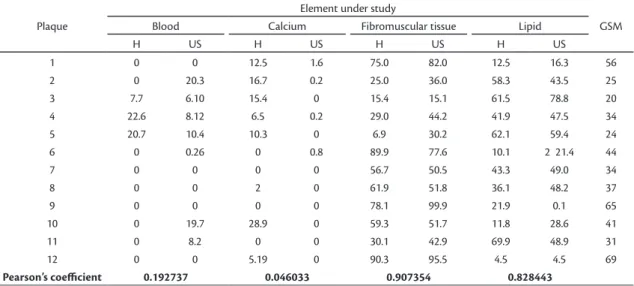
in the study. Table 2 shows the percentages of each component of the atheroma according to the computer analysis and to histology. The correlations
of fat and ibromuscular tissue were signiicant:
R=0.82 and R=0.9. In contrast, calcium and blood
had no signiicant correlations (R=0.04 and R=0.19).
GSM values ranged from 20 to 69 and were in agreement with plaque characteristics in both USVH and conventional histology, as the plaques with a
low GSM were classiied as more lipidic. Mean
GSM of asymptomatic patients was 52.3, and of symptomatic patients, 33.9 (p=0.122); these values are in agreement with those reported in other studies,
material should be classiied and included in future
studies.
Another limitation was the fact that histological analyses were conducted using plaque cross-sections of a micrometric thickness; therefore, a full evaluation of the plaque components is not possible, and only a “picture” of it at a certain point can be obtained. We tried to mitigate this limitation by choosing the point with the best correspondence with the histological section on the ultrasound image. If the entire plaque were analyzed on the longitudinal image, or even on the cross-sectional view of another point in the plaque, results would not have a similarly positive correlation. Therefore, questions are raised about whether histology should remain the criterion standard, or whether the ideal investigation should use the dimensional volume, as in the three-dimensional reconstruction of imaging studies (CT scans).
Currently, an important discussion seems to be which method will be more common in the future. Math values calculated using data about echogenicity of each plaque pixel, such as the grey scale median (GSM), is the technique most often used currently. Other math parameters, such as sample heterogeneity, pixel distribution along a line and the standard deviation of values distributed in a histogram, may also be objectively correlated with
clinical indings and provide information about the
prognosis of patients with carotid disease19,27,28,29. This study also found a possible correspondence between GSM and fat content (Table 2), as plaques Knowing what the lipid content of the atheroma is, as
well as the location of the lipid core and the thickness
of the ibrous cap, may help to make decisions about
the best treatment for asymptomatic patients with stenosis of 70% to 80% of the arterial lumen25, which is one of the topics of debate when discussing current surgical indications. It may also support decisions about which technique (open surgery or angioplasty) to use when treating patients with critical stenosis, as lesions with a higher degree of stenosis and atheromas with a higher lipid content have a greater potential to result in cerebral embolization during angioplasty, even when using cerebral protective devices, such
as ilters, which require that the lesion be crossed
before release. In such cases, the recommendation is to use endarterectomy or cerebral protective devices
with proximal blockage or low reversion through
the internal carotid artery26. Another potential use of in vivo plaque histology is the development of cardiovascular risk tables, which may be used in the
same way as the intima-media thickness, speciically
for the risk of stroke25.
One of the limitations of this study was the fact that the classification developed by Lal et al.20 does not evaluate all the histological structures, and according to the software, there are grey scale “default” intervals that are not assigned to any structures. Most plaques had up to 50% of unclassified material. Histology found a large amount of amorphous material (proteoglycans and
ibrin), which may justify those USVH indings. It
is important to keep in mind that this amorphous
Table 2. Atheroma content (%) according to histological (H) test and the computer-assisted ultrasound (US) exam.
Plaque
Element under study
GSM
Blood Calcium Fibromuscular tissue Lipid
H US H US H US H US
1 0 0 12.5 1.6 75.0 82.0 12.5 16.3 56
2 0 20.3 16.7 0.2 25.0 36.0 58.3 43.5 25
3 7.7 6.10 15.4 0 15.4 15.1 61.5 78.8 20
4 22.6 8.12 6.5 0.2 29.0 44.2 41.9 47.5 34
5 20.7 10.4 10.3 0 6.9 30.2 62.1 59.4 24
6 0 0.26 0 0.8 89.9 77.6 10.1 2 21.4 44
7 0 0 0 0 56.7 50.5 43.3 49.0 34
8 0 0 2 0 61.9 51.8 36.1 48.2 37
9 0 0 0 0 78.1 99.9 21.9 0.1 65
10 0 19.7 28.9 0 59.3 51.7 11.8 28.6 41
11 0 8.2 0 0 30.1 42.9 69.9 48.9 31
12 0 0 5.19 0 90.3 95.5 4.5 4.5 69
Pearson’s coeicient 0.192737 0.046033 0.907354 0.828443
leave in the image. Moreover, calcium produces an acoustic shadow on the US image, which prevents the accurate evaluation of the plaque contents30.
The possibility of using artiicial intelligence
to study the atheroma may facilitate the routine activities of attending physicians. Computers may conduct fully automated analyses, such as the detection and analysis of the artery and the atheroma, or physicians may choose the area of interest and leave the analysis for the computer (semi-automated analysis). This method accelerates the performance and interpretation of the exam, and the histological analysis may become a routine part of exams that evaluate the degree of artery stenosis. Currently, virtual histology is conducted using the
post-processing of stored images, but speciic software
may be included in US scanners already available in the market. Another important area for the dissemination of this method is the standardization of the exam itself and its teaching by associations
involved in the qualiication of vascular ultrasound
specialists.
Several other methods to study the content and the activity of the atheroma have been developed, such
as CT, PET-CT, MRI and scintigraphy using cell
markers3, but US will deinitely have a fundamental role in patient screening because it is easy to use and widely available.
Finally, the computer program used in this study had a few problems and stopped working some
times during the studies. Moreover the classiication
presented here does not apply to all tissues found in an atheromatous plaque. Therefore, there is room for improvement and development of a more user-friendly interface and better discrimination of plaque components. In the medical area, several authors have been working towards the improvement of this technique for the analysis of atheromas, venous thrombi, edema, renal parenchyma and thrombi in the aneurysmal sac to follow up patients after intraluminal corrections of aortic aneurysms31-33.
CONCLUSIONS
There was a correlation between in vivo noninvasive USVH of the carotid plaque and postoperative histology to detect lipids and fibromuscular tissues; there were no significant
correlations in the classiication of blood or calcium.
A more detailed color scale than the one used in this
study, as well as a deinition of standards for USVH
and histological sections, should be developed for additional studies and for the practical use of with lower GSMs were richer in lipids (less
echogenic), which suggests that the histological analysis was actually correct and had the potential to provide more information than the GSM alone. Further studies should be conducted to evaluate the clinical values of USVH in comparison with GSM.
The colorization of the atheroma components, shown in the grey scale B-mode image, may potentially provide a better illustration of the intraplaque disease for the attending physician, whether clinician or surgeon, than the tests currently used, and might facilitate the detection of the lipidic
or necrotic core, the thickness of the ibrous cap and
the areas of hemorrhage. Advances in computer software have also provided opportunities for three-dimensional studies of plaques and their volume, and may potentially provide evaluations of the entire plaque and detect areas of greater interest.
One of the important method limitations was the lack of perfect correlation between ultrasound findings and plaque histology, as ultrasound is performed in vivo and the images are recorded in longitudinal sections that may or may not provide a view of the worst segment of the plaque. Moreover, the atheromatous plaques are often broken and fragmented during surgery. Additionally, in vivo images are taken with a pressurized lumen and the ex vivo histological specimen is analyzed without any luminal pressure expanding the vessel endothelium. Also, a large amount of the soft lipid content is eliminated during surgical manipulation. For the ex vivo histological analysis, the plaque should be
ixed in formaldehyde, decalciied, ixed in parafin,
stained with dyes that remove lipid contents, and prepared as very thin cross-sections. Because of that, several errors are introduced both in the removal and the preparation of the plaque and in the direction of the section under analysis. Lovett et al.13 suggested that the histological study of atheromas should be standardized. In this study, attempts were made to minimize these errors by removing the plaque with as minimal rupture as possible and using the image of the histological slide as the standard reference, choosing the area on the ultrasound image that had the same relation between the lumen and the wall, and studying a section of this region instead of the entire plaque. Therefore, the correlation of lipid
contents and ibromuscular tissue in histology and
USVH analysis was improved.
Both calcium contents and blood in the atheroma
are dificult to evaluate because they are eliminated
14. Wintermark M, Jawadi SS, Rapp JH, et al. High-resolution CT imaging of carotid artery atherosclerotic plaques. Am J Neuroradiol. 2008; 29:875-82. PMid:18272562. http://dx.doi. org/10.3174/ajnr.A0950
15. Farooq MU, Khasnis A, Majid A, Kassab MY. The role of optical coherence tomography in vascular medicine. Vasc Med. 2009; 14:63-71. PMid:19144781. http://dx.doi. org/10.1177/1358863X08095153
16. Sakalihasan N, Michel JB. Functional imaging of atherosclerosis to advance vascular biology. Eur J Vasc Endovasc Sug. 2009; 37:728-34. PMid:19232504.http://dx.doi.org/10.1016/j.ejvs.2008.12.024
17. Gao P, Chen ZQ, Bao YH, Jiao LQ, Ling F. Correlation between carotid intraplaque hemorrhage and clinical symptoms: systematic review of observational studies. Stroke. 2007; 38:2382-90. PMid:17600232. http://dx.doi.org/10.1161/STROKEAHA.107.482760
18. EI-Barghouty N, Geroulakos G, Nicolaides A, Androulakis A, Bahai Nicolaides V. Computer-Assisted Carotid Plaque Characterisation. Eur J Vasc Endovasc Surg. 1995;9:389-95. http:// dx.doi.org/10.1016/S1078-5884(05)80005-X
19. Grønholdt MLM, Nordestgaard BG, Schroeder TV, Vorstrup S, Sillensen H. Ultrasonic Echolucent Carotid Plaques Predict Future Strokes. Circulation. 2001;104:68-3. PMid:11435340. http://dx.doi. org/10.1161/hc2601.091704
20. Lal BK, Hobson RW, Pappas PJ, et al. Pixel distribution analysis of B-mode ultrasound scan images predicts histologic features of atherosclerotic carotid plaques. J Vasc Surg. 2002;35:1210-7. PMid:12042733. http://dx.doi.org/10.1067/mva.2002.122888
21. Menezes FH, Luccas GC, Matsui I, Santos ACOQ, Silveira SAF. Avaliação através da ultra-sonograia duplex de reestenose da carótida interna dos pacientes submetidos à endarterectomia aberta de bifurcação carotídea, com eversão parcial da carótida interna. J Vasc Br. 2005;4(1):47-4.
22. Tan TW, Weyman AK, Barkhordarian S, Patterson RB. Single center experience with modified eversion carotid endarterectomy.
Ann Vasc Surg. 2011;25:87-93. PMid:21172583.http://dx.doi.
org/10.1016/j.avsg.2010.11.004
23. García-García HM, Gogas BD, Serruys PW, Bruining N. IVUS-based imaging modalities for tissue characterization: similarities and diferences. Int J Cardiovasc Imaging. 2011;27(2):215-24. PMid:21327914 PMCid:PMC3078312. http://dx.doi.org/10.1007/ s10554-010-9789-7
24. Mathiesen EB, Bønaa KH, Joakimsen O. Echolucent Plaques Are Associated With High Risk of Ischemic Cerebrovascular Events in Carotid Stenosis : he Tromsø Study. Circulation. 2001;103:2171-5. PMid:11331258. http://dx.doi.org/10.1161/01.CIR.103.17.2171 25. Pedro LM, Fernandes JF, Pedro MM, et al. Ultrasonographic Risk
Score of Carotid Plaques. Eur J Vasc Endovasc Surg. 2002;24:492-8. PMid:12443743. http://dx.doi.org/10.1053/ejvs.2002.1766
26. Biasi GM, Froio A, Diethrich EB, et al. Carotid plaque echolucency increases the risk of stroke in carotid stenting. he Imaging In Carotid Angioplasty And Risk Of Stroke (ICAROS) study. Circulation. 2004;110:756-62. PMid:15277320. http://dx.doi. org/10.1161/01.CIR.0000138103.91187.E3
27. Kakkos SK, Stevens JM, Nicolaides AM, et al. Texture analysis of ultrasonic images of symptomatic carotid plaques can identify those plaques associated with ipsilateral embolic brain infarction. Eur J Vasc Endovasc Surg. 2007;33:422-9. PMid:17161964. http:// dx.doi.org/10.1016/j.ejvs.2006.10.018
28. Russell DA, Wijeyaratne SM, Gough MJ. Changes in carotid plaque echomorphology with time since a neurologic event. J Vasc Surg. 2007;45:367-72. PMid:17264018. http://dx.doi.org/10.1016/j. jvs.2006.09.048
USVH in estimating stroke risk during endovascular procedures and in patient follow-up.
REFERENCES
1. Kwee RM, Van Oostenbrugge RJ, Hofstra L, et al. Indentifying vulnerable carotid plaques by noninvasive imaging. Neurology. 2008;70:2401-9. PMid:18541873. http://dx.doi.org/10.1212/01. wnl.0000314697.76580.cb
2. U-King-Im JM, Young V, Gillard JH. Carotid-artery imaging in the diagnosis and management of patients at risk of stroke. Lancet Neurol. 2009;8:569-80. http://dx.doi.org/10.1016/ S1474-4422(09)70092-4
3. Hermus L, Van Dam GM, Zeebregts CJ. Advanced carotid plaque imaging. Eur J Vasc Endovasc Sug. 2010;39:125-33. PMid:20031452. http://dx.doi.org/10.1016/j.ejvs.2009.11.020
4. Baroncini LAV, Pazin A Fº, Junior LOM, et al. Ultrasonic tissue characterization of vulnerable carotid plaque: correlation between video densitometric method and histological examination. Cardiovasc Ultrasound. 2006;4:32. PMid:16914059 PMCid:PMC1562449. http://dx.doi.org/10.1186/1476-7120-4-32
5. Swinjndregt ADM, Elbers HRJ, Moll FL, Letter J, Ackerstaf RGA. Ultrasonographic characterization of carotid plaques. Ultrasound Med Biol. 1998;24(4):489-93. http://dx.doi.org/10.1016/ S0301-5629(98)00005-2
6. Gao T, Zhang Z, Yu W, Zhang Z, Wang Y. Atherosclerotic carotid vulnerable plaques and subsequent stroke: a high-resolution MRI study. Cerebrovasc Dis. 2009;27:345-52. PMid:19218800 PMCid:PMC2814027. http://dx.doi.org/10.1159/000202011
7. Malgor RD, Wood EA, Lavarone OA, Labropoulos N. Stratifying risk: asymptomatic carotid disease. J Vasc Bras. 2012;11(1):43-52.
8. Executive Committee for the Asymptomatic Carotid Atherosclerosis Study. Endarterectomy for Asymptomatic Carotid Artery Stenosis. JAMA. 1995;273:1421-8. PMid:7723155. http:// dx.doi.org/10.1001/jama.1995.03520420037035
9. Barnett HJM, Taylor DW, Eliasziw M, et al. Beneit of carotid endarterectomy in patients with symptomatic moderate or severe stenosis. N Engl J Med. 1998;339:415-25. PMid:9811916. http:// dx.doi.org/10.1056/NEJM199811123392002
10. European Carotid Surgery Trialists’ Collaborative Group. Randomised trial of endarterectomy of recently symptomatic carotid stenosis: inal results of the MRC European Carotid Surgery Trial (ECST). Lancet. 1998;351:1379-87. http://dx.doi.org/10.1016/ S0140-6736(97)09292-1
11. MRC Asymptomatic Carotid Surgery Trial (ACST) Collaborative Group. Prevention of disabling and fatal strokes by successful carotid endarterectomy in patients without recent neurological symptoms: randomised controlled trial. Lancet. 2004;363:1491-502. http://dx.doi.org/10.1016/S0140-6736(04)16146-1
12. Mantese VA, Timaran CH, Chiu D, Begg RJ, Brott TG, for the CREST Investigators. The carotid revascularization endarterectomy versus stenting trial (CRE ST). Stroke. 2010;41:S31-4. PMid:20876500 PMCid:PMC3058352. http://dx.doi.org/10.1161/ STROKEAHA.110.595330
Correspondence
Fábio Hüsemann Menezes Rua Deusdeti Martins Gomes, 122 CEP 13084-723 - Campinas (SP), Brazil Fone: (19) 35219450 / Fax: (19) 32880202 E-mail: [email protected]
Author information
FHM is assistant professor of Vascular Diseases, Department of Surgery, School of Medical Sciences, Universidade Estadual de Campinas (UNICAMP). TCS is medical student, School of Medical Sciences, Universidade Estadual de Campinas (UNICAMP). SAFS is PhD degree from Universidade Estadual de Campinas, vascular ultrasonographist and vascular physician. SXSC is PhD in radiology and vascular ultrasonography technologist, independent vascular ultrasonography consultant. KM is assistant professor, Department of Pathological Anatomy, School of Medical Sciences, Universidade Estadual de Campinas (UNICAMP). ASCM is chief pathologist, Laboratório de Patologia Menezes.
Author’s contributions*
Conception and design: FHM, TCS, SXSC, SAFS, KM Analysis and interpretation: FHM, TCS, SXSC Data collection: FHM, TCS, KM, ASCM Writing the article: TCS, FHM, SXSC Critical revision of the article: FHM, TCS, SXSC, SAFS, KM, ASCM Final approval of the article**: FHM, TCS, SXSC, SAFS, KM, ASCM Statistical analysis: N/A Overall responsibility: FHM, TCS Obtained funding: he author hiago do Carmo Silveira eceived an undergraduate student scientiic research grant from PIBIC/CNPq
*TCS and FHM share the same authorship responsibilities in the study. **All authors should have read and approved of the inal version of
the article submitted to J Vasc Bras.
29. Mathiesen EB, Johnsen SH, Wilsgaard T, Bønaa KH, Løchen ML, Njølstad I. Carotid plaque area and intima-media thickness in prediction of irst-ever ischemic Sstroke: A 10-year
follow-up of 6584 men and women: The Tromsø Study. Stroke.
2011;42:972-8. PMid:21311059. http://dx.doi.org/10.1161/ STROKEAHA.110.589754
30. Gray-Weale AC, Graham JC, Burnett JR, et al. Carotid artery atheroma: comparison of preoperative B-mode ultrasound appearance with carotid endarterectomy specimen pathology. J Cardiovasc Surg. 1988;29:676-81.
31. Cassou-Birckholz MF, Engelhorn CA, Salles-Cunha SX, et al. Assessment of deep venous thrombosis by grayscale median analysis of ultrasound images. Ultrasound Q. 2011;27(1):55-61. PMid:21343802. http://dx.doi.org/10.1097/ RUQ.0b013e31820e157d
32. Valiente Engelhorn AL, Engelhorn CA, Salles-Cunha SX, Ehlert R, Akiyoshi FK, Assad KW. Ultrasound tissue characterization of the normal kidney. Ultrasound Q. 2012;28(4):275-80. PMid:23149511. http://dx.doi.org/10.1097/RUQ.0b013e318276f12b