Mucoepidermoid carcinoma of the trachea mimicking asthma*
Carcinoma mucoepidermoide da traqueia mimetizando asma brônquica
Ricardo Kalaf Mussi, Ivan Felizardo Contrera Toro, Mônica Corso Pereira
Abstract
In cases of recent asthma in which clinical control with the usual treatment (corticosteroids and bronchodilator) is unsatisfactory, it is important to consider other diagnoses, such as congestive heart failure, gastroesophageal reflux or other forms of airway obstruction. We report the case of a female patient with mucoepidermoid carcinoma of the trachea mimicking asthma. The patient presented cough and wheezing, as well as abnormal spirometry results with an obstructive pattern that was responsive to bronchodilators. One year later, the patient presented clinical and spirometric worsening. The chest X-ray revealed no abnormalities. A CT scan showed a vegetative lesion obstructing the tracheal lumen and located 1 cm from the carina. Fiberoptic bronchoscopy showed a finding similar to a bronchial carcinoid tumor. The anatomopathological diagnosis made after surgical resection was low-grade mucoepidermoid carcinoma, without lymph node involvement. Although the flow-volume curve was not suggestive of upper airway obstruction, the spirometry performed after the surgery showed a significant reduction in the degree of obstruction and greater reversibility after bronchodilator use. There was no evidence of recurrence of the disease or of the symptoms after a two-year follow-up period.
Keywords: Bronchial hyperreactivity; Carcinoma, mucoepidermoid; Trachea; Asthma; Lung diseases, obstructive.
Resumo
Em casos de asma de início recente em que o controle clínico com tratamento habitual (corticosteroide e bronco-dilatador) é insatisfatório, é importante considerar outros diagnósticos, tais como insuficiência cardíaca congestiva, refluxo gastroesofágico ou outras formas de obstrução das vias aéreas. Relatamos o caso de uma paciente do sexo feminino com carcinoma mucoepidermoide da traqueia mimetizando um quadro de asma brônquica. A paciente apresentava tosse e sibilância, bem como espirometria anormal com padrão obstrutivo responsivo a broncodila-tador. Após um ano, apresentou deterioração clínica e espirométrica. Nenhuma anormalidade foi encontrada no radiograma de tórax. A TC revelou lesão vegetativa, a 1 cm da carina, reduzindo a luz traqueal. A fibrobroncoscopia mostrou imagem semelhante a tumor carcinoide brônquico. O diagnóstico anatomopatológico após a ressecção cirúrgica foi carcinoma mucoepidermoide de baixo grau, sem envolvimento linfonodal. Embora a curva fluxo-volume não fosse sugestiva de obstrução de vias aéreas superiores, a espirometria realizada após a cirurgia mostrou redução significativa do grau de obstrução e maior reversibilidade com broncodilatador. Não houve evidência de recidiva da doença ou retorno dos sintomas após dois anos de seguimento.
Descritores: Hiper-reatividade brônquica; Carcinoma mucoepidermoide; Traqueia; Asma; Pneumopatias obstrutivas.
* Study carried out in the Department of Thoracic Surgery of the Universidade Estadual de Campinas – Unicamp, State University at Campinas – Campinas, Brazil.
Correspondence to: Ricardo Kalaf Mussi. Rua Copaíba, 810, Jardim Miriam, CEP 13098-347, Campinas, SP, Brasil. Tel 55 19 3521-9441. E-mail: [email protected]
Financial support: None.
Submitted: 19 October 2007. Accepted, after review: 26 June 2008.
Introduction
Asthma is an affliction that typically appears in childhood. Late onset is less common within the context of the natural history of asthma and should therefore serve to alert physicians to the possibility of differential diagnoses such as heart failure, gastroesophageal reflux and other forms of airway obstruction.
despite receiving appropriate treatment, the patient presented clinical worsening. An addi-tional spirometry test revealed worsening of the obstruction, which was no longer responsive to bronchodilator use (Table 1 and Figure 1b).
We decided to investigate concomitant conditions that might be responsible for the worsening of the clinical profile. A CT scan of the chest showed a vegetative lesion of the trachea, located 1.0 cm from the carina, resulting in significant luminal narrowing (Figure 2). Fiberoptic bronchoscopy revealed a violaceous endobronchial lesion, with well-defined borders and a smooth surface, lodged in the right wall of the trachea and obstructing 90% of the lumen. No biopsy was performed.
The patient was submitted to a right poste-rolateral thoracotomy and resection of 2.5 cm of the trachea, followed by end-to-end anasto-mosis and lymph node drainage. In the frozen section analysis, performed under microscopy, the margins were unaffected.
There were no postoperative complications, and the patient was discharged on postadmis-sion day 7.
carcinoma (MEC). This rare tumor accounts for 0.1-0.2% of lung neoplasms and was previous classified as a bronchial adenoma. However, the World Health Organization currently classifies MEC as an entity distinct from other
carci-nomas of the airways.(1) Individuals of any age
can present MEC,(2) which typically presents as
symptoms of upper airway irritation, such as cough, hemoptysis, atelectasis and
postobstruc-tive pneumonia.(3)
Case report
A 68-year-old female patient presented with a four-year history of cough, wheezing and dyspnea attacks, with progressive worsening. Initially diagnosed as having asthma, the patient presented a satisfactory response to the use of bronchodilators and systemic corticosteroids. The patient described herself as a nonsmoker with no history of lung disease. The chest X-ray was normal. At one year after symptom onset, the patient underwent spirometry, which revealed moderate obstructive lung disease that was responsive to bronchodilator use (Table 1 and Figure 1a). In the two years that followed,
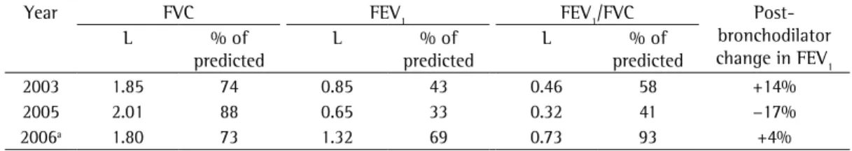
Table 1 - Spirometry findings over the course of the case investigated.
Year FVC FEV1 FEV1/FVC
Post-bronchodilator change in FEV1
L % of
predicted
L % of
predicted
L % of
predicted
2003 1.85 74 0.85 43 0.46 58 +14%
2005 2.01 88 0.65 33 0.32 41 −17%
2006ª 1.80 73 1.32 69 0.73 93 +4%
ªAfter surgery.
Figure 1 - Flow-volume curves.
c b
a
2003 2005 2006
2 2 2
2 00 44 22 44 0
0 00
4 4 4
4
4 4
4 4
-4 -4 -4
-4
to hypoxia or hypercapnia. Wheezing observed during the physical examination is common in individuals with asthma, as well as in those with COPD. Audible wheezing in only one hemithorax or that alters when the patient changes position is indicative of upper airway obstruction, as is evidence of stridor or noisy respiration.
The patient described here, in addition to presenting none of these classical findings suggestive of upper airway obstruction, had a history of asthma, which was a confounding factor. However, the progressive reduction in the response to treatment and the change in the pattern of response to bronchodilator admin-istration during spirometry encouraged us to investigate the differential diagnoses.
Although chest X-ray is the first-line radio-logical test in the evaluation of patients with symptoms of airway obstruction, the results are
rarely conclusive.(4) When there is suspicion of
upper airway damage, a CT scan of the chest or, if necessary, fiberoptic bronchoscopy can be
performed.(5)
In the case presented here, the initial spirom-etry findings indicated moderate obstructive lung disease that was responsive to bronchodilator use (Table 1 and Figure 1a). Due to worsening of the condition of the patient, we performed a second spirometry test, which revealed obstructive lung disease that was more accentuated than in the previous test and no longer responded to the bronchodilator (Table 1 and Figure 1b).
The results of the first spirometry test could easily have belonged either to a patient with asthma or to a COPD patient with a certain degree de bronchial hyperreactivity, whereas the second was suggestive of an individual with an obstruc-tive lung disease presenting low reversibility. In a retrospective analysis of the third spirometry, performed six months after the surgery (Table 1 and Figure 1c), it became clear that the func-tional alteration detected actually translated to obstruction of the upper airways and not to diffuse disease affecting lower airways. In that test, the values were near normality, and the
VEF1 was more than double that observed in the
previous test (Table 1; Figures 1b and 1c). In upper airway obstruction, the impair-ment can be extrathoracic (pharynx, larynx and extrathoracic trachea) or intrathoracic (trachea and main bronchi). The effect of the anatomical lesions or functional abnormalities depends on The anatomopathological study indicated
a well-differentiated, low-grade MEC, without mediastinal lymph node involvement.
No complementary treatments were admin-istered. Weekly endoscopic and tomographic evaluations showed that there was no recurrence during the two-year follow-up period.
Spirometry tests performed at six months after the operation revealed regression of the obstructive profile (Table 1 and Figure 1c).
Discussion
The diagnosis of asthma is made almost exclusively on the basis of clinical findings, and the natural history of the disease includes onset during childhood, remission in adolescence and the return of symptoms in adulthood. However, in some patients, the first clinical manifesta-tions can appear after the fifth decade of life. In such cases, one should consider the differential diagnoses, such as COPD, heart failure, pulmo-nary thromboembolism and gastroesophageal reflux. In addition, if the recommended treat-ments (bronchodilators, inhaled corticosteroids and environmental prophylaxis) are not effica-cious in controlling the symptoms, it becomes imperative to expand the investigation.
Signs and symptoms of upper airway obstruction are not always evident. In tracheal obstruction, dyspnea on exertion appears only when the condition is well advanced and the diameter of the tracheal lumen has been reduced to < 8 mm. In this situation, it is likely that the dyspnea is attributable more to the increased effort required to bring air into the lungs than
it can, when centrally located, be visualized via
fiberoptic bronchoscopy.(9) In this situation, a
lesion can produce symptoms of upper airway
irritation, thereby mimicking asthma.(3,10) A CT
scan of the chest is useful when the tumor is not visible through endoscopy, as well as for the
staging of intrathoracic tumors.(11)
The clinical course of MEC correlates with the histological grade. The five-year survival rate among individuals diagnosed with high-grade tumors is 31%. Low-grade tumors present local-ized growth, rarely affect the lymph nodes and are easily resected. Lymph node involvement has been reported to be an indicator of a worse
prognosis.(1) Complete resection is the
treat-ment of choice for MEC, and five-year survival
after resection can be as high as 80%.(1) The
roles played by radiotherapy and chemotherapy, before or after surgery, have yet to be well estab-lished.(3)
In summary, atypical asthma profiles presenting an unsatisfactory response to treat-ment should be thoroughly investigated, since they can mask other, occasionally rare, diseases. Spirometry findings, the flow-volume curve in particular, constitute a useful tool in making the differential diagnosis, assuming that such find-ings are well understood and carefully evaluated. Nevertheless, simply expanding the investigation can often lead to a definitive diagnosis.
References
1. Vadasz P, Egervary M. Mucoepidermoid bronchial tumors: a review of 34 operated cases. Eur J Cardiothorac Surg. 2000;17(5):566-9.
2. Heitmiller RF, Mathisen DJ, Ferry JA, Mark EJ, Grillo HC. Mucoepidermoid lung tumors. Ann Thorac Surg. 1989;47(3):394-9.
3. Noda S, Sundaresan S, Mendeloff EN. Tracheal mucoepidermoid carcinoma in a 7-year-old child. Ann Thorac Surg. 1998;66(3):928-9.
4. Baldi BG, Fernandes CJ, Salge JM, Takagaki TY. Tracheal polyp. J Bras Pneumol. 2007;33(5):616-20.
5. Chen F, Tatsumi A, Miyamoto Y. Successful treatment of mucoepidermoid carcinoma of the carina. Ann Thorac Surg. 2001;71(1):366-8.
6. Ernst A, Feller-Kopman D, Becker HD, Mehta AC. Central airway obstruction. Am J Respir Crit Care Med. 2004;169(12):1278-97.
7. Sociedade Brasileira de Pneumologia e Tisiologia. Diretrizes para Testes de Função Pulmonar. J Pneumol. 2002;28(Suppl 2):S2-S238.
8. Pellegrino R, Viegi G, Brusasco V, Crapo RO, Burgos F, Casaburi R, et al. Interpretative strategies for lung function tests. Eur Respir J. 2005;26(5):948-68. 9. Devbhandari M, Stamenkovic S, Walker W, Cameron E.
Unusual presentation of mucoepidermoid carcinoma
their location, on the type of obstruction (fixed or variable) and on the extent of the damage.
Functional abnormalities can go unnoticed when the obstruction is minimal. However, as the obstruction increases, the more effort-de-pendent airflows present alterations. Tracheal obstruction that reduces the diameter of the lumen to less than 8 mm typically produces dyspnea on exertion. If the lumen diameter is reduced to less than 5 mm, dyspnea can occur
even at rest, and alterations in the FEV1 can be
seen.(6,7)
Although there is not always a reduction in
FEV1 or FVC, there is typically a pronounced
effect on PEF. However, this parameter is quite effort-dependent, which limits the weight ascribed to it.(8)
The flow-volume curve can be quite sugges-tive of upper airway obstruction, especially if there is a plateau in the inspiratory loop, with or without the same in the expiratory loop. This finding suggests obstruction of the central airway or of the upper airways. However, this variable cannot be reliable unless the test includes three reproducible maneuvers, ensuring that the patient has put forth the maximum effort and that the curve appears similar for all three maneuvers. In contrast, an FEF plateau (in at least three maneuvers) and the absence of a plateau in the forced inspiratory phase suggest variable obstruction of the central or upper airways. Plateaus of similar amplitude in the inspiratory and expiratory loops suggest fixed
obstruction of the central or upper airways.(7,8)
In extrathoracic upper airway obstruction, the maximal inspiratory flow is often reduced. In contrast, in intrathoracic upper airway obstruc-tion, there is little impairment of the maximal inspiratory flow, since the pressure surrounding this region (similar to the intrapleural pressure) is in strong opposition to the intraluminal pres-sure generated by inhalation, which limits the
effect that obstruction has on the airflows.(7,8)
The absence of typical and classical flow-volume curve findings, such as those described above, do not rule out the possibility of upper airway obstruction. In the case reported here, the flow-volume curves demonstrate that the typical findings of intrathoracic obstruction were not present (Figure 1).
11. Kim TS, Lee KS, Han J, Im JG, Seo JB, Kim JS, et al. Mucoepidermoid carcinoma of the tracheobronchial tree: radiographic and CT findings in 12 patients. Radiology. 1999;212(3):643-8.
with recurrent pulmonary embolism. Eur J Cardiothorac Surg. 2002;22(3):482-4.
10. Mehra PK, Woessner KM. Dyspnea, wheezing, and airways obstruction: is it asthma? Allergy Asthma Proc. 2005;26(4):319-22.
About the authors
Ricardo Kalaf Mussi
Attending Physician in the Department of Thoracic Surgery. Universidade Estadual de Campinas – Unicamp, State University at Campinas – Campinas, Brazil.
Ivan Felizardo Contrera Toro
Coordinator of the Department of Thoracic Surgery. Universidade Estadual de Campinas – Unicamp, State University at Campinas – Campinas, Brazil.
Mônica Corso Pereira